Digital Poster Session
Contrast Mechanisms Back to Program-at-a-Glance Back to Program-at-a-Glance
|
Wednesday, 15 May 2019
Digital PosterContrast Mechanisms
3955 -3979 Elastography & Contrast Agents
3980 -4004 CEST, MT & Beyond
4005 -4029 Fat, QSM & CEST |
| |
Elastography & Contrast Agents
Digital Poster
Contrast Mechanisms
Wednesday, 15 May 2019
| Exhibition Hall |
14:30 - 15:30 |
| |
|
Computer # |
|
3955.
 |
101 |
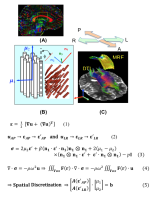
Multi-Excitation MRE in Aging Human Brain: Estimation of Direction-Dependent Mechanical Properties

Nicolas Gallo, Stacey Cahoon, Aaron Anderson, Assimina Pelegri, John Georgiadis
Healthy aging affects the local mechanical properties of the human brain tissue but their estimation remains a challenge, especially in strongly anisotropic white matter regions. This study merges previous MRE aging research involving high-resolution, full-coverage, and multi-excitation MRE imaging with the ability to estimate anisotropic shear moduli locally. We identified important anisotropic differences in the storage modulus in the corpus callosum, which help differentiate old from the young.
|
|
3956.
 |
102 |
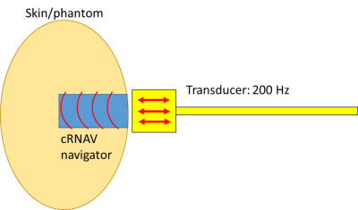
High-resolution one-dimensional shear wave MR elastography at 78 microns: initial results in skin imaging in vivo.
Marian Troelstra, Alessandro Polcaro, Peter Worsley, Torben Schneider, Jurgen Runge, Ralph Sinkus
Imaging the biomechanical properties of thin structures with MR is challenging due to the generally low spatial resolution of MR elastography. In this study, we discuss a novel approach for imaging these structures using high-resolution motion sensitised pencil beam MRE at 78 microns to determine transient shear wave propagation. Initial results describe the application of this approach in phantom and skin imaging.
|
|
3957.
 |
103 |
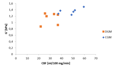
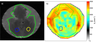
In vivo Assessment of Gray Matter Stiffness and Perfusion in the Human Brain
Siri Svensson, Grethe Løvland, Wibeke Nordhøy, Ralph Sinkus, Omar Darwish, José De Arcos, Kyrre Emblem
MR Elastography (MRE) is an emerging imaging technique used to quantify the stiffness and viscosity of tissue. Few studies have investigated the relationship between these biomechanical properties and brain perfusion. If understood, this could provide valuable new insight in diseases that alter biomechanical properties or functionality. In our study we assess the cerebral blood flow and the tissue stiffness of gray matter in healthy volunteers. We find that in cortical gray matter, both the cerebral blood flow and the shear storage modulus G’ was higher than in deep gray matter.
|
|
3958.
 |
104 |
Characterization of fibril aggregates in ex vivo rat brain with multi-frequency MR Elastography – Preliminary results
Mathilde Bigot, Fabien Chauveau, Ralph Sinkus, Olivier Beuf, Simon Lambert
Fibrils are biomarkers for early stages of dementia. In this work, α-synuclein fibrils were injected in rat striatum. Brains were imaged ex-vivo using multi-frequency MR Elastography. Estimation of the real part kr of the complex wave number was made for each studied frequency in ROIs surrounding the inclusion, a contralateral control injection and the whole brain. Exponent of the frequency power law was derived from kr maps acquired at different frequencies. No difference was observed between krvalues for the different ROIs, but the exponent was more important at the fibrils location, potentially indicating that multi-frequency MRE can detect fibrils.
|
|
3959.
 |
105 |
Real-time magnetic resonance elastography: application for monitoring lower leg muscle function during plantar flexion
Carsten Warmuth, Felix Schrank, Jürgen Braun, Ingolf Sack
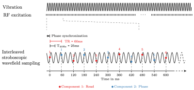
Real-time elastography is well established in ultrasound but has not yet been achieved in MRI. Real-time elastography would be desirable for studying processes that are not easily repeatable or reproducible such as skeletal muscle function. Therefore, we developed a new concept in magnetic resonance elastography (MRE) based on stroboscopic wave sampling and single-shot spiral-k-space acquisition to push temporal resolution of MRE towards real-time elastography. Application of the new method is demonstrated in the lower leg muscles of healthy volunteers.
|
|
3960.
 |
106 |
In-Vivo Quantification of Aortic Stiffness using a Multi-Slice Spin-Echo Echo-Planar Imaging Sequence: A Comparison to a Gradient-Recalled Echo Sequence
Huiming Dong, Ning Jin, Prateek Kalra, Richard White, Arunark Kolipaka
Aortic stiffness is an important biomarker of cardiovascular diseases. Magnetic Resonance Elastography (MRE) is a non-invasive tool for measuring in-vivo aortic stiffness. Gradient-Recalled Echo (GRE) MRE sequences are widely employed for aortic MRE. However, GRE MRE sequences are sensitive to T2* decay, leading to signal loss and lower Signal-to-Noise Ratio (SNR). In this work, a cardiac-gated Spin-Echo Echo-Planar Imaging (SE-EPI) MRE sequence was developed and validated against GRE MRE. Similar aortic stiffness was observed between the two techniques. Moreover, shorter scan time, higher first-harmonic amplitude, and Octahedral Shear Strain-Based SNR (OSS-SNR) were achieved using SE-EPI MRE (p<0.05).
|
|
3961
 |
107 |
The contribution of cells and extracellular matrix to liver viscoelasticity: Histological analysis combined with multifrequency MR elastography and diffusion weighted imaging in rat liver specimens
Video Permission Withheld
Ingolf Sack, Heiko Tzschätzsch, Baptiste Polchlopek, Paul Janmey, Jürgen Braun, Angela de Schellenberger
The effect of freezing/thawing at -20°C and -80°C on liver tissue was analyzed by diffusion-weighted imaging (DWI), multifrequency MR elastography (MRE) and histological methods. Freezing-induced deterioration of cell membranes, detachment of sinusoidal endothelial cells and disorganization of sinusoidal collagen together with reduced water diffusion and macroscopic shear modulus and affected tissue viscosity. Histological analysis revealed that collagen structures at the space of Disse are better preserved at -80°C than -20°C which was mirrored by tissue viscosity indicating the sensitivity of multifrequency MRE to microstructural changes of soft biological tissues.
|
|
3962.
 |
108 |
Analysis and Maximization of SNR in MR Elastography Inversion
Cemre Ariyurek, Bilal Tasdelen, Alireza Sadeghi-Tarakameh, Yusuf Ider, Ergin Atalar
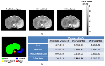
Reconstructed elastography maps suffer from noise due to derivative operations in the inversion algorithms. Considering the noise on the complex MRI signal, SNR of the reconstructed elastogram is derived analytically for k-MDEV inversion. Verified SNR derivations by Monte Carlo Simulations, an SNR-weighted k-MDEV inversion was proposed to maximize SNR of the elastogram. Quality of the proposed reconstruction was assessed by estimation error, noise performance and edge response analyses. SNR of the elasticity improved twice as the conventional method. Results on phantom and human liver data are also provided. As future work, this analysis will be performed for other inversion methods.
|
|
3963
 |
109 |
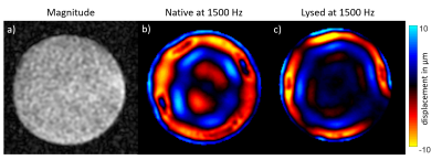
Tabletop MR Elastography (MRE): Preliminary Results Towards an Assessment of Frozen Tissue Bank Samples.
Video Permission Withheld
Rolf Reiter, Martina Guidetti, Marco Zampini, Shreyan Majumdar, Harish Palnitkar, Bernd Hamm, Thomas Royston, Dieter Klatt
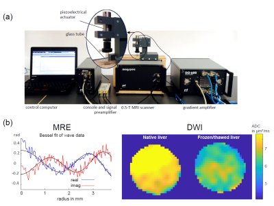
Tabletop magnetic resonance elastography (MRE) at 0.5 Tesla was recently introduced as a low-cost technique for assessing mechanical properties of small tissue samples. Frozen tissue banks potentially facilitate access to large amounts of specimens. Therefore, we aimed to characterize changes of mechanical properties of fresh ex vivo (native), and frozen and thawed (lysed) porcine muscle, liver and kidney specimens. Frequency dependent mechanical properties at 500, 1000, 1500 and 2000 Hz decreased uniformly after lysis (ratio lysed/native ranging from 0.46 to 0.60). Rheological fitting was more complex and motivates further investigation of the rheological behavior in native and lysed states.
|
|
3964.
 |
110 |
In Vivo Cardiac MR Elastography with a Gravitational Transducer
Ayse Sila Dokumaci, Torben Schneider, Daniel Fovargue, Emma Burnhope, Myrianthi Hadjicharalambous, Stefan-Heinz Hoelzl, Jelizaveta Sudakova, Adela Capilnasiu, Marian Troelstra, Sweta Sethi, Tevfik Ismail, Ralph Sinkus, David Nordsletten
MR Elastography is beneficial in measuring the stiffness values of tissues which can change in disease. Nevertheless, cardiac MR Elastography is not only challenging from the acquisition side but also from the reconstruction side because of the higher stiffness values expected in the cardiac muscle compared to the other organs in which MR Elastography is commonly applied. Here, we present data in healthy subjects and a patient acquired with the gravitational transducer with a novel synchronising strategy in combination with a single-shot SE-EPI-MRE sequence employing second-order motion compensated motion encoding gradients with and without ZOnal Oblique Multislice Imaging.
|
|
3965
 |
111 |
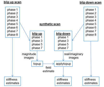
Blip-Up Blip-Down Correction for Head MR Elastography
Video Permission Withheld
Roger Grimm, Kevin Glaser, Richard Ehman
Geometric distortion in echo planar imaging-spin echo (EPI-SE) images caused by a non-uniform B0 field remains an issue in clinical imaging. In elastography, this geometric distortion leads to systematic errors in stiffness estimates. While blip-up blip-down correction is routinely used in diffusion imaging in the head, this correction is not used in MR elastography. The purpose of this work is to assess the effect of applying distortion correction on elastography scans and the resulting stiffness estimates. We show that the corrections provide reduced variability in stiffness maps with no additional acquisition time required.
|
|
3966.
 |
112 |
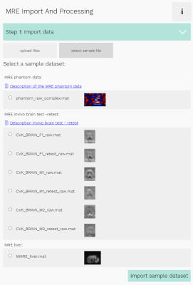
Online platform for extendable server-based processing of magnetic resonance elastography data.
Tom Meyer, Heiko Tzschätzsch, Jürgen Braun, Prateek Kalra, Arunark Kolipaka, Ingolf Sack
The variety of reconstruction algorithms throughout the MRE community makes it difficult to compare the validity and strengths of these methods. We propose an extendable online platform, combining multiple processing methods to facilitate a direct comparison. The platform is implemented as a Java web application, designed to run every command line program. Processing is done on the server, configured through the easy-to-use web-interface. Currently, state-of-the-art MRE reconstruction algorithms, multiple pre-processing methods, automated data import as well as sample data of phantom-MRE and in-vivo MRE are included. We aim at extension of the platform and an automation of adding new methods.
|
|
3967.
 |
113 |
Cluster based analysis of fMRE data: computing heterogeneous regions of visual activation through correlation analysis.
Reihaneh Forouhandehpour, Carlos Girard, Guillaume Gilbert, Kevin Whittingstall, Elijah Van Houten
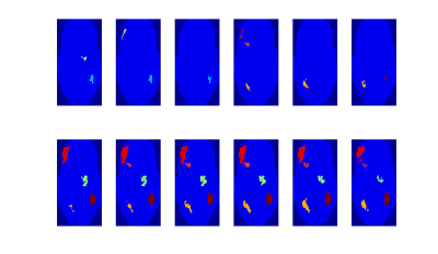
A method for more completely assessing the regions of activation in functional Magnetic Resonance Elastography is presented. This method allows the full size of the regions of activation to be estimated and also provides information on the region substructure. Results are presented in conjunction with Time of Flight cerebrovascular data, showing the relationship between arterial dilation, viscoelastic changes, and region substructure. This method has excellent potential to expand our understanding of mechanical and structural changes in the active brain.
|
|
3968
 |
114 |
Magnetic resonance shear wave elastography using transient acoustic radiation force excitations and sinusoidal displacement encoding
Video Permission Withheld
Lorne Hofstetter, Henrik Odéen, Bradley Bolster, Jr., Allison Payne, Dennis Parker
A novel elastography approach that uses sinusoidal motion encoding gradients to encode the displacement time-history following a single transient acoustic radiation force excitation is presented. This technique may be particularly well suited for performing elastography before, during, and after MR-guided focused ultrasound treatments since the same device used for therapy is used as an excitation source for elastography.
|
|
3969.
 |
115 |
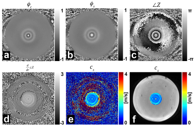
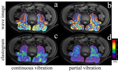
Partial Vibration MR Elastography: Zero-MR phase image MRE
Tomokazu Numano, Daiki Ito, Kazuyuki Mizuhara, Toshikatsu Washio, Tetsushi Habe, Keisuke Igarashi, Takamichi Ueki, Toshiki Maeno, Masaki Misawa, Naotaka Nitta
We developed a new technique for MR elastography (MRE) without using MR phase images (Zero-MR phase image MRE). A general MRE uses MR phase images as a wave image. Proposed technique was used MR magnitude images instead of MR phase images as a wave image by using the partial vibration. Proposed technique (partial vibration MRE technique) performance was comparable to that of a continuous vibration (conventional) MRE. Since partial vibration MRE need an only few second vibration, it could dramatically eliminated the patients' vibration-related discomfort.
|
|
3970.
 |
116 |
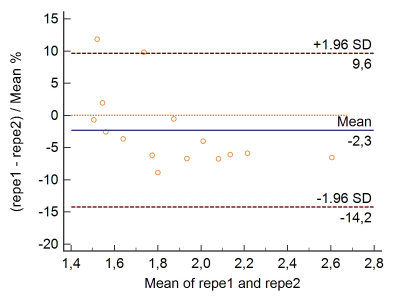
Effect of breath hold mode on the storage modulus of the liver in healthy individuals studied with a gravitational transducer at magnetic resonance elastography
Philippe Garteiser, Gwenael Page, Aimé-Pacifique Manzi, Valérie Vilgrain, Ralph Sinkus, Bernard Van Beers
Although MR elastography (MRE) is a preferred diagnostic tool for hepatic fibrosis, MRE-derived mechanical properties are affected by factors such as portal pressure. Since portal pressure is modulated across the breathing cycle, we sought to evaluate the effect of breathing condition by acquiring MRE in end expiration and end inspiration in 19 healthy volunteers. A gravitational transducer operating at 50Hz was used, which yielded a satisfactory repeatability. Storage modulus values were significantly higher at end inspiration than at end expiration, and the amplitude of the difference was significantly correlated to the end inspiration storage modulus values.
|
|
3971.
 |
117 |
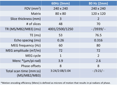
Fast Brain MR Elastography Using a Simultaneous Multislice EPI Acquisition on a Compact 3T Scanner
Yi Sui, Ziying Yin, Phillip Rossman, Matthew Murphy, Joshua Trzasko, Jonathan Scott, Kevin Glaser, Kiaran McGee, Matt Bernstein, Richard Ehman, John Huston III
In this study, we developed a fast MR elastography (MRE) pulse sequence using a simultaneous multislice (SMS)-EPI acquisition. The feasibility of SMS-EPI-MRE was demonstrated on a compact 3T scanner in a pilot volunteer study using 2 different head-coil arrays. Our results show that the overall scan time for 60-Hz 3-mm isotropic and 80-Hz 2-mm isotropic whole-brain MRE acquisitions were greatly reduced to 1-2 minutes (depending on the multiband acceleration) and 3:21 minutes, respectively. The wave images and stiffness maps from the SMS acquisitions were comparable to conventional MRE results.
|
|
3972.
 |
118 |
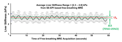
Repeatability and Clinical Performance of Non-gated, Free-breathing, MR Elastography (MRE) of the Liver
Jiahui Li, Bogdan Dzyubak, Kevin J. Glaser, Ziying Yin, Jun Chen, Alina Allen, Sudhakar K. Venkatesh, Armando Manduca, Vijay Shah, Richard L. Ehman, Meng Yin
While conventional MRE is easily performed during suspended respiration, a free breathing MRE technique would be advantageous in pediatric and other patients. We assessed the repeatability of free-breathing MRE in comparison to conventional breath held acquisitions in volunteers and the clinical performance of free-breathing MRE in a cohort of 56 patients with chronic liver diseases. Results demonstrated comparable repeatability and excellent agreement of the averaged liver stiffnesses measured using the two techniques. In summary, free-breathing MRE provides highly repeatable and accurate liver stiffness values.
|
|
3973.
 |
119 |
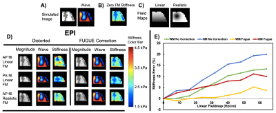
Quantitative Effects of Geometric Distortion On Brain Mechanical Property Estimation In Magnetic Resonance Elastography
Grace McIlvain, Matthew McGarry, Curtis Johnson
Geometric distortion in MRI arises from magnetic field-inhomogeneity and susceptibility differences at air/tissue/bone interfaces, and this distortion is greater at long readout times used in high-resolution imaging. Distortion may particularly impact magnetic resonance elastography (MRE), which calculates brain mechanical properties by imaging shear wave displacements then solving applying a nonlinear inversion algorithm. In this study, we systematically examine effects of distortion and correction algorithms on MRE data through a series of simulations and in vivo experiments, and find that geometric distortion limits the quantitative accuracy of brain MRE.
|
|
3974.
 |
120 |
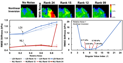
Low-Rank Denoising of Magnetic Resonance Elastography Images
Grace McIlvain, Ariel Hannum, Anthony Christodoulou, Matthew McGarry, Curtis Johnson
Magnetic resonance elastography (MRE) is a phase contrast-based MRI technology that can create whole brain mechanical property maps in vivo. MRE involves the solution of an inverse problem to estimate mechanical properties. Data noise degrades the accuracy of the recovered property images (elastograms). Most MRE acquisitions aim to achieve signal-to-noise ratio (SNR) above a certain threshold, though low SNR is a common issue. We propose a new method to denoise MRE data through spatiotemporal modeling. By approximating MRE data as low-rank, large improvements in SNR can be achieved, which can lead to the ability to salvage MRE data that otherwise would be unusable.
|
|
3975.
 |
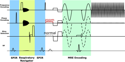
121 |
Multi-frequency magnetic resonance elastography with spiral readout, respiratory navigator and stroboscopic wave sampling for cardiac stiffness mapping with high spatial and temporal resolution.
Felix Schrank, Carsten Warmuth, Lars-Arne Schaafs, Heiko Tzschätzsch, Thomas Elgeti, Jürgen Braun, Ingolf Sack
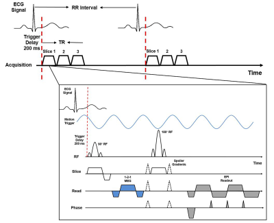
Cardiac magnetic resonance elastography (MRE) promises a quantitative mapping of the mechanical function of the myocardium. However, the implementation of cardiac MRE is challenging due to fast motion, high heterogeneity and temporally varying intrinsic properties of the heart. To overcome these limitations, we introduce a novel cardiac MRE sequence with spiral readout, respiratory navigator and stroboscopic wave sampling. It allows acquiring 100 wave fields with 45ms temporal resolution, 1.6x1.6mm² in-plane resolution at three frequencies within 9 minutes. Preliminary data in 8 healthy volunteers show a substantial variation of myocardial shear-wave velocity from 1.5±0.2m/s during diastole to 2.1±0.2m/s during systole.
|
|
3976.
 |
122 |
Correcting portal-venous partial-volume effects in dynamic contrast-enhanced MRI of the liver
Susmita Basak, David Longbotham, Ashley Guthrie, Daniel Wilson, Raj Prasad, Steven Sourbron
We investigated the effect of partial-volume corrections on the liver perfusion and function measured from DCE-MRI data using dual input two compartment uptake model. Following the convection principle in the human body, partial-volume error for hepatic portal vein is determined by taking the ratio of time integrals of venous and arterial input function. We found moderate correlation between the corrected and uncorrected venous plasma flow and a poor correlation between corrected and uncorrected extracellular volume and uptake rate. This indicates that partial-volume effect is significant and should be corrected for in the determination of perfusion and liver function.
|
|
3977.
 |
123 |
Magneto-Caloric Materials as Symmetric Reverse-Contrast Switchable MRI Labels
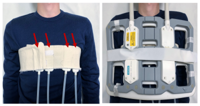
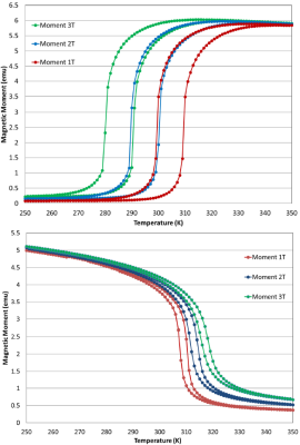
Mladen Barbic, Hatem ElBidweihy, Stephen Dodd, H Douglas Morris, Alan Koretsky
We describe the use of magneto-caloric materials as symmetric reverse-contrast switchable MRI labels at physiological temperatures. We present physical and temperature tunable MRI measurements on two different magneto-caloric materials, Iron-Rhodium (Fe-Rh) and Lanthanum-Iron-Silicon (La-Fe-Si) that both have sharp first-order magnetic phase transitions at the same bias DC magnetic field of 1 Tesla and physiological temperature of 37°C (310K), but with simultaneously positive and negative slope of magnetization vs. temperature, respectively, and therefore reverse image contrast in MRI. Thus, we show that different magneto-caloric materials provide an opportunity for the development of versatile multi-functional high differential contrast ratio switchable MRI labels.
|
|
3978.
 |
124 |
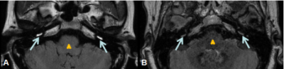
Labyrinthine lymph MR imaging of patients with vertigo disease: an imaging comparison study with intratympanic or intravenous gadolinium injection
Jinye Li, Lixin Sun, Hui Zhao, Weiqiang Dou, Jing Tian, Na Hu, Ruozhen Gong
In this study, we aimed to determine whether intratympanic (IT) or intravenous (IV) injection of gadolinium is more suitable for labyrinthine lymph imaging of patients with vertigo disease. The corresponding MR images with IV and IT injection were thus systematically compared. While comparable detection of endolymphatic hydrops were shown using both injection methods, higher image contrast between affected and unaffected sides of perilymph region were found using IV method than using IT method. We therefore, demonstrated that IV method can be further applied in clinical diagnosis for patients with vertigo disease.
|
|
3979.
 |
125 |
Effects of Multiple Contrast Agents in Multi-Modality Imaging for Liver Embolizations: A Preliminary MR Analysis
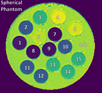
Carson Hoffman, Paul Laeseke, Oliver Wieben, Diego Hernando
Transarterial embolization (TAE) is a standard treatment for liver tumors and results in stasis and an accumulation of iodinated contrast agent in the liver. This study demonstrated that when iodinated contrast agents are present in MR, a T1 and T2 shortening effect can occur. We completed a preliminary analysis investigating pre- and post-TAE MR signal changes in the presence of iodine and gadolinium based contrast agents in-vivo (pilot swine study) and in phantoms. Understanding variations in MR images caused by contrast agents used for other modalities could help reduce errors in multimodality imaging studies.
|
|
| Top |
CEST, MT & Beyond
Digital Poster
Contrast Mechanisms
Wednesday, 15 May 2019
| Exhibition Hall |
14:30 - 15:30 |
| |
|
Computer # |
|
3980.
 |
126 |
Quantitative Magnetization Transfer: A Comparison of State-of-the-Art Acquisitions
Fritz Bayer, Peter Jezzard, Alex Smith
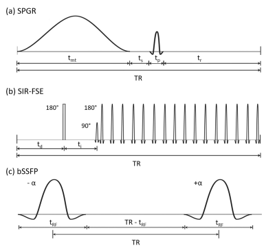
As development of quantitative magnetization transfer (qMT) has progressed, different sequences have been developed and independently optimised. Here, we present a comparison of the three main qMT acquisition sequences: spoiled gradient echo (SPGR), selective inversion recovery with fast spin echo (SIR-FSE) and balanced steady-state free precession (bSSFP). A novel comparison is accomplished by optimising each protocol using Cramér-Rao lower bounds (CRLB) theory. Our results indicate that SPGR qMT may introduce additional uncertainty due to the low sensitivity surrounding T2f, while bSSFP may provide a robust alternative for efficient 3D acquisitions.
|
|
3981.
 |
127 |
Optimizing Neuromelanin-sensitive Turbo Spin Echo sequences using the extended phase graph formalism including magnetization transfer effects
Rita Nunes, Rui P Teixeira, Shaihan Malik, Joseph Hajnal
Neuromelanin(NM)-sensitive imaging detects contrast differences due to magnetization transfer (MT) effects. We aimed to evaluate the value of adding preparation inversion pulses (IR) to multi-slice turbo spin echo (MS-TSE) sequences, using the recently introduced extended phase graphs with MT effects framework. Simulation results were in agreement with experimental phantom data. The MT contrast could be increased using IR MS-TSE sequences and these results were confirmed in vivo, with improved detectability of the substantia nigra. The next step will be to test the use of this type of sequences in the clinic, finding the optimal tradeoff between contrast and signal-to-noise ratio.
|
|
3982.
 |
128 |
Neural network for fast quantitative magnetization transfer imaging
Donghyun Kim, Jae-Woong Kim, Seung Hong Choi, Sung-Hong Park
Quantitative magnetization transfer (qMT) imaging provides quantitative information of macromolecular properties of tissues, but requires a long scan time. In this study, a neural network is proposed for the acceleration of qMT imaging. The network was trained to output the full 12 MT images (6 offset frequencies, 2 RF powers) from an input of only 4 MT images (2 offset frequencies, 2 RF powers). The qMT imaging with the neural network showed results comparable to those from the conventional qMT imaging with the full 12 MT images, indicating reduction in scan time by a factor of 3.
|
|
3983.
 |
129 |
Estimation of Semi-solid Saturation in Brain White Matter after Composite MT Pulses by Means of Bloch Simulation
Yicun Wang, Peter van Gelderen, Jeff Duyn
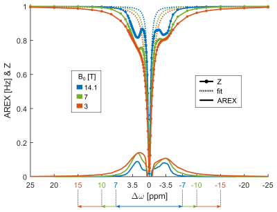
The magnetization level of macromolecular protons (MPs) affects that of water protons through magnetization transfer, a phenomenon that represents a confounding factor in quantitative MRI. Here we investigated whether MP spectral lineshape can be sufficiently accurately modelled by a single Lorentzian, which would facilitate estimating MP magnetization and saturation levels from Bloch simulations and a 2-pool MT model. We compared simulated effects of 10 different composite MT pulses and experimental results from 11 healthy volunteers at 7T. Results demonstrate a good fitting to the observed water signal evolution. Deviation of the simulated initial MP saturation from calculated levels was observed, indicating potentially unaccounted factors such as inhomogeneous MT effects.
|
|
3984.
 |
130 |
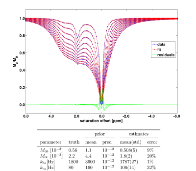
Bayesian fitting of Chemical Exchange Saturation Transfer (CEST) imaging data with an analytical model for multiple CEST pools
Aaron Kujawa, Eleni Demetriou, Mina Kim, Moritz Zaiss, Xavier Golay
A Bayesian fitting algorithm was combined with an analytical solution of the Bloch-McConnell equations for multiple CEST pools. The performance of the suggested fitting approach was evaluated in simulated and phantom experiments. Specifically, the application to Taurine-Creatine solutions which exhibit overlapping resonance peaks in the Z-spectrum was investigated. Although the analytical solution introduced biases into the estimated parameters, the estimated exchange rates correlated with the pH of the solutions. Hence, this approach can be applied to detect pH differences based on exchange rate estimates in the case of overlapping resonance peaks in the Z-spectrum.
|
|
3985.
 |
131 |
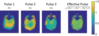
Parallel Transmission Based Alternating Saturation Pulse Design for CEST Imaging
Zhipeng Cao, Zhongliang Zu, Kristin O'Grady, Jun Ma, Seth Smith, William Grissom, John Gore
A novel saturation pulse design method for ultra-high field human CEST imaging is presented. By alternating complementary saturation patterns using parallel transmission, the method achieves homogeneous CEST saturation in the whole brain volume.
|
|
3986.
 |
132 |
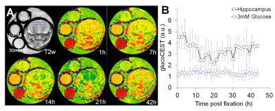
GlucoCEST MRI detects metabolic degradation in the mouse brain after rapid microwave fixation
Chao-Hsiung Hsu, Stephen Lin, Tirone Johnson, Ke-Yi Wu, Liang-Ron Chen, Paul Wang, Joseph Scafidi, Tsang-Wei Tu
The study of neurometabolites and the detection of glucose in the brain has relied on anesthetized animals. Prolonged periods of anesthesia required for these studies may affect these metabolites and the ability to interpret glucose detection in the brain. However, performing post-mortem studies of neurometabolites and glucose detection in the brain is difficult to interpret because of rapid degradation. In this study, we utilized focal beam microwave irradiation (FBMI) without anesthesia to collect brain tissue and determine whether there is postmortem degradation of neurometabolites and glucose using 1H-NMR and glucoCEST.
|
|
3987.
 |
133 |
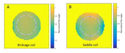
A custom-designed 1H saddle coil for CEST imaging at 14.1 Tesla
Claudia Zanella, Elise Vinckenbosch, Jérémie Clément, Masoumeh Dehghani, Bernard Lanz, Rolf Gruetter
We propose an alternative RF method for small animal CEST imaging at ultra-high magnetic field by using a 1H saddle coil instead of a quadrature birdcage coil. The coils create similar homogeneous B1 maps with an 2.2x increase of SNR for the saddle coil. Both coils show a similar degree of precision when tested for in vitro glycoCEST imaging. In rodent skeletal muscle the saddle coil offers a good performance with respect to the sensitivity for glycoCEST imaging.
|
|
3988.
 |
134 |
Towards glycoCEST in the liver at 7T with a multi-transmit system
Aline Xavier, Vitaliy Khlebnikov, Jeanine Prompers , Marcelo Andia, Catalina Arteaga de Castro
The purpose of this work was to optimize and evaluate different parameters of the CEST acquisition protocol at 7 Tesla to be able to detect the glycoCEST signal in the human liver. The so far optimized parameters were determined as 65 frequency offsets, 60 saturation pulses of 25 ms duration and an effective B1 of 1.5uT. As conclusion, we have demonstrated that in-vivo liver glycoCEST can be detected in the human liver by using a CEST acquisition at 7T. The parameters can be further optimized to obtain a stronger glycoCEST effect.
|
|
3989.
 |
135 |
Imaging of hydrogel biomaterials with Magnetization Transfer MRI
Vitaliy Khlebnikov, Klaus Neef, Annette van der Toorn, Rick Dijkhuizen, Caroline van Heijningen, Patricia Dankers, Carlijn Bouten, Steven Chamuleau, Dennis Klomp, Jeanine Prompers
We developed and validated MT-MRI protocols for imaging of hydrogel biomaterials and their pH-switchable behavior. Given that all hydrogels are expected to have an MT effect, the proposed MT-MRI protocols may be applicable for imaging of other hydrogel biomaterials as well.
|
|
3990.
 |
136 |
Tuning the ratio of exchangeable protons in iodinated agents to improve the accuracy and detection range of pH mapping under a single low power saturation
Presentation Not Submitted
Quan Tao, Peiwei Yi, Zhifeng Chen, Guojing Wei, Yingjie Mei, Yanqiu Feng
Ratiometric chemical exchange saturation transfer MRI (acidoCEST) using two distinct exchangeable protons in iodinated agents has been widely developed for in vitro and in vivo pH mapping. The accuracy of acidoCEST and pH detection range greatly relies on the applied B1 saturation pulse and chemical properties of labile protons. In this study we try to make full use of different chemical property of two labile protons. Through finely adjusting their ratio, the accuracy and pH detection range under a single low-power B1 saturation has shown to be improved
|
|
3991.
|
137 |
Thermal sensitive pH imaging using CEST
Yuki Kanazawa, Daiki Chiba, Masafumi Harada, Tosiaki Miyati, Mitsuharu Miyoshi, Hiroaki Hayashi, Yuki Matsumoto, Takashi Abe, Akihiro Haga
We performed phantom study with CEST and clarified pH change depending on temperature. Linear regression analysis shows a strong significant correlation between temperature and MTRasym at 2.0 ppm in all egg white albumin samples (R2 > 0.78, P < 0.05). As pH changed with temperature, we found that the offset frequency of MTRasym was more sensitive at 2.0 ppm than that at 3.5 ppm.
|
|
3992.
 |
138 |
Comparison of Different CEST Metrics for Brain Tumor Grading with Semi-Automatic ROIs
Ruibin Liu, Xianlong Wang, Zhibo Wen, Tingting Liu, Jinyuan Zhou, Dan Wu, Yi Zhang
The performance of three CEST metrics including CESTR, CESTRnr, MTRRex at various frequency offsets was evaluated for human brain tumor grading. Regions of interest were created semi-automatically based on an initial radiologist-drawn contour and automatic histogram analysis in order to minimize subjective bias. Thirty-nine patients with 23 confirmed high-grade and 16 confirmed low-grade gliomas were included. CESTRnr metric at 3 ppm were superior to other frequency offsets for grading human brain gliomas, with the maximum area under the curve value of 0.88.
|
|
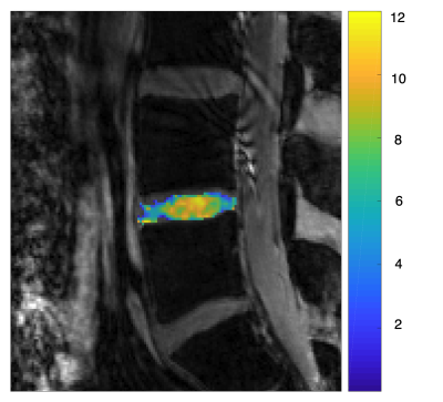
3993.
 |
139 |
GagCEST of human intervertebral lumbar discs compared to 23Na MRI at 7 Tesla
Stephan Gruber, Esaú Poblador Rodríguez, Alena Svatkova, Thomas Stulnig, Siegfried Trattnig, Wolfgang Bogner, Lenka Minarikova
Six healthy volunteers were measured to assess the feasibility and differences of 23Na-MRI and gagCEST in human intervertebral lumbar discs at 7T. MTRasym values from GagCEST data have shown to have similar potential as normalized 23Na-MRI, but were acquired in half the time and without the need for special hardware.
|
|
3994.
 |
140 |
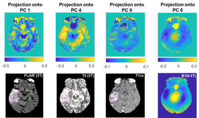
Data-Driven Spectral Feature Extraction in 9.4T CEST MRI data of the human brain
Mark Schuppert, Anagha Deshmane, Kai Herz, Klaus Scheffler, Moritz Zaiss
Model-based extraction of features, e.g. Lorentzian fitting of Z-spectra, in CEST MRI can be limited by the underlying model assumptions. Here we analyzed high spectral resolution Z-spectra acquired at 9.4T in five healthy subjects and one tumor patient using principal component analysis, a purely data-driven statistical procedure. Projection of Z-spectra onto principle components from a group of healthy subjects provides several relevant contrasts which reveal anatomical detail and correlate with Gadolinium uptake signatures in a brain tumor patient.
|
|
3995.
 |
141 |
Development of whole-brain 3D snapshot CEST MRI at 3T
Sebastian Mueller, Anagha Deshmane, Kai Herz, Klaus Scheffler, Moritz Zaiss
In its application Chemical exchange saturation transfer (CEST) suffers from the drawback of long acquisition times. To approach this issue we demonstrate the feasibility of a 3D gradient echo based CEST sequence for 3T that provides whole brain (WB) coverage without additional measurement time. In addition, the suggested post processing helps to improve the determination of the CEST effects from the WB data. CEST contrasts derived from the presented method are of equal quality as those of commonly used methods.
|
|
3996.
 |
142 |
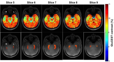
Feasibility of Partial 3D GluCEST in Healthy Human Adults at 7.0T
Ravi Prakash Reddy Nanga, Abigail Cember, Deepa Thakuri, Dushyant Kumar, Neil Wilson, Hari Hariharan, Mark Elliott, Cynthia Epperson, Ravinder Reddy
GluCEST imaging at 7.0T has been promising in clinical applications as demonstrated in small subset of temporal lobe epilepsy subjects and in early psychosis. However, these studies employed 2D GluCEST limiting to single slice and limited coverage of anatomy of interest. Here, we tried to implement the partial 3D GluCEST in order to capture the glutamate contrast from the entire hippocampus as opposed to single slice in the same amount of time.
|
|
3997.
 |
143 |
Pulsed Saturation Transfer in Glioblastoma at 1.5T
Rachel Chan, Sten Myrehaug, Greg Stanisz, Arjun Sahgal, Angus Lau
The purpose of this study was to quantify the effects of saturation transfer (CEST and MT) measurements in glioblastoma (GBM) patients at 1.5T. Data from GBM patients (n=10), treated with intensity modulated radiation therapy with a dose of 2 Gy per session with concurrent temozolomide, were analyzed before and after 10 treatment fractions. MT data were fitted to the Bloch-McConnell (BM) equations using a two-pool model with extended phase graphs (EPG) incorporated into the model. Results showed differences between normal and tumor regions and demonstrated promise for the application of CEST at 1.5T.
|
|
3998.
 |
144 |
Measuring the pH of human blood using CEST
Andrew Carradus, Olivier Mougin, Hans Hoogduin, Penny Gowland
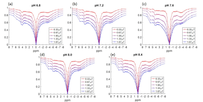
Iopamidol has been previously used as a CEST based contrast agent to measure pH in mouse kidneys. Here we aim to move this work towards clinical translation by calibrating the pH of ex vivo human blood to the ratiometric values calculated from the two Iopamidol CEST peaks. This is performed in the physiological range for human blood, between pH 6.8 and 8.4.
|
|
3999.
 |
145 |
Measuring the water exchange rate for various pools in the z-spectrum of the human brain
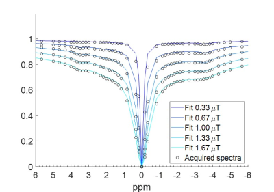
Andrew Carradus, Olivier Mougin, Hans Hoogduin, Penny Gowland
True quantification of MT, CEST and NOE pools is difficult to achieve, as the exchanging pool size and exchange rate cannot be readily separated. Here we use a Particle Swarm Optimisation algorithm to solve this problem, which we show to be capable of quantifying pool size, exchange rate, and apparent T2s of exchanging pools without the need for initial guesses. We apply this to z-spectra acquired in vivo from the human brain, and quantify the exchanging pools in grey and white matter.
|
|
4000.
 |
146 |
Initial CEST study in a microcavity array based 3D cell culture system
Dennis Kleimaier, Steffen Goerke, Cordula Nies, Eric Gottwald, Lothar Schad
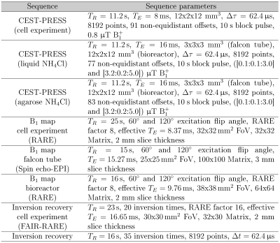
Chemical exchange saturation transfer (CEST) is a powerful method for the investigation of biomolecules. An MR-compatible bioreactor, on the other hand, creates a well-controlled environment for human cells. The combination of both methods should make it possible to extract cellular signals from the bioreactor. Therefore, we investigated the possibility of performing CEST measurements in an actively-perfused bioreactor with HepG2 cells. We could show that perfusion had no effect on the CEST signals and that the main signals originated from the cells. However, the sub-optimal shim condition in the bioreactor led to a reduction in CEST signals.
|
|
4001.
 |
147 |
Producing a Synthetic Magnetization Transfer Reference Volume with T1 Maps Acquired using a Different Pulse Sequence: Practical Considerations.
Sofia Chavez, Kimberly Desmond
Macromolecular proton fraction (MPF) mapping can be achieved using only two acquired volumes, one with and one without a magnetization transfer (MT) pulse, in addition to T1, B1 and B0 maps. Further scan time reduction is possible if the T1 and B1 maps required for MPF computations are used to generate a synthetic reference image, eliminating the reference image acquisition. This has been shown to work when the images used to generate T1 were acquired from the same sequence and parameters as the MT. We investigate if we can use a different sequence with a calibration to achieve this.
|
|
4002.
 |
148 |
Positive Contrast Chemical Exchange Imaging using RACETE
Fabian Gutjahr, Peter Jakob
The RACETE sequence is a novel method for imaging chemical exchange. As it refocuses the magnetization transferred while stored in the longitudinal state, it yields a positive contrast. By creating a refocused sequence other properties of standard MR sequences, such as excitation pulse phase variation, can be exploited.
|
|
4003
 |
149 |
Microfluidics preparation of liposome containing alginate hydrogel microbeads for CEST MRI
Video Permission Withheld
P. Xiao, X. Han, J. Huang, J. Li, Raymond Lam, Kannie Chan
Image-guided cell therapy plays an important role in monitoring and adjusting the treatment regimen. We have demonstrated that cell viability could be monitored by CEST pH-nanosensors in alginate hydrogel microbeads. Here, we investigated the use of microfluidics to generate liposome containing microbeads and examined their CEST properties after loading with the iodine CT contrast agents Iohexol and Iopamiro. We have successfully demonstrated that the microbead size has greatly reduced from 300-400 μm to 20 μm. Moreover, iodine agents loaded liposomal microbeads generated 4% CEST contrast at 4.2 ppm at 3T. These findings demonstrate a robust platform for the fabrication of liposome containing microbeads under CEST MR guidance.
|
|
4004.
 |
150 |
Extended phase graph framework for "inhomogeneous" MT systems.
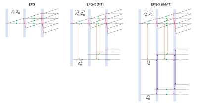
Daniel West, Jacques-Donald Tournier, Shaihan Malik
Recently, the extended phase graph with exchange (EPG-X) formalism has been introduced for modelling magnetization transfer (MT) effects during arbitrary pulse sequences. Here, we present a further extension to include a dipolar order component of the macromolecular (i.e. semisolid) pool, as has been proposed to understand the “inhomogeneous” MT (ihMT) contrast mechanism observed in ordered structures such as myelin and muscle. Preliminary results demonstrate the flexibility of the developed simulation tool for ihMT contrast optimisation and the investigation of steady-state sequence dynamics.
|
|
| Top |
Fat, QSM & CEST
Digital Poster
Contrast Mechanisms
Wednesday, 15 May 2019
| Exhibition Hall |
14:30 - 15:30 |
| |
|
Computer # |
|
4005.
 |
151 |
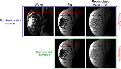
Eliminating chemical shift artefact using simultaneous, separate water and fat excitation combined with CAIPIRINHA
Beáta Bachratá, Bernhard Strasser, Albrecht Schmid, Wolfgang Bogner, Siegfried Trattnig, Simon Robinson
Imaging of many body regions is adversely affected by the chemical shift between fat and water. We propose a new method for simultaneous, separate imaging of fat and water in which multiband pulses are used to simultaneously excite fat and water at their characteristic resonance frequencies, and CAIPIRINHA in combination with parallel imaging to reconstruct separate images of the two species. The fat image is corrected for chemical shift displacement and either recombined with the water image or evaluated separately. The proposed method achieves reliable water-fat separation and complete elimination of the chemical shift artefact in musculo-skeletal and breast imaging.
|
|
4006.
 |
152 |
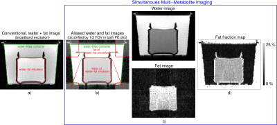
Fat quantification using simultaneous, separate water and fat excitation combined with CAIPIRINHA
Beáta Bachratá, Radim Korínek, Bernhard Strasser, Albrecht Schmid, Martin Krššák, Wolfgang Bogner, Siegfried Trattnig, Simon Robinson
Fat quantification by MRI plays an important role in assessing hepatic steatosis and bone marrow changes. We propose a new method for fat quantification in which multiband pulses are used to separately but simultaneously excite fat and water. CAIPIRINHA encoding allows overlapping images of the two species to be separated. Fat fraction maps can be calculated from fat and water images with appropriate consideration of relaxation times. The proposed method is validated on phantoms with different fat concentrations, where it yields similar fat fraction values to the Dixon approach and single-voxel spectroscopy.
|
|
4007.
 |
153 |
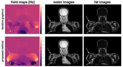
Accelerated Single-Min-Cut Graph-Cut Algorithm Using a Variable-Layer Graph Construction Improves Field-Mapping in Water–Fat Regions
Christof Böhm, Maximilian Diefenbach, Axel Haase, Dimitrios Karampinos
Accurate field-mapping is critical in body imaging for both robust water–fat separation and accurate quantitative susceptibility mapping. The present work proposes a single-min-cut field-mapping method to improve field-map results in water–fat regions. The proposed method based on a variable layer graph construction, solves the phase-wrapping problem and yields directly into non-wrapped field-maps. In challenging areas with low SNR or strongly varying field-maps the accuracy is significantly increased, while the computational cost is drastically reduced.
|
|
4008.
 |
154 |
Separation of Water and Fat during Spin-Lock Magnetic Resonance Imaging with Multifrequency Fat Spectrum Modeling
Weitian Chen, Baiyan Jiang, Maximilian Diefenbach, Dong Liang, Dimitrios Karampinos
Spin-lock technologies are useful for probing macromolecular environment of tissue. The conventional spin-lock approaches cannot lock water and fat spins simultaneously due to chemical shift effect of fat, which can lead to artifacts in tissues with fatty infiltration. Based on the latest development of the spin-lock technique, we propose spin-lock Dixon methods to perform water-fat separation in spin-lock MRI. Our methods may offer a solution to perform reliable spin-lock MRI in tissues with fatty infiltration.
|
|
4009.
 |
155 |
Transition region extraction algorithm for Two Point Dixon Imaging
Hao Peng, Chao Zou, Wenzhong Liu, Chuanli Cheng, Yangzi Qiao, Qian Wan, Changjun Tie, Xin Liu, Hairong Zheng
Purpose: To propose a transition region extraction algorithm for two point fat-water separation. Methods:In the proposed method, fat-water transition region is first extracted as initial information. Remaining pixels are determined based on transition region in units of sub-region instead of pixels to improve calculation efficiency and avoid the vulnerability to the growing path.
Results:Fat-water separation were successfully achieved by the proposed method in the datasets tested.
Conclusion: A method based on fat-water transition region extraction is proposed for two point fat-water separation.
|
|
4010.
 |
156 |
B1 and B0 Insensitive Uniform Fat-saturation for Joint Imaging
Venkata Veerendranadh Chebrolu, Sinyeob Ahn, Peter Kollasch, Fei Han, Xiaodong Zhong, John Grinstead, Vibhas Deshpande
In this work we propose B1 and B0 insensitive, uniform, and robust fat suppression for joint imaging using dual repetition spectrally selective high bandwidth Shinnar–Le Roux RF pulses. The optimal flip angle combinations for B1 insensitivity were calculated using Bloch simulations. Efficacy of the novel fat-sat mechanism in achieving B1 robust uniform fat-sat was demonstrated in the knee at 3T.
|
|
4011.
 |
157 |
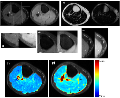
Sub-voxel Estimation of Fat Infiltration in Degenerative Muscle Disorders using Multi-T2 Analysis – a Quantitative Disease Biomarker
Jannette Nassar, Yann Le Fur, Dvir Radunsky, Tamar Blumenfeld-Katzir, David Bendahan, Noam Ben-Eliezer
T2 relaxation is an effective biomarker for muscle pathologies including inflammation, necrosis, or fatty infiltration. Accurate quantification of T2 values is hampered due to the inherent bias of fast multi-echo spin-echo (MESE) protocols by stimulated echoes. The echo-modulation curve (EMC) algorithm overcomes this problem and provides accurate T2 values which are stable across scanners and scan-settings. In this work, we present an extension of the EMC algorithm for T2-based fat/water quantification, alongside two new quantitative biomarkers of disease state. The technique is demonstrated on a calf muscle of a patient with Dysferlinopathy, and compared to conventional Dixon analysis.
|
|
4012.
 |
158 |
Robust Chemical Exchange Spin-lock performed using adiabatic pulse with Toggling Inversion Preparation at 3.0T
Baiyan Jiang, Tao Jin, Jian Hou, Weitian Chen
The measurement of R1rho (1/T1rho) spectrum and its asymmetry have several advantages over Chemical Exchange Saturation Transfer (CEST) to probe chemical exchange effect. However, these approaches suffer from B1 Radiofrequency (RF) and B0 field inhomogeneities. In this work, we proposed a new approach to perform robust R1rho asymmetry measurement using adiabatic spin-lock RF pulse and demonstrated its potential to probe metabolites.
|
|
4013.
 |
159 |
Relaxation-compensated APT and rNOE CEST-MRI of human brain tumors at 3 T
Steffen Goerke, Yannick Soehngen, Anagha Deshmane, Moritz Zaiss, Johannes Breitling, Philip Boyd, Kai Herz, Ferdinand Zimmermann, Karel Klika, Mark Ladd, Peter Bachert
In this study, the established acquisition protocol for relaxation-compensated APT and rNOE CEST-MRI at 7 T has been successfully transferred to a clinically relevant magnetic field strength of 3 T. This opens up the door to clinical trials with a large number of participants, thus enabling a comprehensive assessment of the clinical relevance of relaxation-compensation in CEST-MRI. The presented CEST sequence is currently part of the clinical routine acquisition protocol for brain tumor patients at our institute.
|
|
4014.
 |
160 |
Deep Neural Network for Single-Point Dixon Imaging with Flexible Echo Time
Jong Bum Son, Marion Scoggins, Basak Dogan, Ken-Pin Hwang, Jingfei Ma
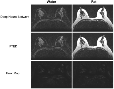
Dixon imaging generally requires multiple input images acquired at varying echo-times for robust phase-correction and water and fat separation. However, multi-echo Dixon imaging suffers from relatively long scan-time and is more susceptible to motion related artefacts and inflexible in choosing scan-parameters. Recently, it was reported that deep neural networks can help separate water and fat from two-point, or multi-point Dixon images. In this work, we present a deep learning based method that can achieve water and fat separation from a single image acquired at a flexible echo-time and therefore can help alleviate the limitations of multi-point Dixon imaging.
|
|
4015.
 |
161 |
Detecting the presence of fat using PLANET-based parameter mapping
Yulia Shcherbakova, Cornelis van den Berg, Soraya Gavazzi, Chrit Moonen, Lambertus Bartels
The PLANET method was introduced to simultaneously map T 1, T 2, banding free magnitude, local off-resonance, and RF phase using phase-cycled bSSFP data. All these parameters are estimated from a linear least squares fitting of an ellipse to complex data. The PLANET method is based on a Lorentzian single-component relaxation model, which results in symmetric bSSFP magnitude profile and elliptical behavior of the complex transverse magnetization. However, when a second or more components with different frequency distributions are present within a voxel, the complex signal might not lie on an ellipse anymore. Hence, PLANET post-processing would return erroneous quantitative parameters.
In this study we show that the sensitivity of PLANET to the presence of multiple components can be exploited to map the spatial distribution of voxels in which multiple spectral peaks are present. We demonstrate that this feature of PLANET can be used to create fat-only map.
|
|
4016.
 |
162 |
deepCEST: 9.4 T spectral super resolution from 3 T CEST MRI data - optimization of network architectures
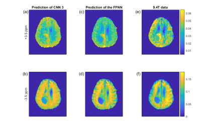
Moritz Zaiss, Florian Martin, Felix Glang, Kai Herz, Anagha Deshmane, Benjamin Bender, Tobias Lindig, Klaus Scheffler
Different neural network architectures for predicting 9T CEST contrasts from 3T spectral data are investigated as well as the influence of different training data sets on the quality of resulting predictions. Although optimized convolutional neural network (CNN) architectures perform well, the best results were reached with a simpler feedforward neural network (FFNN). As CNNs have many hyperparameters to tune, this work forms a basis for CNN architecture optimization for the proposed super-resolution CEST application.
|
|
4017.
 |
163 |
A Potentially New Class of Redox-Sensitive CEST Agents: Preliminary Thiol-CEST Studies with Glutathione and Cysteine
Johnny Chen, Nirbhay Yadav, Abhishek Gupta, Tim Stait-Gardner, William Price, Gang Zheng
In this study, we show that thiol-water proton exchange can generate MRI contrast by performing proton chemical exchange saturation transfer (1H-CEST) experiments on glutathione (GSH) and cysteine (Cys). The thiol proton exchange was quantified at various pHs, with Cys thiol exhibiting faster and more base-catalyzed exchange than GSH. However, both GSH and Cys thiol exchange were too fast to generate contrast at physiological pH. Potential applications of thiol compounds as redox-sensitive CEST agents are suggested.
|
|
4018.
 |
164 |
Characterization of B1 calibration curves with respect to durations for myo-Inositol CEST ( MICEST) in the healthy human brain at 7.0T
Deepa Thakuri, Ravi Prakash Reddy Nanga, Abigail Cember, Dushyant Kumar, Neil Wilson, Hari Hariharan, Ravinder Reddy
myo-Inositol (MI) is one of the important brain metabolites. In the recent years, MICEST technique has emerged as a neuroimaging biomarker and its application was shown in an Alzhemer’s mouse model. In this study, we tried to explore and evaluate the behavior of MICEST at different saturation parameters in healthy human volunteers at 7.0T with an aim to understand and set optimal saturation parameters for the human studies.
|
|
4019.
 |
165 |
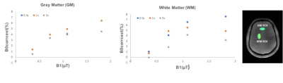
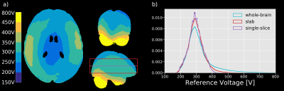
Optimized B1/B0 correction for whole-brain CEST imaging using 3D-EPI at 7T
Suzan Akbey, Philipp Ehses, Rüdiger Stirnberg, Moritz Zaiss, Tony Stöcker
Compared to conventional single-slice or small volume measurements, whole-brain CEST acquisitions present new challenges, in particular at ultra high-field (UHF). In a first step to account for the broader B1 variation, we analyzed the distribution of reference voltages in the brain of nine subjects. Based on these results, we repeated a whole-brain CEST experiment with seven different reference voltages to optimize the correction for B1 inhomogeneities in the CEST contrast maps. Additionally, we extended the saturation-offset list to compensate for the higher variation of the static magnetic field for whole-brain experiments compared to examinations with smaller FOVs.
|
|
4020.
 |
166 |
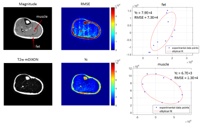
Validation of a tool for body fat and skeletal muscle quantification using MRI and application in the assessment of body composition in patients with colorectal and lung cancer
Naomi Sakai, Timothy Bray, Jagadish Kalasthry, Margaret Hall-Craggs, Stuart Taylor
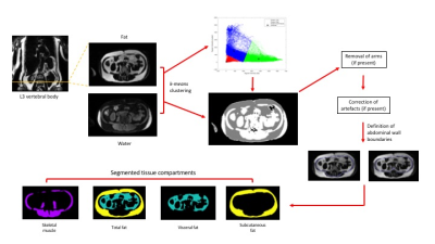
Body composition analysis holds considerable promise in clinical prognostication. For example, sarcopenia (particularly sarcopenic obesity) is associated with poor outcomes in cancer patients. To date, most clinical studies using body composition analysis have used data derived from volumetric CT; however, whole-body MRI (WB-MRI) is increasingly used in cancer staging and analysis of these MRI data could be an alternative means to calculate body composition. In this study, we describe and validate a simple, practical tool for quantification of body fat and skeletal muscle from WB-MRI and demonstrate its application to two cohorts of patients with lung and colon cancer.
|
|
4021.
 |
167 |
An Automated Approach for the Optimised Estimation of Breast Density with Dixon Methods
Rosie Goodburn, Evanthia Kousi, Alison Macdonald, Veronica Morgan, Erica Scurr, Martin Leach, Mamatha Reddy, Louise Wilkinson, Elizabeth O'Flynn, Romney Pope, Robin Wilson, Steven Allen, Maria Schmidt
In Breast MRI, Dixon fat-water separation techniques have been invaluable for the measurement of breast-density in studies of breast cancer risk. A fundamental source of error in Dixon methods arises from differences in the signal intensity from water and fat, associated with differences in proton density and relaxation times. We propose an automated method to introduce a scaling factor that minimizes these errors. We demonstrate our method in a group of 14 subjects, imaged at 3T with different levels of T1-weighting.
|
|
4022.
 |
168 |
Thermal measurement in fatty tissues with Z-Spectrum Imaging
Alessandro Scotti, Li Li, Fred Damen, Weiguo Li, Victoria Gil, Wenzhen Zhu, Chong Wee Liew, Kejia Cai
Z-spectrum imaging (ZSI) has been recently introduced as a method to quantify fat-water fraction based on the direct saturation of both water and fat. Here we demonstrate that ZSI can also assess temperature in fatty tissues, by measuring the relative fat-water chemical shift (FWCS) change due to the shift of water resonance at increased temperature. The protocol was tested on a whipped cream phantom and showed FWCS changing linearly with temperature. The protocol was also tested on healthy mice and subjects, paving the way for the study of fat metabolism, in particular of brown adipose tissue.
|
|
4023.
 |
169 |
A quantitative tool for the speciation of mineralized iron in the brain
Lucia Bossoni, Laurens Boers, Andrew Webb, Louise van der Weerd
We implemented and optimized an Off-Resonance Saturation (ORS) pulse sequence on a 7T- preclinical scanner, with the aim of quantifying iron present in brain phantoms doped with ferritin-bound and magnetite-bound iron. These mineralized iron forms are known to be involved in ageing, and also in toxic cellular pathways in neurodegenerative diseases. We show that the ORS method offers the possibility to quantify, and possibly differentiate, iron ions in brains affected by neurodegenerative diseases which are characterized by disturbed iron homeostasis. Additionally, our results are discussed in the light of susceptibility values of the same minerals.
|
|
4024.
 |
170 |
CEST MRI with dual-echo readout for the functional assessment of transplanted kidney in vivo: a preliminary study
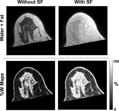
Julia Stabinska, Alexandra Ljimani, Eric Bechler, Christian Tell, Helge Zöllner, Hans-Jörg Wittsack, Anja Müller-Lutz
In this study we present the first application of dual-echo CEST MRI technique in renal transplant recipients. Our goal was to optimize the CEST-MRI acquisition and analysis for kidney graft imaging at 3T, which remains technically challenging and volatile due to large B0 inhomogeneity and presence of fat. Strong lipid signals upfield from the water resonance may lead to erroneous CEST contrast. In this preliminary work we demonstrate that the combination of dual-echo CEST with Dixon provides effective fat signal removal, allowing for more accurate quantification of the CEST effects in the transplanted kidney.
|
|
4025.
 |
171 |
Analysis of different phase unwrapping methods to optimize QSM in the abdomen
Eric Bechler, Helge Jörn Zöllner, Hans-Jörg Wittsack
We evaluated six different phase unwrapping algorithms for their use in QSM in the abdomen. Therefor a numerical phantom of the abdomen was simulated using a forward model. The resulting unwrapped phases and susceptibility maps were compared with the ground truth and root mean squared error maps were calculated. Results suggest that the “graph-cuts” algorithms should be used for phase unwrapping in the abdomen, as it was the most robust around the lungs and bones and further showed the smallest difference to the ground truth susceptibility.
|
|
4026.
 |
172 |
Correlation between CT and QSM in the brain
Sonoko Oshima, Yasutaka Fushimi, Tomohisa Okada, Akira Yamamoto, Satoshi Nakajima, Gosuke Okubo, Hikaru Fukutomi, Yusuke Yokota, Kaori Togashi
Quantitative susceptibility mapping (QSM) is a technique which can provide quantitative values of magnetic susceptibility, and thus is a useful tool for differentiating hemorrhage from calcification. Computed tomography (CT) is also used for evaluation of hemorrhage and calcification by using CT values. However, the correlation between CT and QSM has not been much investigated. In this study, we found positive correlations between CT values and susceptibility in the globus pallidus and hemorrhagic lesions. Negative correlations were observed in the choroid plexus and calcified lesions. Our results may help radiologists revisit the CT values based on the susceptibility values calculated from QSM.
|
|
4027.
 |
173 |
MRI Susceptibility Mapping Shows Decreased Venous Oxygen Saturation in Sickle Cell Anaemia
Russell Murdoch, Hanne Stotesbury, Jamie Kawadler, Fenella Kirkham, Karin Shmueli
Ischemic stroke is a common and severe occurrence in Sickle Cell Anaemia (SCA) but no accurate screening measures currently exist for adults. Changes in venous oxygen saturation (Yv) have been suggested as a potential biomarker but measuring Yv in vivo is challenging. This work explores the potential of MRI susceptibility mapping to measure Yv in SCA subjects. Susceptibility-mapping-based measures of Yv were compared between 25 SCA subjects and 15 healthy controls. Significantly lower Yv was measured in the superior sagittal sinus in the SCA group compared to healthy controls, showing that QSM is sensitive to changes in Yv in SCA.
|
|
4028
 |
174 |
Deep Quantitative Susceptibility Mapping by combined Background Field Removal and Dipole Inversion
Video Permission Withheld
Stefan Heber, Christian Tinauer, Steffen Bollmann, Stefan Ropele, Christian Langkammer
Deep learning based on u-shaped architectures has been successfully used as a means for the dipole inversion crucial to Quantitative Susceptibility Mapping (QSM). In the present work we propose a novel deep regression network by stacking two u-shaped networks and consequently both, the background field removal and the dipole inversion can be performed in a single feed forward network architecture. Based on learning the theoretical forward model using synthetic data examples, we show a proof-of-concept for solving the background field problem and dipole inversion in a single end-to-end trained network using in vivo Magnetic Resonance Imaging (MRI) data.
|
|
4029.
 |
175 |
Achieving Real-Time QSM Reconstruction Using Deep Neural Network
Hoeseong Kim, Jaeyeon Yoon, Jongho Lee
Conventional QSM reconstruction algorithms impose long computation time, which inhibits their adoption for real-time clinical use. In this work, we propose a method that replaces conventional iterative algorithms for background removal and dipole inversion with two deep neural networks. The reconstruction results demonstrate comparable performance to the previous outcomes while the new method takes only 3 seconds (up to 106 times faster!), which is unparalleled to conventional methods.
|
|
Back to Program-at-a-Glance |  Back to Top Back to Top |
| The International Society for Magnetic Resonance in Medicine is accredited by the Accreditation Council for Continuing Medical Education to provide continuing medical education for physicians. |
