Digital Poster Session
Body: Breast, Chest, Abdomen, Pelvis Back to Program-at-a-Glance Back to Program-at-a-Glance
|
Wednesday, 15 May 2019
Digital PosterBody: Breast, Chest, Abdomen, Pelvis
4030 -4054 Pelvis: Rounding Out the Bottom
4055 -4077 Fetal & Placental Imaging
4078 -4102 Lung: Proton-Based Imaging
4103 -4126 Breast: Technical
4127 -4150 Lung: Xenon |
| |
Pelvis: Rounding Out the Bottom
Digital Poster
Body: Breast, Chest, Abdomen, Pelvis
Wednesday, 15 May 2019
| Exhibition Hall |
15:45 - 16:45 |
| |
|
Computer # |
|
4030.
 |
1 |
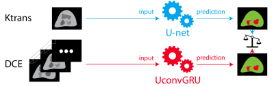
Do we still need mathematical modeling in the age of deep learning? A case-study comparison of the Tofts model versus end-to-end deep learning in prostate cancer segmentation

Alessandro Guida, Peter Lee, Steve Patterson, Thomas Trappenberg, Chris Bowen, Steven Beyea, Jennifer Merrimen, Cheng Wang, Sharon Clarke
The rise in popularity of deep learning is revolutionizing the way biomedical images are acquired, processed, analyzed. Just a few years ago, extracting high-level understanding from biomedical images was a process restricted to highly trained professionals often requiring multidisciplinary collaborations. In the work presented, we showcase a study that compares the performance of a model trained end-to-end using a novel deep learning architecture, versus a model trained on its corresponding state-of-the-art mathematically engineered feature. Results show that end-to-end deep learning significantly outperforms the mathematical model, suggesting that feature engineering will play a less important role in the coming years.
|
|
4031.
 |
2 |
Comparison of SENSE-accelerated PROPELLER and Single-shot Turbo Spin-Echo Sequences for Visualization of the Prostatic Urethra during MRI-based Radiotherapy Treatment Planning
Kristen Zakian, Andreas Wibmer, Hebert Vargas Alvarez, Eveline Alberts, Mo Kadbi, Borys Mychalczak, Marisa Kollmeier, Daniel Gorovets, Sean McBride, Margie Hunt, Michael Zelefsky, Neelam Tyagi
At our institution, a Foley catheter is inserted during prostate SBRT simulation to ensure visualization of the prostatic urethra. Our goal was to find an optimal motion-robust T2-weighted pulse sequence that would allow for accurate catheter-free visualization and segmentation of the urethra. We compared Multivane XD (MVXD), a PROPELLER-based multislice TSE sequence with SENSE acceleration to single-shot TSE with SENSE (SSTSE). Two expert GU radiologists scored urethra visibility in MVXD and SSTSE series of equivalent spatial resolution and duration. Urethra visibility scores were significantly higher in the MVXD series and SNR was found to be superior in all MVXD series.
|
|
4032
 |
3 |
Comparison of Stretched-Exponential, IVIM and Monoexponential Diffusion-Weighted Imaging Mathematical Models for Assessment of Tumor Aggressiveness in Prostate Cancer at 3T
Video Permission Withheld
Tsutomu Tamada, Ayumu Kido, Hidemitsu Sotozono, Akihiko Kanki, Akira Yamamoto, Yu Ueda
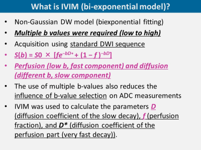
We compared monoexponential and non-Gaussian models including intravoxel incoherent motion imaging (IVIM) and stretched-exponential model in diffusion-weighted imaging (DWI) for characterization of prostate cancer. ADCmean in monoexponential model, D in IVIM, and DDC in stretched model showed high discrimination ability with AUC>0.80 between GS=3+3 and GS≥3+4 tumors as well as GS≤3 + 4 and GS≥4 + 3 tumors, but no significant difference among these metrics. Our study did not show a clear clinical value of non-Gaussian models including IVIM and stretched model compared with standard monoexponential model for assessment of tumor aggressiveness in prostate cancer.
|
|
4033.
 |
4 |
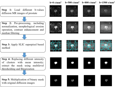
Optimal b-Value for Superpixel based Automatic Prostate Gland Segmentation using Diffusion-Weighted Imaging
Dharmesh Singh, Sayantan Bhattacharya, Virendra Kumar, Chandan Das, Anup Singh, Amit Mehndiratta
Accurate segmentation of the prostate gland is a challenging task due to the high variability of prostatic anatomic structures. The diagnostic accuracy of diffusion-weighted-imaging (DWI) to detect prostate cancer (PCa) is well established. Proper delineation using DWI of the prostate gland can take an essential part in the computer-aided-diagnosis of PCa. The purpose of this study is to automatic segmentation of prostate-gland using superpixel-based segmentation in DWI and select the optimal b-value of DWI for segmentation. Experimental results provide a close match to radiologist manual delineation and able to determine the optimal b-value of DWI for superpixel-based prostate-gland segmentation.
|
|
4034.
 |
5 |
Ensemble learning-based analysis of T1-weighted and T2-weighted magnetic resonance (MR) radiomics for the prediction of prostate cancer grades with small-scale cohort
Huipeng Ren, Zhuanqin Ren
Traditionally, PCA diagnosis and classification are based on prostate specific antigen (PSA) levels, ultrasound and biopsy. This study combines Adasyn and XGBOOST methods to compare the predictive effects of T1-weighted, T2-weighted and T1-T2 co-registration MR images on prostate cancer. The results showed that the integrated learning algorithm using XGBoost can effectively predict prostate cancer grade based on T2WI radiomics features,and T2WI has a better prognostic performance compared with T1-T2 fusion images.
|
|
4035.
 |
6 |
Cascading Classifiers Improve Prostate Segmentation
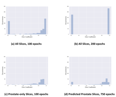
Ronald Nowling, John Bukowy, Sean McGarry, Andrew Nencka, Jay Urbain, Allison Lowman, Alexandar Barrington, Mark Hohenwalter, Anjishnu Banerjee, Kenneth Iczkowski, Peter LaViolette
We evaluated the U-Net segmentation model on prostate segmentation using data from 39 patients, achieving a Dice score of 73.9%. We improved segmentation performance by applying a convolutional neural network (CNN) to determine whether slices have prostates. Images with prostates are then forwarded to a U-Net model for segmentation. Our two-phase approach achieves a higher Dice score of 85.2%.
|
|
4036.
 |
7 |
Evaluating effect of Combination and Number of Multiple b-values in IVIM Analysis of Prostate
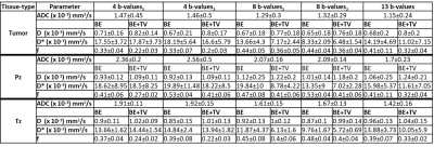
Archana Malagi, Chandan Das, Kedar Khare, Fernando Calamante, Amit Mehndiratta
Optimization of b-values in DWI-IVIM is necessary for precision and reproducibility of parameter estimation. The IVIM biexponential model with Total variation correction (BE+TV) method was used here, which reduces sudden changes in parameter values and removes non-physiological heterogeneity in maps. Various b-value combinations were utilized in prostate: 4, 8 and 13 b-values. 8 and 13 b-values showed similar trend in coefficient of variation (CV), which was confirmed using Bland-Altman plots, including comparable qualitative maps; in contrast, 4 b-values showed variable CV and noisy maps. In prostate, 8 b-values can be used with Total Variation method for faster acquisition protocol and low variability in parameter maps.
|
|
4037.
 |
8 |
Potential utility of T1 rho imaging for diagnosis of cystic tumor
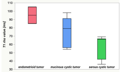
Hiroyuki Morisaka, Katsuhiro Sano, Taiki Seno, Yasuo Sakurai, Tomoaki Ichikawa
T1 rho imaging is sensitive to the tissue macromolecular inclusion such as proteoglycan of meniscus cartilage. In this study, we demonstrated T1 rho values were differ in ovarian cystic tumors and not in hepatic solid tumors.
|
|
4038.
 |
9 |
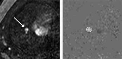
Value of MR placental protusion sign in predicting postpartumhemorrhage in patients with placenta previa
Presentation Not Submitted
Jiping Wang, Bei Zhang
Objective: To explore the MRI findings of placental protusion sign in predicting postpartum hemorrhage in pa—tients with placenta previa. Methods: Totally 35 4 placenta previa patients with whole clinical data underwent MR scaning 2weeks before operation.Association of postpartum hemorrhage and placental protusion sign was analyzed.
Results: Among354 patients with placental previa,the age of the pregnant women(#?4.34,P?0.04),gestational age at delivery(z2—5.19,P?0.02)and the number of cesarean sections(Z2=44.85,P<O.01)had associated with postpartum hemorrhage.Eight cases had placental protusion sign in MRI,while 6 cases occurred postpartum hemorrhage.The incidence of postpar—tum hemorrhage was 75.00%(6/8)and 12.72%(44/346)in patients with placenta accreta and with placental abruption?respectively(3[2—20.14,P<O.01).The sensitivity,specificity,odds ratio(95%confidence interval)and positive likeli—hood ratio of predicting postpartum hemorrhage was 12.00%(6/50),99.34%(302/304),20.59(4.03,105.23)and15.68,respectively.
Conclusion MRI placental protrusion sign has important clinical reference value in predicting postpar—tum hemorrhage.
|
|
4039.
 |
10 |
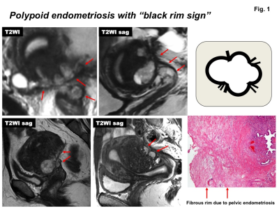
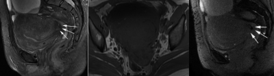
MR manifestations of polypoid endometriosis: Clinical feasibility of conventional and advanced MR techniques
Mayumi Takeuchi, Kenji Matsuzaki, Masafumi Harada
Polypoid endometriosis (PE) is a rare variant of benign endometriosis and may mimic malignant tumors. MR manifestations of five women with pathologically proven PE were retrospectively evaluated. PE was associated with pelvic endometriosis (4/5) with “black rim sign” (3/5), and with adenomyosis (5/5). PE showed high intensity on T2WI (5/5), high intensity hemorrhagic foci on T1WI (3/5), hemorrhagic signal voids on susceptibility-weighted sequences (2/2), high intensity on DWI (4/4) with relatively high ADC values (mean, 1.67 x 10⁻³ mm2/s), gradually increasing time-intensity curve pattern on DCE-MRI (4/4) with intense contrast-enhancement (4/5) or weak contrast-enhancement (1/5, with malignant transformation).
|
|
4040.
 |
11 |
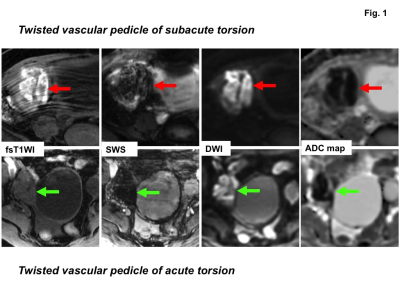
Clinical utility of susceptibility-weighted MR sequences for the evaluation of adnexal torsion
Mayumi Takeuchi, Kenji Matsuzaki, Masafumi Harada
Because venous thrombus within the twisted vascular pedicle is a specific pathological finding for adnexal torsion, to reveal the thrombus by MRI may be diagnostic. Surgically proven five benign ovarian masses with torsion were retrospectively evaluated. High intensity venous thrombus within the twisted vascular pedicle on fat-saturated T1WI were detected in four lesions (80%) but not in one acute torsion, whereas signal voids on susceptibility-weighted MR sequences (SWS) were detected in all five lesions (100%). SWS also revealed hemorrhagic infarction of all five ovarian masses with torsion, whereas fat-saturated T1WI could demonstrate high intensity hemorrhagic infarction in four lesions.
|
|
4041.
 |
12 |
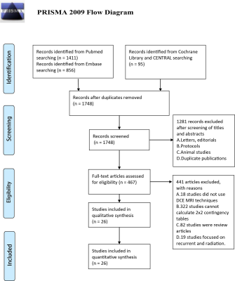
Accuracy of dynamic contrast-enhanced magnetic resonance imaging in the diagnosis of prostate cancer: meta-analysis
Presentation Not Submitted
Zhiqiang Chen, Yi Zheng
The goals of this meta-analysis were to assess the effectiveness of dynamic contrast-enhanced magnetic resonance imaging (DCE-MRI) in patients with prostate carcinoma (PCa) and to explore the risk profiles with the highest benefit. Compared with T2-weighted imaging (T2WI), DCE was statistically superior to T2. In conclusion, DCE had relatively high specificity in detecting prostate cancer (PCa) but relatively low sensitivity as a complementary functional method. DCE-MRI might help clinicians exclude cases of normal tissue and serve as an adjunct to conventional imaging when seeking to identify tumor foci in patients with PCa.
|
|
4042.
 |
13 |
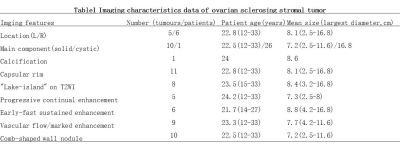
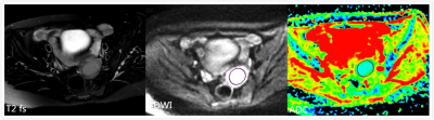
Imaging characteristics of ovarian sclerosing stromal tumor
Gangqiang Hou, Zhifeng Zhou
Sclerosing stromal tumor (SST) is an extremely rare benign sex cord-stromal tumor of the ovary. Due to its clinical presentation and imaging findings are similar to those of borderline or malignant epithelial tumors and other sex cordstromal tumors, accurate preoperative clinical diagnosis can be very difficult. This study evaluated magnetic resonance imaging and computed tomography features and pathological findings of SST of ovary and the correlation between them. The results demonstrated that MRI examination with dynamic and delayed enhancement could play a critical role in making appropriate diagnosis preoperatively when combined with clinical information.
|
|
4043.
 |
14 |
Evaluation of DWI with lymphovascular space invasion and without lymphovascular space invasion in the stage I endometrial Carcinoma
Chengyan Wang, Mei-yu Sun, Yanan Wu, Lizhi Xie
Endometrial cancer is one of the most commonly diagnosed gynecologic malignancies in developed countries. The fundamental extent of the operation involves hysterectomy and bilateral salpingo-oophorectomy for the endometrial carcinoma. The presence of LVSI is associated with lymph node metastases. However, Lymphadenectomy can’t improve the survival rate and has a reported higher rate of postoperative complications in endometrial cancer patients. So lymphadenectomy is a controversial issue in recent years. LVSI is a crucial factor in determining the lymphadenectomy and cannot be detected on traditional MR imaging.
|
|
4044
 |
15 |
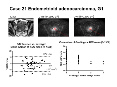
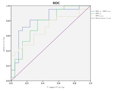
ADC mean based on a b-value combination of 0 and 1500 s/mm2 would be appropriate as a clinical biomarker for uterine endometrial lesion
Video Permission Withheld
Masamitsu Hatakenaka, Naomi Koyama, Koichi Onodera, Naoya Yama, Maki Onodera, Hiroyuki Takashima, Mitsuhiro Nakanishi
Among several combinations of b-values and ADC histogram metrics, ADC mean calculated with a b-value combination of 0 and 1500 s/mm2 showed high repeatability and a significant correlation with grading of endometrioid adenocarcinoma.
|
|
4045.
 |
16 |
Could Whole-Tumor Histogram and Texture Analysis of conventional DWI and DKI Identify Histologically Features of Rectal Cancer? A Preliminary Study
Presentation Not Submitted
Sun Yiqun, Tong Tong, Fu Caixia, Xin Chao, Cai Sanjun, Yan Xu, Peng Weijun, Gu Yajia
We investigate the potential of the whole-tumor histogram and texture analysis of the conventional DWI and DKI derived parameters to predict the histology of rectal cancer. Our study demonstrated that the whole-tumor histogram features of ADC and DKI maps could depict the histology of rectal cancer.
|
|
4046.
 |
17 |
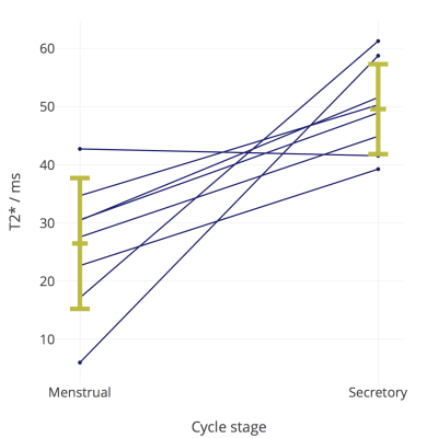
DCE-MRI and T2* measurement of the endometrium in the menstrual and secretory phases: preliminary data in women with normal menstrual bleeding
Lucy Kershaw, Jane Reavey, Scott Semple, Hilary Critchley, Jackie Maybin
Heavy menstrual bleeding (HMB) affects approximately 1 in 4 menstruating women per year in the UK and has a severe impact on sufferers’ quality of life. Medical treatments are available but side effects are common and the underlying physiological mechanisms of HMB remain unclear. We imaged eight women with normal menstrual bleeding during the menstrual and secretory phases of the cycle, measuring endometrial T2*, microvascular plasma flow and microvascular plasma volume. We saw a shorter T2* at menstruation, adding to the evidence from other techniques that hypoxia may be important in cessation of bleeding and endometrial repair.
|
|
4047.
 |
18 |
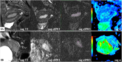
Value of the heterogeneity of intravoxel diffusion (a) in diagnosing stage IA and IB of endometrial carcinoma
Meng-yao Wang, Mei-yu Sun, Xu Han, Rui Fan, Lizhi Xie
Preoperative information about depth of myometrial invasion is essential in tailoring the surgical approach for endometrial carcinoma (EC) patients in stage I. To evaluate the value of stretched-exponential models of intravoxel incoherent motion (IVIM) in the differential diagnosis between stage IA and IB of endometrial carcinoma. In this work, we found it is feasible that IVIM sequence derived α value can be applied in the differential diagnosis between stage IA and IB of endometrial carcinoma, which can provide detailed information for clinical treatment.
|
|
4048.
 |
19 |
MR features including ADC values of Uterine Carcinosarcoma
Meng-yao Wang, Mei-yu Sun, Xu Han, Rui Fan
Uterine carcinosarcoma(UCS) is a rare and aggressive tumor with universally poor prognosis. To reinforce the knowledge of uterine carcinosarcoma(UCS) and improve the preoperative diagnostic accuracy on magnetic resonance imaging(MRI). A retrospective imaging review was performed of the MR images of 12 patients with uterine carcinosarcoma. In this study, we summarized its MRI features.
|
|
4049.
 |
20 |
MRI changes of pubic symphysis injuries in primiparous women
Piaoe Zeng, Jianyu Liu, Yan Zhou
Objective: MRI changes of pubic symphysis injuries of primiparas and its relationship with pubic symphysis pain and mode of delivery. Methods: 20 primiparas with MRI, were divided into cesarean, normal vaginal delivery and high-risk vaginal delivery. MR changes were observed. Results : pubic symphysis fibroadenoma avulsion was significantly correlated with pubic symphysis pain. Width of pubic symphysis of the primiparas was wider. primiparas had pubic bone marrow edema, which was not associated with pubic symphysis pain. 1 case pubic fracture. MRI changes were not associated with mode of labor. Conclusion: The severy pubic symphysis pain may be related to the symphysis pubis fibrocartilage avulsion and was not related to the pubis bone marrow edema.
|
|
4050
 |
21 |
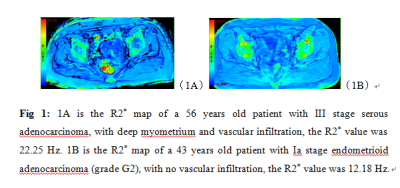
The value of R2* in quantitative evaluation the biological behavior of endometrial carcinoma: a preliminary study
Video Permission Withheld
Shifeng Tian, Ailian Liu
R2* value can be used as a MR non-enhancement quantitative index for evaluating the biological behavior of EC.
|
|
4051.
 |
22 |
DCE-MRI texture analysis in differentiating adenocarcinoma and squamous cell cancer of cervix
Presentation Not Submitted
Xie Yuanliang, Wang Xiang, Du Dan, Jiang Yanping, Sun Jianqing
Adenocarcinoma comprises 25% of cervical cancers and has a bad prognosis and poor outcome of radiotherapy and chemical treatment in the advanced stage. Here, we report a radiomics method with multi-class texture features from semi-quantitative DCE-MRI maps to distinguish adenocarcinoma from squamous cell cancer. Multivariate models were trained on the training cohort and their performance was evaluated on the 5-fold cross-validation cohort using the area under ROC curve (AUC), accuracy, specificity and sensitivity. Our results showed the mean sensitivity, specificity, PPV, NPV and AUC were 0.96, 0.889, 0.967, 0.889 and 0.967 respectively in diagnosing adenocarcinoma of cervix.
|
|
4052.
 |
23 |
Exploring analytical models of the diffusion MR signal in non-pregnant cervical tissue in 3T
Elisenda Bonet-Carne, Jana Hutter, Paddy Slator, Lucilio Cordero-Grande, Daniel Alexander, Andrew Shennan, Mary Rutherford, Eleftheria Panagiotaki, Lisa Story
Preterm Birth is a major healthcare problem and its appropriate prediction remains a challenge. Extensive research utilising advanced MR protocols has been undertaken on the cervical malignancy, but cervical structure and remodelling in term and preterm pregnancies is little studied. As part of a larger project, this work aims to explore model-based diffusion MRI microstructural models along the non-pregnant cervix. We compare multi-compartment microstructure models to analyse the diffusion signal. Initial results reveal that more complex models than the Apparent Diffusion Coefficient are required to characterise the cervical tissue microstructure.
|
|
4053.
 |
24 |
Cervix heterogeneity: recommendations for scanning the non-pregnant cervix
Elisenda Bonet-Carne, Jana Hutter, Daniel Alexander, Andrew Shennan, Mary Rutherford, Eleftheria Panagiotaki, Lisa Story
Imaging the non-pregnant cervix requires some unique considerations. The cervix has numerous functions including ensuring the fetus is maintained within the uterine cavity until the onset of labour. Alterations in the integrity of cervical tissue are purported to be one of the mechanisms which may contribute to preterm delivery. Quantitative MRI techniques can be used to characterise tissue microstructure however this has not been applied to date to the non-pregnant cervix in the absence of malignancy. This works aims to highlight some limitations and aspects to consider prior to scanning a non-pregnant cervix.
|
|
4054.
 |
25 |
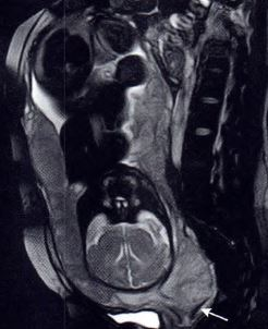
Pelvic endometriosis: The added value of the R2*MFGRE sequence in magnetic resonance imaging
Yifan Xu, Qing Wang
Purpose: The diagnostic accuracy of endometriosis by conventional magnetic resonance imaging (MRI) sequences is not satisfactory. Minimum endometriosis is particularly challenging to diagnose, which may lead to recurrence or incomplete relief of symptoms after surgery. The R2* multi-fast gradient echo (R2*MFGRE) sequence with the highest sensitivity of hemorrhage in endometriosis may help to address this problem. Methods: We selected 54 adenomyosis or endometriosis patients and referred them to MRI including the R2*MFGRE sequence and analyzed the results.
Conclusion: The analysis revealed that the R2*MFGRE sequence may provide useful information to help diagnosing endometriosis. |
|
| Top |
Fetal & Placental Imaging
Digital Poster
Body: Breast, Chest, Abdomen, Pelvis
Wednesday, 15 May 2019
| Exhibition Hall |
15:45 - 16:45 |
| |
|
Computer # |
|
4055.
 |
26 |
Fetal cardiac 4D phase-contrast MRI using Doppler ultrasound gating to visualize fetal hemodynamics in utero: preliminary results
Fabian Kording, Björn Schönnagel, Christian Ruprecht, Manuela Tavares de Sousa, Jin Yamamura, Daniel Giese
In this study 2D and 4D phase contrast MRI angiography was performed in six foetuses (gestational week 30-35) with direct cardiac gating using Doppler ultrasound at 1.5T. The fetal vasculature and hemodynamics could be visualised in detail including the ductus arteriosus. Blood flow was similar between 2D and 4D PCA. This method may provide an important additional tool for prenatal diagnosis of CHD such as coartation of the aorta.
|
|
4056.
 |
27 |
Feasibility of free-breathing T1-weighted 3D radial VIBE for fetal MRI in various anomalies
Taotao Sun, Ling Jiang, Zhongshuai Zhang, Mengxiao Liu, Marcel Nickel, Zhaoxia Qian
Traditional breath-hold, fat-suppressed T1-weighted sequences (T1W images) may be degraded by motion artifacts arising from both the mother and fetus. Images obtained with the radial VIBE sequence had a higher overall image quality score than Cartesian sampled 3D VIBE T1W images and less motion artifact in various fetal regions. Fetal lesions presented in radial VIBE images had good lesion conspicuity and edge sharpness.
|
|
4057.
 |
28 |
The reduction of perfusion fraction in placenta increta --a pilot study of placental perfusion using IVIM MRI
Presentation Not Submitted
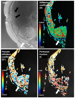
Ting Liu, Le Cao, Cao Jin, Xianjun Li, Hui Zhang, Xiaocheng Wei, Jian Yang
Placenta increta, a serious threat to maternal and fetal life, is difficult to distinguish by traditional MRI. In this study, IVIM imaging was used for the first time to detect the perfusion level of some placenta implanted, and it was found that the perfusion fraction was significantly lower than that of the normal placenta. That is help for establishing a new method for clinical diagnosis of placental implantation, especially placenta non-percreta.
|
|
4058.
 |
29 |
Diffusion weighted imaging evaluation of cerebral changes in twin pregnancies with one fetus demise
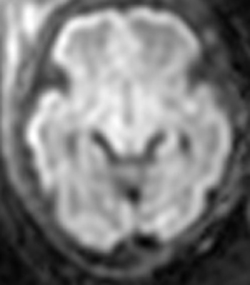
Ying Liu, Ran Huo, Huimin Xu, Zheng Wang, Huishu Yuan
Diffusion-weighted imaging (DWI) has been applied to fetal MRI and provides important information in evaluating the microstructures. Apparent diffusion coefficient (ADC) value is derived from DWI sequence. The aim of this study is to compare ADC values of brain in surviving fetus of one fetus demise with normal fetuses using diffusion weighted imaging and to find the differences and underlying cerebral microstructure changes. Decrease of ADC values were detected in surviving fetus of one twin demise. DWI and ADC values are very useful for detecting underlying changes, even changes of signal are not visible on conventional sequences.
|
|
4059.
 |
30 |
Joint placental angiography with perfusion and apparent oxygenation mapping
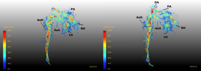
Laurence Jackson, Jana Hutter, Anthony Price, Alison Ho, Laura McCabe, Thomas Roberts, Maria Deprez, Lucy Chappell, Mary Rutherford, Enrico De Vita, Joseph Hajnal
In this work we combine two novel methods for imaging the oxygen transfer system between maternal and fetal circulations, 1) a motion robust angiography acquisition and 2) a combined perfusion and oxygenation acquisition. This enables the matching of functional regions of placenta to the vessels that supply them in utero. This may provide further insight into the vascular origins of major obstetric diseases such as fetal growth restriction and pre-eclampsia.
|
|
4060.
 |
31 |
Multislice and Multidimensional Blood Flow Mapping in Fetal Congenital Heart Disease Using Highly Undersampled Phase-Contrast MRI
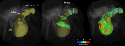
Datta Singh Goolaub, Eric Schrauben, Davide Marini, Christopher Roy, Mike Seed, Christopher Macgowan
In this study, multislice and multidimensional fetal blood flow mapping was performed using highly undersampled radial phase contrast MRI. The goal of this work was to visualize intracardiac flow in human fetuses with congenital heart disease, where complex flow patterns are difficult to capture with conventional, 2D phase contrast techniques. Reconstructions were performed using compressed sensing, with retrospective motion correction and image-based cardiac gating. The developed technique allowed whole fetal heart coverage in reasonable time and provides insight into multi-directional intracardiac flows. We present results from 5 human fetuses with congenital heart disease and demonstrate the first multi-directional intracardiac flows obtained by MRI.
|
|
4061.
 |
32 |
Changes in placental oxygenation in subclinical uterine contractions in the third trimester
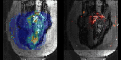
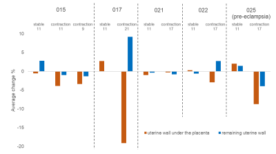
Neele Dellschaft, Simon Shah, Christopher Bradley, Lopa Leach, Nia Jones, Richard Bowtell, Penny Gowland
Subclinical uterine contractions in the third trimester have been detected with MRI in recent years and we regularly observe these contractions in 10 minute longitudinal scans. In a small pilot study, single echo-planar imaging T2* weighted scans and associated quantitative susceptibility maps suggest that the uterine contractions are localised to the placenta and that the placenta is more oxygenated after the contraction. We hypothesise that the contractions have the function of mixing of blood in the placenta to aid transport through what is otherwise a low flow system.
|
|
4062.
 |
33 |
Model-Driven Registration for 3D Placental Diffusion-Weighted MRI
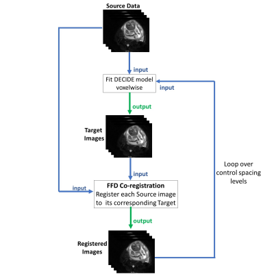
Dimitra Flouri, Rosalind Aughwane, Magdalena Sokolska, Nada Mufti , David Atkinson, Giles Kendall, Alan Bainbridge, Tom Vercauteren, Sebastien Ourselin, Anna David, Andrew Melbourne
The placenta is essential for successful pregnancy outcome. Placental imaging is however challenging due to maternal breathing motion and fetal movements, making motion correction important for subsequent analysis. This study introduces an iterative model-based registration technique which incorporates a joint placenta-specific signal model for diffusion and relaxation data into the motion correction process. We compared our method to a pre-existing method used for contrast-enhanced data making use of principal component analysis. Our results suggest that the proposed method significantly improves alignment of free-breathing placenta relaxation and DW-MRI data and evidence that the precision of markers of function obtained in placenta can be improved.
|
|
4063.
 |
34 |
Fetal dynamic phase-contrast angiography using Doppler ultrasound gating and comparison with Doppler ultrasound measurements
Jin Yamamura, Fabian Kording, Roland Fischer, Gerhard Adam, Christian Ruprecht, Kai Fehrs, Manuela Tavares de Sousa, Björn Schönnagel
In this prospective study 2D PC-MRI angiography was performed in nine human fetuses (gestational age 32.2 ± 2.4 weeks) using a novel MR compatible DUS device for gating of the fetal heart at a 1.5T MR scanner. The novel MR compatible DUS device for fetal cardiac gating allowed successful application of PC-MRI angiography and quantification of hemodynamics in the fetal AoD. This new technique for direct gating of the fetal heart demonstrates the diagnostic potential of PC-MRI for the quantification of fetal hemodynamics.
|
|
4064.
 |
35 |
Pattern Analysis of Placental Blood Flow Distribution using ASL MRI
Eileen Hwuang, Nadav Schwartz, Walter Witschey, John Detre, Dylan Tisdall
Arterial Spin Labeling MRI is a promising approach to assess blood flow to the placenta. Although previous studies have largely attempted to quantify global perfusion, we believe that the unique physiology of blood flow through the placental intervillous space rather than through capillaries warrants regional pattern analysis. We present an image analysis framework leveraging a spline-based transform of the image coordinates, watershed segmentation, and clustering analysis. We report Bayesian statistics to quantify features of blood flow distribution and the degree of uncertainty at the uteroplacental interface.
|
|
4065.
 |
36 |
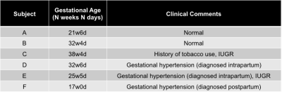
Measurement of Uterine Artery and External Iliac Artery Pulse Wave Velocity using 4D Flow MRI
Eileen Hwuang, Nadav Schwartz, Dylan Tisdall, John Detre, Walter Witschey
Maintenance of blood supply to the placenta is believed to be dependent on the geometric and hemodynamic properties of the uteroplacental vasculature. In this study we present an MRI method for measuring pulse wave velocity in the uterine arteries. In 6 healthy pregnant subjects, we measured path length and time-to-peak of the velocity waveforms in the uterine arteries and external iliac arteries. The uterine arteries have lower pulse wave velocity than the external iliac arteries (5.5±2.5 vs. 12.9±4.6 m/s, p=3x10⁻⁵), indicating possible biomechanically greater compliance.
|
|
4066.
 |
37 |
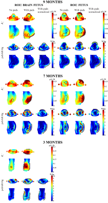
Optimized High-Permittivity Pads Can Reduce SAR and Increase Transmit Field Homogeneity in Fetal Imaging at 3T
Jeroen Gemert, Wyger Brink, Rob Remis, Andrew Webb
One of the main concerns in fetal MRI is the amount of power that is deposited both in the fetus and the mother. In this work, we show that a well-designed high-permittivity pad can tailor the energy of the RF field such that the transmit efficiency increases and the field becomes more homogeneous. The increase in transmit efficiency subsequently enables a substantial reduction in the SAR in both the mother and the fetus for models of the third, seventh, and ninth months of gestation.
|
|
4067.
 |
38 |
Classification and prognosis of fetal hepatic hemangioendothelioma using MRI
Cong Sun, Xinhong Wei, Xin Chen, Yufan Chen, Jinxia Zhu, Ruiqin Shan, Guangbin Wang
The aim of this study was to investigate the MRI features and classification of 23 fetal hepatic hemangioendothelioma (HAE) and to compare the prognosis of HAE-I and HAE-II. The results showed that fetal HAE-I tends to be more vascular in the lesion, and the larger the focus, the more prone to complications. Once complications occur, the prognosis is poor; however, in HAE-II, the lesions are always smaller, there is a lack of vascularity, they are homogeneous, and the prognosis is good. Our study demonstrated that the MRI features and classification of HAE can effectively predict and guide prognosis.
|
|
4068.
 |
39 |
Assessment of placental microcirculation by joint analysis of flow compensated and non-flow compensated intravoxel incoherent motion data
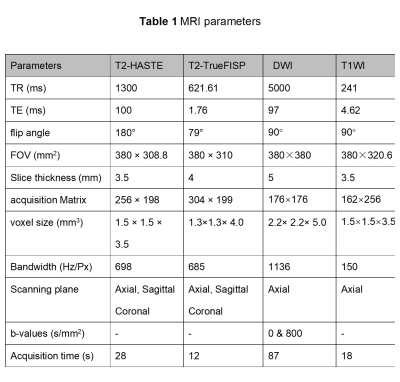
Esra Abaci Turk, Borjan Gagoski, Jeffrey Stout, S. Mazdak Abulnaga, Natalie Copeland, Drucilla Roberts, Polina Golland, Lawrence Wald, Elfar Adalsteinsson, William Barth Jr, P. Ellen Grant, Yogesh Rathi
Using diffusion weighted imaging and the intravoxel incoherent motion model (IVIM) of blood flow in capillaries, we can measure placental properties relating to maternal and fetal blood flow and perfusion. In this study, we focused on improving the accuracy and precision of the estimated parameters in IVIM imaging by using joint analysis of flow compensated and non-flow compensated diffusion data. With flow compensation, we observed strong re-phasing, approximately cancelling the blood flow effect and allowing more accurate and consistent estimation of diffusion and perfusion measures.
|
|
4069
 |
40 |
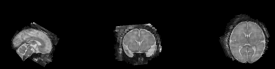
Fetal Brain-Heart-Placental Interactions with Acute Hypoxia Challenge in Genetic Mouse Models of Hypoplastic Left Heart Syndrome with in utero 4D Dynamic MRI
Video Permission Withheld
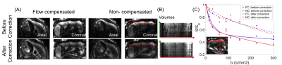
Anthony Christodoulou, George Gabriel, Cecilia Lo, Yijen Wu
The objective of this study is to develop 4D time-resolved high-resolution in utero MRI for simultaneous interrogation of placenta, heart and brain development and function in genetic mouse models of hypoplastic left heart syndrome. The structural and functional imaging of the fetal heart, brain, and placenta can be correlated with genotype. In addition, temporal and spatial characteristics of BOLD responses to acute hypoxia challenge can be derived.
|
|
4070.
 |
41 |
Comparison of fetal lung diffusivity in cases of pulmonary hypoplasia to normative MRI data
Clemente Velasco-Annis, Ali Gholipour, Judy Estroff, Richard Parad, Terry Buchmiller, Carol Barnewolt, Sila Kurugol, Simon Warfield, Onur Afacan
Typically the fetal lungs are measured using structural magnetic resonance (MRI) or ultrasound images, but improvements in fetal MR imaging have made diffusion weighted imaging (DWI) feasible for even fetal patients. Previously it was shown that lung diffusion could be reliably measured for fetuses with normal lung development and that diffusivity increased with gestational age (GA). In this study we applied the same techniques to measure lung diffusivity in patients indicated for congenital diaphragmatic hernias. We then compared diffusivity measurements for both the herniated-side, left lung and right lung to normative lung diffusivity data.
|
|
4071.
 |
42 |
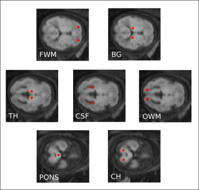
Fetal Brain Tissue Characterization at 1.5 T using STAGE
Feifei Qu, Taotao Sun, Yongsheng Chen, Brijesh Yadav, Ling Jiang, Zhaoxia Qian, Ewart Haacke
MRI-based tissue properties such as T1 and proton spin density are known to be important biomarkers of fetal brain maturation. However, fetal motion poses a big challenge in MRI due to long scan durations. Henceforth, there is lack of knowledge about the MRI properties of the human fetal brain tissue. To address these issues, in this study we use a rapid multiparametric method referred to as Strategically Acquired Gradient Echo (STAGE) imaging to obtain T1 and proton density maps of the fetal brain and generate T1-weighted images with enhanced tissue contrast in two minutes.
|
|
4072.
 |
43 |
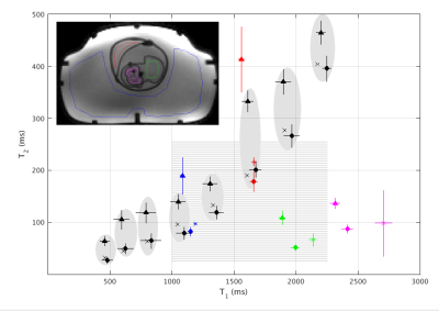
T1 and T2 mapping of the placenta and fetal brain with magnetic resonance fingerprinting (MRF) during maternal hyperoxia
Jeffrey Stout, Congyu Liao, Esra Abaci Turk, Borjan Gagoski, P. Grant, Lawrence Wald, Elfar Adalsteinsson
Quantitative MRI may improve diagnosis and monitoring of placental disease by characterizing baseline oxygen content and dynamic oxygen transport during hyperoxia. MRF permits fast quantitative imaging of the adult brain and heart, but use during pregnancy targeting the placenta and fetal brain presents challenges due to motion and large B1+ variation. We tested the accuracy of MRF in phantoms and then scanned pregnant mothers, estimating T1 and T2 of the placenta and fetal brain. MRF-based T1and T2 mapping is a promising technique to determine placental oxygenation at baseline and oxygenation changes in the placenta and fetal brain after hyperoxia.
|
|
4073
 |
44 |
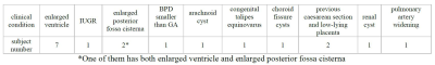
Reliable assessment of apparent diffusion coefficient in normal and ventriculomegaly fetal brains
Video Permission Withheld
Maria Giovanna Di Trani, Lucia Manganaro, Amanda Antonelli, Michele Guerreri, Riccardo De Feo, Mattia Borrelli, Giacomo Pratesi, Carlo Catalano, Silvia Capuani
Ventriculomegaly (VM) is one of the most common disease in fetuses; however, DWI studies during gestation are scarce. In this work, DWI was performed in normal and VM fetal brains at different gestational ages (GA) and ADC was calculated. Denoising and artifacts corrections were used to improve signal-to-noise ratio and the reliability of measures. We found significant variations in ADC as GA increases, reflecting microstructural changes due to brain maturation. Moreover, ventricles dilatation measured on DWIs provided an accurate classification of VM. DWI with artifacts corrections could be a powerful tool to evaluate brain abnormalities and accurately diagnose VM.
|
|
4074
 |
45 |
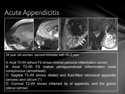
Why MRI? Evaluating Acute Abdominal and Pelvic Pain During Pregnancy.
Video Permission Withheld
Elyssa Cohen, Frank Miller, Courtney Moreno, Thomas Estes, Pardeep Mittal
A variety of pathological processes can be the cause of abdominopelvic pain during pregnancy, such as acute appendicitis, acute cholecystitis, hepatic adenoma, spontaneous adrenal hemorrhage, ovarian mature cystic teratoma, ectopic pregnancy, cystic degeneration of an intramural fibroid, and the placenta accreta spectrum. Standard MRI protocols without gadolinium contrast are discussed, as well as protocols for fluid sensitive sequences with and without fat suppression, T1-weighted sequences, and thin slice sequences. These MR imaging techniques are valuable in determining an accurate diagnosis and subsequent management plan for the mother and fetus.
|
|
4075.
 |
46 |
Fetal Brain Response to an Auditory Stimulus
Estee Goldberg, Charles McKenzie, Barbra de Vrijer, Roy Eagleson, Sandrine de Ribaupierre
Functional magnetic resonance imaging (fMRI) was used in conjunction with an auditory task to assess fetal activation in the primary auditory cortex. Activation was found in the fetal right Heschl’s gyrus in response to an auditory task. This is a first report of fetal brain response to an “internal” auditory stimulus.
|
|
4076.
 |
47 |
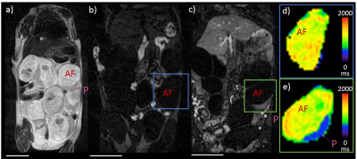
Novel Nanoparticle-enhanced T1-Mapping Enables Estimation of Placental Fractional Blood Volume in a Pregnant Mouse Model
Andrew Badachhape, Laxman Devkota, Igor Stupin, Mayank Srivastava, Prajwal Bhandari, Poonam Sarkar, Ketan Ghaghada, Eric Tanifum, Ananth Annapragada
Greater than 60% of placentae from low birth weight infants show signs of hypoxic or ischemic injury from vascular hypo-perfusion. Placental fractional blood volume (FBV) is indicative of perfusion and may be used as a marker of local ischemia. Non-invasive methods for the estimation of placental FBV are therefore of interest in the study of placental pathology. In this pre-clinical study, we investigated contrast-enhanced magnetic resonance imaging (MRI) for the estimation of placental FBV in a pregnant mouse model. A high T1 relaxivity blood-pool liposomal-gadolinium (liposomal-Gd) contrast agent, which does not permeate placental barrier in rodents, was used to calculate placental FBV.
|
|
4077.
 |
48 |
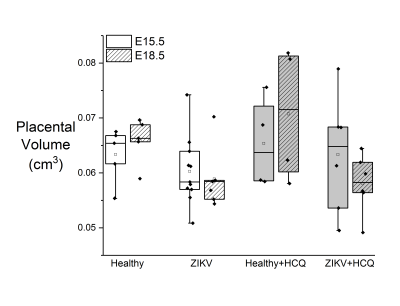
Functional and Developmental Effects of Zika Virus Infection and Hydroxychloroquine Treatment in Mouse Placenta
Kelsey Meinerz, Brooke Liang, James Quirk, Indira Mysorekar, Joel Garbow
Functional and developmental effects of Zika virus (ZIKV) infection and hydroxychloroquine (HCQ) treatment on the murine placenta were measured using MRI and oxygen enhancement to assess placental volume and oxygenation. HCQ treatment helps to prevent adverse placental volume outcomes earlier in gestation, but late-gestation results are similar to those of ZIKV-infected dams. R1 and R2* measurements show that HCQ has a significant effect on placental oxygenation throughout gestation, whereas ZIKV does not significantly affect these metrics.
|
|
| Top |
Lung: Proton-Based Imaging
Digital Poster
Body: Breast, Chest, Abdomen, Pelvis
Wednesday, 15 May 2019
| Exhibition Hall |
15:45 - 16:45 |
| |
|
Computer # |
|
4078.
|
51 |
Comparison of Quantitative Pulmonary Ventilation Imaging using 3D-UTE under breath-holding and free-breathing in patients with structural lung disease at 3 T.
Julius Heidenreich, Andreas Weng, Corona Metz, Lenon Mendes Pereira, Thomas Benkert, Josef Pfeuffer, Thorsten Bley, Herbert Köstler, Simon Veldhoen
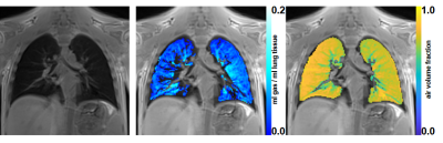
UTE-MRI allows morphologic examination of lung parenchyma and acquisition of different breathing states enables determination of quantitative pulmonary ventilation (QV). In 4 patients with lung disease imaging with a prototypical UTE Spiral VIBE sequence during breath-holding was compared with a free-breathing 3D-UTE koosh-ball technique. Different breathing states were acquired, and QV values and the air volume fraction (AVF) were calculated. Both techniques yielded clinically sufficient image quality and comparable results for QV and AVF values. While the breath-hold approach acquires the data in a fraction of time but is dependent on patient compliance, the self-gated free-breathing technique overcomes that problem with the drawback of a longer acquisition time.
|
|
4079.
 |
52 |
Validation of Fourier Decomposition Lung MR Perfusion using Breath-hold Correction
Maria Liljeroth, Maria Duvaldt
Co-registration of free breathing coronal lung images in Fourier decomposition (FD) lung MRI is a crucial part of the processing pipeline for this technique. It is vital that the registration works on the correct parts of the image and does not interfere with the data in an unfavourable way. This work aims to evaluate the extent to which registration affects the perfusion, and by association the ventilation results of this technique, using a breath-hold acquisition as a gold standard. Results encourage the generation of confidence maps to aid interpretation of FD lung MR results, particularly in regions affected by registration.
|
|
4080.
 |
53 |
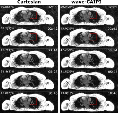
Free-Breathing Self-Gated 4D-Lung MRI with wave-CAIPI
Julian Richter, Tobias Wech, Andreas Weng, Manuel Stich, Simon Veldhoen, Stefan Weick, Thorsten Bley, Herbert Köstler
The wave-CAIPI technique was applied to self-gated dynamic Lung MRI during free breathing. To prevent synchronization between respiratory motion and phase-encoding, the order of the phase-encoding steps was randomized using non-uniformly distributed, two-dimensional random numbers which oversample the k-space center. Breathing motion was gated using an additionally recorded DC signal. A healthy volunteer was investigated using the proposed method and a standard Cartesian scan for reference. For a scan time of 10:46 min, both methods exhibit comparable image quality. However, for accelerated scans, the wave-CAIPI technique clearly shows reduced residual image artifacts after applying parallel imaging.
|
 |
4081.
 |
54 |
Real-Time Imaging during Free Breathing for Patient-Friendly V/Q Scan of the Whole Lung in One Minute at 3T
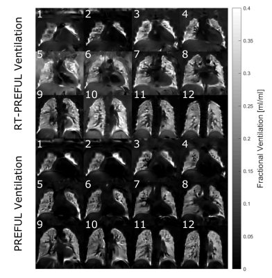
Andreas Voskrebenzev, Till Kaireit, Marcel Gutberlet, Filip Klimes, Lea Behrendt, Christoph Forman, Frank Wacker, Jens Vogel-Claussen
MRI techniques like phase-resolved functional lung imaging (PREFUL) offer the possibility for pulmonary ventilation (V) / perfusion (Q) imaging without ionizing radiation or contrast agents. One major drawback of such methods is the long acquisition time, if a whole lung coverage is required. In this study, five healthy volunteers were scanned with a highly accelerated 2D multi-slice real-time PREFUL (RT-PREFUL) acquisition with compressed sensing and parallel imaging reconstruction to establish a V/Q scan of the whole lung in one minute. The comparison with conventional PREFUL technique showed very similar ventilation results and comparable perfusion results with 9-fold acceleration.
|
|
4082.
 |
55 |
Magnetization transfer and diffusion-weighted imaging in differential diagnosis of solid pulmonary masses: A comparison study
Presentation Not Submitted
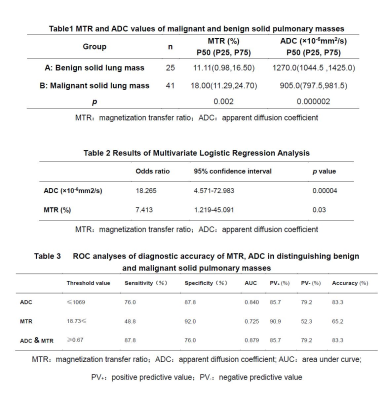
Shan Dang, Haifeng Duan, Dong Han, Nan Yu, Qi Yang, Shaoyu Wang
In this study, magnetization transfer (MT) and diffusion-weighted imaging(DWI) techniques were used to differentiate the solid pulmonary masses. It was found that magnetization transfer can be used to distinguish benign and malignant pulmonary masses. Combining magnetization transfer and diffusion-weighted imaging, the accuracy of differentiation can be further improved.
|
|
4083.
 |
56 |
Imaging functional and microstructural changes in the lungs of children born prematurely
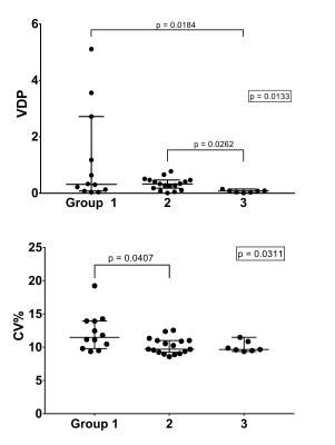
Jim Wild, Alberto Biancardi, Ho-Fung Chan, Laurie Smith, Jody Bray, Helen Marshall, Paul Hughes, Guillem Collier, Graham Norquay, Andrew Swift, Kylie Hart, Michael Cousins, Sailesh Kotecha
Preterm-births account for 10% of worldwide births, and those born at 32 weeks gestation or less have deterioration in their lung function in childhood and beyond. Here we assessed pulmonary micro-structural and functional changes using hyperpolarized 129Xe gas ventilation and diffusion-weighted MRI in three groups of children: (1) those born preterm with FEV1<=85%, (2) born preterm with normal lung function and (3) those born full term with normal lung function. Significant differences in ventilation heterogeneity and the kurtosis of the distribution of mean alveolar dimension were observed between groups.
|
|
4084.
 |
57 |
A Comprehensive Pipeline for non-contrast enhanced Pulmonary Vascular 1H MRI
Alexander Matheson, Dante Capaldi, Fumin Guo, David McCormack, Grace Parraga
Structure-function relationships are central to our understanding of pulmonary disease and play an important role in evaluating novel therapies. Many chronic lung diseases, such as asthma and chronic obstructive pulmonary disease (COPD), have vascular abnormalities that are not accounted for in current image analysis platforms. Recent MRI developments provide complementary pulmonary vascular structural and functional information and include ultra-short echo time and Fourier-decomposition MRI. To facilitate clinical translation, we developed a comprehensive structure-function pipeline to visualize and measure pulmonary vascular vessel trees and perfusion for patients with severe asthma, a demanding clinical target.
|
|
4085.
 |
58 |
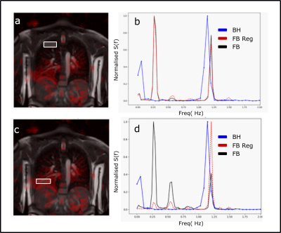
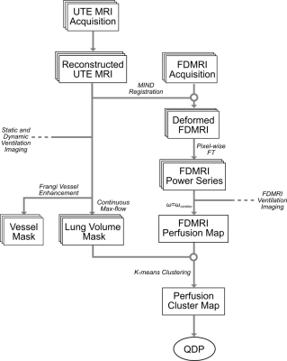
Flow-Compensated Ventilation and Perfusion Fourier-decomposition Pulmonary MRI
Alexander Matheson, Dante Capaldi, Fumin Guo, Grace Parraga
Fourier-decomposition pulmonary MRI ventilation and perfusion maps are subject to substantial flow artifacts stemming from cardiac frequencies which makes lung perfusion measurements complex and in some regions of interest, impossible to acquire. We eliminated flow artifacts by shimming only on cardiac regions, instead of the entire field-of-view. Flow compensation improved vessel resolution and segmentation around the heart but also increased the incidence of off-resonance banding artifacts around the diaphragm. MRI ventilation defect percent was greater in flow-compensated versus uncompensated images as well as hyperpolarized gas MRI, demonstrating that ventilation images are highly sensitive to changes in field inhomogeneities.
|
|
4086.
 |
59 |
Self-Gated 2D UTE Lung Ventilation Imaging
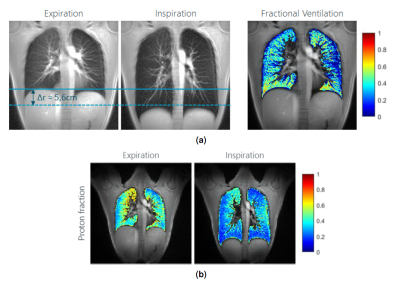
Anke Balasch, Patrick Metze, Kilian Stumpf, Tobias Speidel, Volker Rasche
Deriving functional lung data by MRI is challenging, but initial studies have proven the applicability of multi-respiratory stage data for deriving fractional lung ventilation data. In this study a two-dimensional ultra-short TE protocol was used to acquire breathhold (expiration and inspiration) and free breathing date of the lung parenchyma. Lung proton density changes and respective fractional ventilation could be assessed with either techniques. Due to the shallow breathing, in the free-breathing approach differences resulted less pronounced as in the breathhold approach.
|
|
4087.
 |
60 |
Comparison of 1.5T and 3T 2D UTE Breathhold Lung Imaging
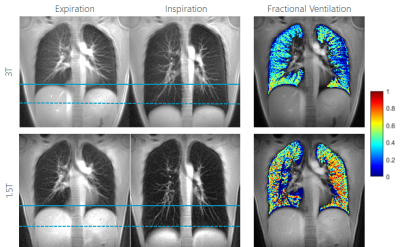
Anke Balasch, Patrick Metze, Kilian Stumpf, Tobias Speidel, Volker Rasche
In this study a breathhold two-dimensional ultra-short TE (UTE) protocol was compared for imaging of the lung parenchyma and ventilation at 1.5T and 3T field strength. The resulting SNR values and parameter maps indicate a clear advantage for 3T. Proton fraction and SNR changes (anterior to posterior, expiration vs. inspiration) can be clearly assessed with both field-strength. Different resulting values for the lung and proton fractions need further attention.
|
|
4088.
 |
61 |
2D UTE BH lung ventilation imaging at 3T – influence of the repetition time
Anke Balasch, Patrick Metze, Kilian Stumpf, Tobias Speidel, Volker Rasche
In this study a two-dimensional ultra-short TE protocol was used to get breath holding (expiration and inspiration) images. It was investigated whether resulting SNR and ventilation and proton density maps can be improved by optimization the TR under given breathhold constraints for multi-slice single breathhold lung imaging. Even though the SNR in the resulting images can clearly be improved by performing an interleaved multi-slice acquisition instead of acquiring slices sub-sequentially, the impact on the resulting parameter maps was not obvious.
|
|
4089.
 |
62 |
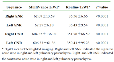
MultiVane Technique at 1.5T Improved the Quality of Lung MRI: Comparison with Routine T2-Weighted MRI and CT Imaging
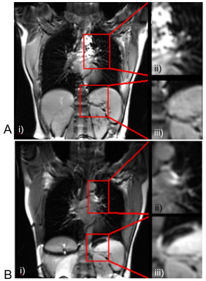
Liya Wang, Zhou Liu, Guangle Shen, Jianhua Li, Yuzhong Zhang, Lanjin Lai, Hui Mao
Radiation-free MRI with MultiVane motion correction technique improved the lung MR imaging quality with satisfactory quality compared to routine T2 weighted imaging or CT imaging. In this study, MultiVane lung MRI protocol provided diagnostic performance acceptable in detection of several common lung abnormalities with a good agreement to the findings obtained in routine clinical CT examinations, especially to identify caseation or liquefactive necrosis. However, MRI is less desirable to detect the nodules with calcification. The current lung MRI with non-radiation is feasible. It offers a promising alternative to the clinical standard CT in diagnosis and characterization of pulmonary diseases.
|
|
4090.
 |
63 |
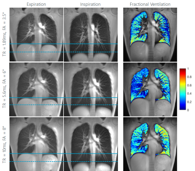
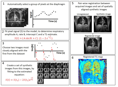
Free breathing lung T1 mapping in idiopathic pulmonary fibrosis
Laura Saunders, Andy Swift, James Eaden, James Wild
A method for free breathing Look-Locker T1 mapping using image registration is presented. This method uses a fitted model inversion recovery signal modulated with respiratory motion to create a set of synthetic images, to which all acquired free-breathing Look-Locker images are registered. The method was implemented in 9 patients with idiopathic pulmonary fibrosis (IPF) and 9 healthy volunteers and the results compared to T1 maps acquired with the same Look-Locker sequence acquired in an inspiratory breathold state for patients with IPF, and inspiratory and expiratory breath hold states for healthy volunteers. Patients with IPF were found to have a lower T1 during free breathing and a higher standard deviation of regional T1 values in the lung when compared to volunteers.
|
|
4091.
 |
64 |
Pseudo-diffusion effects in lung MRI
Lukas Buschle, Christian Ziener, Thomas Kampf, Volker Sturm, Ke Zhang, Sabine Heiland, Martin Bendszus, Heinz-Peter Schlemmer, Felix Kurz
Magnetic resonance imaging of lung tissue is strongly influenced by susceptibility effects between spin-bearing water molecules and air-filled alveoli. The measured lineshape, however, also depends on the blood-flow around alveoli that can be approximated as pseudo-diffusion. Both effects are quantitatively described by the Bloch-Torrey-equation, which was so far only solved for dephasing on the alveolar surface. In this work, we extend this model to the whole range of physiological relevant air volume fractions. The results agree very well with in vivo measurements in human lung tissue.
|
|
4092.
 |
65 |
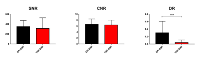
Intravoxel incoherent motion diffusion-weighted Imaging of lung: comparison between turbo spin-echo and echo-planar imaging
Qi Wan, Xinchun Li, Qiang Lei, Tianjing Zhang, Zhongping Zhang
The image quality of single-shot echo-planar imaging (EPI)-DWI (SS-EPI-DWI) is usually deteriorated by susceptibility artefacts, especially in lung, because EPI sequence is vulnerable to magnetic inhomogeneity and phase error accumulation. This could lead to inabilities of diagnosis or inaccuracy of measurements. In this study, lung DWI were acquired using both single-shot turbo spin-echo (SS-TSE) and SS-EPI techniques. Significantly changes were observed in distortion ratio (DR) between both techniques. SS-TSE-DWI has better image quality and almost zero distortion compared to SS-EPI-DWI.
|
|
4093.
 |
66 |
Assessment of scan parameter effects on time resolved bSSFP acquisition for Fourier Decomposition Pulmonary MR
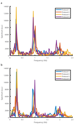
Efe Ilicak, Jascha Zapp, Lothar Schad, Frank Zoellner
Pulmonary functions play an important role in diagnosis of pulmonary diseases. One method for functional lung MRI is Fourier Decomposition technique, which obtains ventilation and perfusion weighted images from dynamic free-breathing acquisitions. Although this method is validated against well-established methods, its robustness depends on scan parameters and temporal resolution. In this work, we have investigated the effects of common scan parameters on image quality. In vivo results are presented to demonstrate the performance differences in protocols.
|
|
4094.
 |
67 |
Quantitative Pulmonary Ventilation Imaging in Patients with Cystic Fibrosis using breath-hold 3D-UTE at 3T
Simon Veldhoen, Julius Heidenreich, Andreas Weng, Tobias Wech, Florian Segerer, Helge Hebestreit, Thomas Benkert, Josef Pfeuffer, Thorsten Bley, Herbert Köstler
We demonstrate first results of breath-hold 3D-UTE MRI for ventilation imaging in CF patients. Ten patients underwent 3D-UTE MRI of the lungs. Data were acquired in breath-hold for 5 different breathing states, in order to obtain information regarding the ventilation. 3D-UTE MRI provided sufficient morphologic image quality for the detection of structural lung disease and site-resolved information regarding pulmonary ventilation. Ventilation measurements correlated well with parameters of conventional lung function testing and were in accordance with pulmonary ventilation measured with techniques published recently. The technique could be an innovative method able to enhance diagnosis and monitoring of structural lung disease.
|
|
4095.
 |
68 |
Free-Breathing Dynamic Contrast-Enhanced Magnetic Resonance Imaging of the Lung Using Compressed-Sensing VIBE
Presentation Not Submitted
Yu Zheng, Shi-hong Li, Guang-wu Lin, Cai-xia Fu, Marcel Nickel
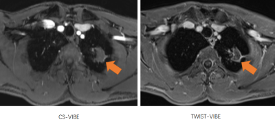
The purpose of this study was to compare the image quality of free-breathing CS-VIBE and TWIST-VIBE in patients with lung disease. In patients who were able to hold their breath well, the images quality of CS-VIBE and TWIST-VIBE were comparable (P>0.05). In patients who failed to hold their breath during the required period, CS-VIBE allowed for superior image quality, lesion delineation, artifact level, and diagnostic confidence compared to TWIST-VIBE. Our study demonstrated that free-breathing CS-VIBE is feasible for lung imaging for patients who have difficulties holding their breath.
|
|
4096
 |
69 |
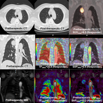
Comparison of Therapeutic Effect Prediction among Dynamic Perfusion Area-Detector CT, Dynamic Perfusion MR Imaging and FDG-PET/CT in Small Cell Lung Cancer Patients with Limited Disease
Video Permission Withheld
Yoshiharu Ohno, Masao Yui, Shigeharu Ohyu, Yasuko Fujisawa, Shinichiro Seki, Takeshi Yoshikawa, Katsusuke Kyotani, Takamichi Murakami
No direct comparisons of therapeutic effect prediction have been performed among dynamic perfusion ADCT, dynamic perfusion MRI and FDG-PET/CT in small cell lung cancer (SLCLC) patients. We hypothesize that multiparametric approach by dynamic CE-perfusion ADCT, MRI and FDG-PET/CT can improve therapeutic effect prediction capability in SCLC patients, when compared with each modality assessment. The purpose of this study was to directly compare the capability for therapeutic outcome prediction among dynamics CE-perfusion ADCT and CE-perfusion MRI assessed by same mathematical method and FDG-PET/CT in SCLC patients assessed as limited disease (LD).
|
|
4097
 |
70 |
Comparison of the Capability for Distinguishing Malignant from Benign Pulmonary Nodules among DWI with FASE Sequence at a 3T System, DWIs with EPI Sequence at 1.5T and 3T Systems and FDG-PET/CT
Video Permission Withheld
Yoshiharu Ohno, Masao Yui, Yoshimori Kassai, Takeshi Yoshikawa, Shinichiro Seki, Katsusuke Kyotani, Takamichi Murakami
No one have been compared the capability between DWI obtained by EPI and FASE at 3T or 1.5T systems and PET/CT in this setting. We hypothesized that DWI obtained by FASE sequence at 3T system are more useful than that by EPI sequence at 1.5T and 3T systems and PET/CT for diagnosis of SPN. The purpose of this study was to directly and prospectively compare the quantitative capability for diagnosis of SPN among DWI with FASE and EPI sequences at 3T system, DWI with EPI sequence at 1.5T system and FDG-PET/CT.
|
|
4098
 |
71 |
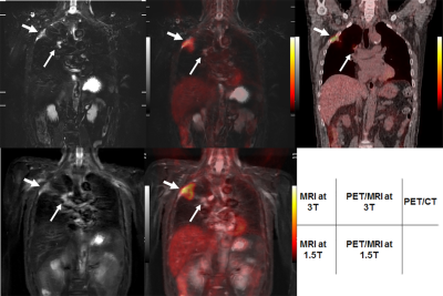
Whole-Body MRI and PET/MRI at 1.5T and 3T MR System: Comparison of TNM Stage Assessment Capability with PET/CT in Non-Small Cell Carcinoma (NSCLC)
Video Permission Withheld
Yoshiharu Ohno, Takeshi Yoshikawa, Masao Yui, Kota Aoyagi, Yoshimori Kassai, Shinichiro Seki, Katsusuke Kyotani, Takamichi Murakami
No one reported direct comparisons for TNM staging capability among whole-body MRI and PET/MRI at 1.5T and 3T systems and PET/CT in NSCLC patients. We hypothesize that whole-body MRI and PET/MRI at 1.5T and 3T MR systems have better potential for TNM stage assessment than whole-body FDG-PET/CT in NSCLC patients. The purpose of this study was to prospectively and directly compare TNM stage classification capability among whole-body MRI and PET/MRI at 1.5 and 3T MR systems and PET/CT in NSCLC patients.
|
|
4099.
 |
72 |
Free-Breathing Magnetic Resonance Elastography of the Lung: A Repeatability and Reproducibility Study.
Faisal Fakhouri, Stephan Kannengiesser, Josef Pfeuffer, Arunark Kolipaka
Lung stiffness is altered in many diseases making it an important biomarker of understanding lung mechanics. Many patients struggle to hold their breath during scanning due to shortness of breath. We have developed a free-breathing spin-echo-spiral magnetic resonance elastography (MRE) sequence, and a feasibility study was performed on 5 healthy volunteers. It was found that the shear stiffness of the lung during normal breathing was 1.9±0.51kPa and 8.87±1.44kPa with and without considering lung density correction, respectively. Also the measurements were repeatable and reproducible.
|
|
4100.
 |
73 |
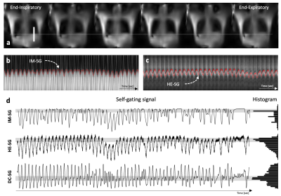
Half-echo respiratory navigator for motion suppressed 3D ultrashort echo time image for Lung imaging
Pan Ki Kim, Young-Jung Yang, Yoo Jin Hong, Chul Hwan Park, Dong Jin Im, Donghyun Hong, Byoung Wook Choi
In this study, we proposed a new self-gating signal to obtain a high-resolution 3D lung UTE image with suppressed respiratory motion artifacts.This half-echo self-gating (HE-SG) signal can monitor the position of the diaphragm with high spatial and temporal resolution acquired for every TR without any increasing the scan time. Also, this HE-SG can automatically estimate the respiratory motion of the subject so that it can be reconstructed seamlessly for high-quality lung images or time-resolved lung images.
|
|
4101.
 |
74 |
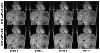
Dynamic 3D lung MRI using the stack-of-stars sequence with SI navigation
Shuo Li, Xi Chen, Kang Yan, Xudong Chen, Xiaomao Gong, Jian Xu, Qi Liu, Quan Chen, Huajun She, Yiping Du
Lung MRI has great potential in clinical early lung cancer screening and staging as it has a high detection rate for pulmonary nodules measuring. However, conventional lung MRI was limited by the physiological motion including respiration and heart beating. In this study, a free-breathing 3D golden-angle radial stack-of-stars pulse sequence with superior-inferior (SI) navigation was proposed for 3D dynamic lung MRI. Lung images in different respiratory stages were obtained. The proposed method provided higher SNR, CNR (between pulmonary vessels and the neighboring background in lung) and fewer motion artifacts than that without SI navigation.
|
|
4102.
 |
75 |
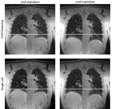
UTE-SENCEFUL with a coil clustering algorithm for automatic DC gating
Lenon Mendes Pereira, Andreas Weng, Tobias Wech, Simon Veldhoen, Thorsten Bley, Herbert Köstler
In this work UTE-SENCEFUL is combined with a technique for automatic DC gating to produce ventilation weighted maps of the lung in 3D and in high-resolution.
|
|
| Top |
Breast: Technical
Digital Poster
Body: Breast, Chest, Abdomen, Pelvis
Wednesday, 15 May 2019
| Exhibition Hall |
15:45 - 16:45 |
| |
|
Computer # |
|
4103.
 |
76 |
Investigating the feasibility of performing quantitative DCE-MRI in an abbreviated breast examination
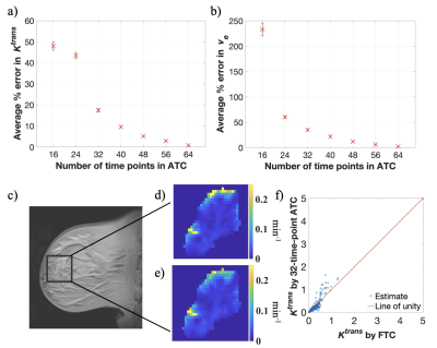
Kalina Slavkova, Julie DiCarlo, Anum Syed, John Virostko, Anna Sorace, Thomas Yankeelov
MRI is the most sensitive imaging modality for detecting breast cancer, but its use as a screening tool is limited. There has been recent interest in developing an “abbreviated” breast MRI protocol as a screening protocol, which involves a significantly shorter breast MRI exam that does not compromise diagnostic accuracy. In this work, we evaluate the accuracy of estimating perfusion parameters from dynamic contrast-enhanced MRI data by truncating the data into a series of abbreviated-time courses and comparing the corresponding parameter estimates to the original, full-time course parameter estimates.
|
|
4104.
 |
77 |
Quantitative assessment of changes in breast background parenchymal enhancement following tamoxifen treatment
Milica Medved, Keiko Tsuchiya, Gregory Karczmar, Hiroyuki Abe
Our study introduces a standardizing model of background parenchymal enhancement (BPE) in DCEMRI sequences that can be used to quantitatively and objectively describe changes in BPE rates following preventative tamoxifen treatment. This is in contrast with the current practice of using 4 subjective categories to describe BPE. Decreased BPE rates post-treatment agree with earlier results showing that BPE, like breast density, correlates with breast cancer risk. Standardization for imaging parameters and contrast agent relaxivity increases the observed effect size, giving evidence of increased sensitivity to treatment-induced changes and the potential as a tool for individual breast cancer risk management.
|
|
4105.
 |
78 |
Sparse Radial VIBRANT: High Spatiotemporal Resolution Dynamic Contrast Enhanced MRI with Compressed Sensing in Breast
Meredith Sadinski, Li Feng, Kang Wang, Ty Cashen, Ali Ersoz, Maggie Fung, Ersin Bayram, Natsuko Onishi, Eun Sook Ko, Elizabeth Sutton, Elizabeth Morris, Ricardo Otazo
High temporal resolution dynamic contrast enhanced (DCE) MRI is a promising technique for breast cancer detection and diagnosis. We present here Sparse Radial VIBRANT, a high spatial and temporal resolution DCE-MRI method for breast imaging using golden angle radial sampling and temporal compressed sensing, and compare with a view sharing method in breast cancer patients.
|
|
4106.
 |
79 |
Classifying HER-2 expression level in breast cancer with types of MRI time-signal intensity curve
zhou jing, BAI YAN, LIN YUSONG, WANG MEIYUN
Breast cancer is one most common cancer in women worldwide. Human epidermal growth factor receptor-2(HER-2)is an important biomarker whose expression status closely relates to the disease outcomes in patients with breast cancer. This study retrospectively explored the difference of MRI time-signal intensity curve types between patients with HER-2 overexpression and patients without HER-2 overexpression. The results indicated the type of washout time-signal intensity curve(TIC) and Non-tumor strengthening is more common in HER-2 amplification or overexpression. The MRI TIC can assess Her-2 status of amplification or overexpression non-invasively before surgery ,and this can help to guide clinician treatment decision and prognosis evaluation.
|
|
4107.
 |
80 |
Feasibility of 1 mm isotropic 3D T2-weighted breast imaging accelerated using Compressed SENSE
Yajing Zhang, Jiazheng Wang, Yishi Wang
We demonstrated the feasibility of 5.3 times acceleration for 3D T2-weighted breast imaging with a 1 mm isotropic resolution at 1.5 T, using Compressed SENSE. This technique showed either better image quality or enhanced imaging speed when compared to traditional SENSE with different imaging schemes.
|
|
4108.
 |
81 |
Diagnostic value of intravoxel incoherent motion (IVIM) in benign and malignant breast lesions
Presentation Not Submitted
Aodan Zhang, Jie Bian , Jiawen Luo , Honghai Chen, Bing Wu
In recent years, incoherent motion in voxel has been initially applied to the diagnosis and differential diagnosis of breast lesions. Traditional single-index model DWI can not completely reflect the biological characteristics of tissue diffusion. IVIM bi-exponential model and stretched-exponential model can separate the pseudo-diffusion produced by blood flow in microvessels from the real water molecular diffusion, and accurately evaluate the benign and malignant tumor. This study intends to reflect the characteristics and changes of microstructure in breast tissues at molecular level by quantitative parameters of combined lVIM bi-exponential model and stretched-exponential model, which provides an effective basis for early detection and accurate diagnosis of breast cancer
|
|
4109.
 |
82 |
Parenchymal Enhancement in Peritumoral Breast Tissue During DCE-MRI Reflects Response to Neoadjuvant Therapy
John Virostko, Erin Higgins, Chengyue Wu, Anna Sorace, Thomas Yankeelov
DCE-MRI induced enhancement of fibroglandular parenchyma surrounding breast tumors reflects response to neoadjuvant therapy. Longitudinal MRI performed over the course of NAT demonstrates progressive declines in fibroglandular parenchymal enhancement, with more pronounced declines in patients achieving favorable response to therapy. The degree of parenchymal enhancement is affected by patient age and increases with both tumor enhancement and proximity to the tumor.
|
|
4110.
 |
83 |
Clinical Feasibility of Reduced Field-of-View Diffusion-weighted Magnetic Resonance Imaging with Computed Diffusion-weighted Imaging Technique in Breast Cancer Patients
Presentation Not Submitted
Eun Cho, Jin Hwa Lee
We evaluated the clinical feasibility of the reduced field-of-view diffusion weighted imaging (rFOV DWI) with computed DWI technique in patients with breast cancer by performing a comparison and analysis of the inter-method agreement among the acquired rFOV DWI (rFOVA), rFOV DWI with computed DWI technique (synthetic rFOV DWI; rFOVS), and dynamic contrast-enhanced (DCE) magnetic resonance imaging (MRI) in patients with breast cancer. We found that the rFOV DWI and rFOV DWI with computed DWI technique showed nearly equivalent level of image quality required for analysis of the tumors and lesion conspicuity compared with DCE MRI.
|
|
4111.
 |
84 |
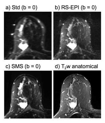
Simultaneous multi-slice single-shot DWI compared to routine read-out-segmented DWI for evaluation of breast lesions
Wendelien Sanderink, Jonas Teuwen, Linda Appelman, Ioannis Sechopoulos, Nico Karssemeijer, Ritse Mann
The aim of this study was to compare a prototype simultaneous multi-slice single-shot echo planar imaging (SMS-ss-DWI-EPI) sequence with conventional readout-segmented echo-planar imaging (rs-DWI-EPI) for diffusion-weighted imaging of the breast at 3T magnetic resonance imaging (MRI). A reader study was conducted to evaluate image quality, lesion conspicuity and BI-RADS® score. Our results show that although the image quality with the conventional rs-DWI-EPI is superior, malignant lesions have improved visibility with the SMS-ss-DWI-EPI sequence.
|
|
4112
 |
85 |
Diffusion-weighted imaging with apparent diffusion coefficient (ADC) mapping in non-mass enhancing breast tumors: influence of region-of-interest placement in diagnostic accuracy and intra-/inter-reader agreement
Video Permission Withheld
Daly Avendano, Maria Adele Marino, Doris Leithner, Blanca Bernard-Davila, Maxine Jochelson, Elizabeth Morris, Pascal Baltzer, Thomas Helbich, Katja Pinker
Non-mass enhancing (NME) breast tumors are a diagnostic challenge and limited data exists for characterization with DWI with ADC mapping. We aim to investigate the influence of ROI placement and different ADC metrics on diagnostic accuracy of DWI and to assess intra and inter-reader agreement of ADC measurements with DWI in NME breast lesions. Two readers independently assessed DWI using three different ROI´s approaches for ADC measurements. Data indicate that diagnostic accuracy of DWI with ADC mapping is limited in lesions presenting as NME on DCE-MRI with only moderate inter-reader agreement
|
|
4113.
 |
86 |
Determine Breast Density from T1-weighted Images using a 3D Convolutional Neural Network
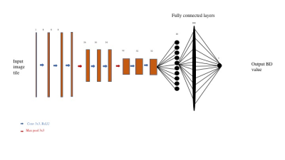
Mario Serrano-Sosa, Jie Ding, Karl Spuhler, Chuan Huang
Using MRI-based breast density (BD) measurements, we trained a convolutional neural network to take a T1-weighted MRI and output a BD value. Our CNN yielded a 3.1% error for the entire data set and 4.1% error in the testing set. Having a CNN work on T1-weighted MRI to quantify BD will allow BD to be a part of every practice.
|
|
4114.
 |
87 |
The feasibility of using computerized analysis features of magnetic resonance imaging to predict the outcomes of neoadjuvant chemotherapy for breast cancer
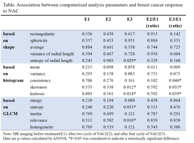
JINGYU ZHOU, HUIMING SHAN
This study was based on mathematical calculation software to calculate the imaging features of dynamic enhanced magnetic resonance images(DCE-MRI). The feasibility of outcome prediction of neoadjuvant chemotherapy(NAC) for breast cancer was tested by three kinds of computerized analysis features: morphological features, grayscale statistical features, and texture features. The results indicated that DCE-MRI computerized analysis features before and after 2-cycles of NAC can not predict the degree of pathological remission of breast cancer. DCE-MRI computerized analysis features after 4-cycles of NAC can evaluate the degree of pathological remission of breast cancer. In other words, DCE-MRI computerized analysis features has the potential to be a new noninvasive method of evaluating NAC outcomes.
|
|
4115.
 |
88 |
Radiomic Analysis of Breast Cancer Peritumoral Tissue on Contrast Enhanced MRI Prior to Neoadjuvant Chemotherapy
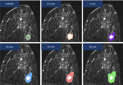
Eva Gombos, Carlos Aramayo, Jayender Jagadeesan
This study set out to identify key radiomic features in peritumoral breast tissue associated with a high response to neoadjuvant chemotherapy, defined as a >90% reduction of tumor cells. For each case, a tumor label map and 5 distinct masks at 2.5, 5, 10, 15, and 20mm beyond the tumor border were created using pretreatment DCE-MRIs. Fifty-seven metrics were then computed for each mask using the 3D Slicer HeterogeneityCAD module. Using final pathology reports, cases were separated into “low-response” and “high-response” groups. Statistical correlation using univariate Mann-Whitney tests to compare the two response groups ultimately yielded 6 radiomicfeatures.
|
|
4116.
 |
89 |
Adaptation of a Computer Vision Blur Metric to Objectively Compare High Resolution DWI Strategies in in vivo Breast Imaging
Jessica McKay, Gregory Metzger, Steen Moeller, An Church, Michael Nelson, Patrick Bolan
DWI has shown promise for detecting and characterizing breast cancers but is limited by the low spatial resolution of standard spin-echo EPI techniques. Several strategies have been proposed to generate high resolution DWI, including reduced field-of view, steady-state imaging, readout-segmented EPI, and simultaneous multi-slice imaging (SMS). In this work we adopt the Crété-Roffet blur metric to objectively compare resolution of three DWI strategies, including standard SE-EPI, and RO-segmented EPI and SMS-EPI high resolution approaches. In this comparison, both high-resolution DWI methods showed an improvement in in-plane resolution over the standard technique. The Crété-Roffet blur metric appears to be a robust and objective means of comparing effective resolution.
|
|
4117.
 |
90 |
Correction of B0 Inhomogeneities-Induced Artifacts in EPI for Breast Imaging
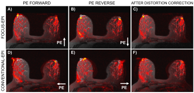
Ana Rodríguez-Soto, Helen Park, Dominic Holland, Hauke Bartsch, Haydee Ojeda-Fournier, Anders Dale, Rebecca Rakow-Penner
Echo-planar imaging (EPI) is prone to B0 inhomogeneities that arise from susceptiblity effects resulting in spatial distortions more pronounced in the phase-encoded (PE) direction. Reduction and correction of such distortion artifacts are performed in both acquisition and post-processing stages. The present work evaluates the combined effect of a field-of-view (FOV) optimized and constrained undistorted single-shot (FOCUS) EPI and a distortion correction approach to that exploits the symmetry of the artifact on EPI images acquired with opposite PE polarities. After distortion correction good agreement was found between corrected EPI datasets and anatomical images.
|
|
4118.
 |
91 |
Feature Engineering for the Subtype Classification of Breast Cancer: A Model Incorporating DCE and DWI Images
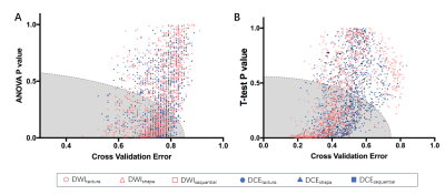
Zhe Wang, Boyu Zhang
For the 4-IHC classification task, the best accuracy of 78.4% was achieved based on linear discriminant analysis (LDA) or subspace discrimination of assembled learning in conjunction with 25 selected features, and only small dependent emphasis of Kendall-tau-b for sequential features based on the DWI images (DWIsequential) with the LDA model yielding an accuracy of 53.7%. The subspace discriminant of ensembled learning using eight features yielded the highest accuracy of 91.8% for comparing TN to non-TN cancers, and the maximum variance for DWIsequential alone together with a linear support vector machine (SVM) model achieved an accuracy of 83.6%.
|
|
4119.
 |
92 |
Semi-automatic ROI registration and texture feature extraction for pre- and post- neoadjuvant chemotherapy contrast-enhanced MR in predicting response of mass-like breast cancers
Kun Cao, Bo Zhao, Ying-Shi Sun
In mass-like breast cancers, semi-automatic ROI registration from pre- to post-treatment MR images, and combined with usage of texture feature extraction for automatically segmentation of enhanced areas, the ability for predicting residual tumors after neoadjuvant chemotherapy can be improved significantly and cost lesser time in imaging reading.
|
|
4120.
 |
93 |
Focal areas of NME undergoing MRI-guided VABB: outcomes and frequency of malignancy
Satoko Okamoto, James Covelli, Shutian Chen, Bruce Daniel, Debra Ikeda
We evaluated the positive predictive values (PPVs) of internal enhancement and kinetic data in focal areas of non-mass enhancement (NME) in patients undergoing MRI-guided vacuum-assisted breast biopsy (MRI-VABB). Our results show the PPVs of clumped and heterogeneous internal enhancement were similar (PPV=25% vs 21%) suggesting that heterogeneous internal is similar to clumped enhancement. The most predictive kinetic pattern of malignancy was fast initial phase without any tendency for cancer using the delayed phase.
|
|
4121.
 |
94 |
A comparative study of multi-shot diffusion-weighted imaging (DWI) using multiplexed sensitivity encoding (MUSE) and conventional single-shot echo-planar imaging (EPI) DWI in breast cancer
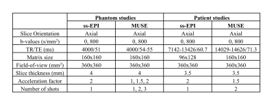
Gabrielle Baxter, Ramona Woitek, Amy Frary, Gavin Houston, Martin Graves, Fiona Gilbert, Andrew Patterson
MUSE (multiplexed sensitivity encoding) is a multi-shot echo-planar imaging (EPI) technique used in diffusion-weighted imaging (DWI) that reduces distortion and blurring compared to single-shot EPI. Experiments using a breast phantom aimed to investigate the effect of varying parallel acceleration and number of shots on geometric distortion and quantification of the apparent diffusion coefficient (ADC). A refined protocol was applied to a small cohort of patients to comparatively assess improvements in image quality.
|
|
4122.
 |
95 |
Threshold isocontouring on high b-value diffusion weighted images in magnetic resonance mammography
Fangrong Zong, Sebastian Bickelhaupt, Tristan Kuder, Wolfgang Lederer, Heidi Daniel, Anne Stieber, Heinz Schlemmer, Xiaoliang Zhang, Petrik Galvosas, Frederik Laun
Quantitative evaluation in clinical practice is important for accurate diagnosis, which is often difficult in breast diffusion measurements where large variability of the diffusion metrics is present. Motivated by similar appearance of malignant breast lesions in high b-value diffusion-weighted imaging (DWI) and positron emission tomography (PET), we propose a threshold isocontouring approach commonly used in PET to analyse breast DWI data with minimal inter-observer variability. Our results support that using a relative threshold level of 0.85 almost completely suppresses the intra- and inter-individual variability. This study suggests that the proposed approach is advantageous in the breast DWI data analysis and quantitative evaluation.
|
|
4123.
|
96 |
Identifying metastatic axillary lymph node of breast cancer by quantitative parameters with histogram and texture features on pharmacokinetic modeling dynamic contrast-enhanced MRI: A Pilot Radiomics Study
Presentation Not Submitted
Hong-Bing Luo, Yuan-Yuan Liu, Shao-Yu Wang, Jing Ren, Peng Zhou
This study aimed to investigate the discriminative performance of pharmacokinetic quantitative parameters with histogram and texture features on dynamic contrast-enhanced magnetic resonance imaging (DCE-MRI) in predicting axillary lymph nodes metastasis in breast cancer. Results showed that 6 pharmacokinetic quantitative and their histogram parameters,53 texture features of DCE-MRI were statistically difference between the positive and negative group. In cross-validation, the accuracy of the classifier obtained 90% for identifying the metastatic axillary lymph node. The radiomic features based on quantitatively pharmacokinetic DCE-MRI demonstrated promising application in discriminating between metastatic positive and negative axillary nodes of breast cancer.
|
|
4124.
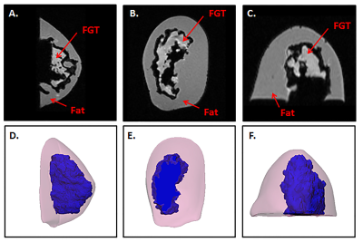 |
97 |
Design of a 3D Printed Patient-Specific Dual Compartment Breast Phantom
Nicole Wake, Carlotta Ianniello, Ryan Brown, Jerzy Walczyk, Linda Moy, Christopher Collins, Henry Rusinek
Presented here is the design of a 3D printed anthropomorphic breast phantom that includes the key breast tissue compartments which can be filled with desired tissue-mimicking fluids. The breast phantom can be used for validating MRI, coil development, safety evaluation and pulse sequence evaluation.
|
|
4125.
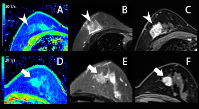 |
98 |
Whole-Tumor Histogram and Textural Analysis of Model-based T2 Mapping for the Ki-67 Labeling Index of Breast Cancer
Presentation Not Submitted
Tianwen Xie, Qiufeng Zhao, Caixia Fu, Robert Grimm, Tobias Kober, Tom Hilbert, Yajia Gu, Weijun Peng
Recently, there has been increased interest in quantitative MR parameters for assessing tumor proliferation. In this study, we proposed the use of whole-tumor histogram texture features using the model-based T2 mapping method GRAPPATINI to differentiate the positive and negative Ki-67 status of breast cancer. Classification performed between Ki-67-positive and Ki-67-negative groups resulted in an area under the ROC curve of 0.808.
|
|
4126.
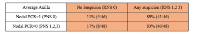 |
99 |
MRI Staging of Axillary Lymph Node Post Neoadjuvant Chemotherapy Predicts Pathological Complete Response
Renee Cattell, Julie Leong, Thomas Ren, James Kang, Pauline Huang, Ashima Muttreja, Haifang Li, Ankita Katukota, Nikita Katukota, Priya Mukhi, Lea Baer, Jules Cohen, Cliff Bernstein, Sean Clouston, Roxanne Palermo, Timothy Duong
This study evaluated a radiological nodal staging metric for accurate identification of diseased axillary lymph nodes (aLNs) on MRI associated with breast cancer. Comparison was made with pathological nodal staging as the ground truth. Radiological scoring of aLNs identifies diseased nodes as suspicious more correctly than it identifies normal nodes as normal. Overall, radiological scoring of the axillary lymph nodes based on morphological characteristics showed moderate accuracy in distinguishing disease or no-disease in the nodes based on standard clinical breast MRI data. Improvement in image signal-noise-ratio (SNR), spatial resolution and contrast of the axillary lymph nodes is needed.
|
|
| Top |
Lung: Xenon
Digital Poster
Body: Breast, Chest, Abdomen, Pelvis
Wednesday, 15 May 2019
| Exhibition Hall |
15:45 - 16:45 |
| |
|
Computer # |
|
4127.
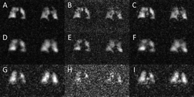 |
101 |
R2* decay rates of hyperpolarized dissolved-phase 129Xe as a novel biomarker of fibrotic lung injury
Jeff Kammerman, Andrew Hahn, Sean Fain
Spectroscopic imaging of the hyperpolarized xenon-129 dissolved in the pulmonary tissues and blood can probe regional gas exchange within the lung. However, estimates of the frequency shifts and decay rates of the dissolved-phase signal from whole-lung spectroscopy are confounded by the large magnetic field inhomogeneities of the lung. In this work, we develop a regularized, model-based reconstruction that estimates the spectral properties of pulmonary HP 129Xe to improve image quality. Importantly, we show that the estimated r2* decay rates of dissolved-phase compartments differ substantially from those estimated in whole lung spectroscopy and are a significant biomarker of gas-exchange.
|
|
4128.
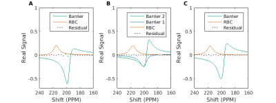 |
102 |
A magnetic susceptibility model of hyperpolarized 129Xe spectroscopy in alveolar tissues
Jeff Kammerman, Annelise Malkus, Sean Fain
Hyperpolarized 129Xe MRI has been used to characterize gas exchange in the lung by measuring xenon dissolved in the red blood cells and pulmonary barrier tissues. However, recent work identified a potential third component of the dissolved-phase spectrum, observed as a splitting of the barrier peak. We propose to explain the appearance of this peak using a signal model that considers the susceptibility differences at the air tissue interface of the alveoli. Integrating this model into whole lung HP 129Xe spectroscopy measures in 8 healthy subjects and 16 patients with idiopathic pulmonary fibrosis, we find improved correlations between the RBC-to-barrier ratio and DLCO %predicted compared to conventional modeling of the dissolved-phase spectrum.
|
|
4129
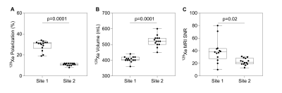 |
103 |
Two-site Reproducibility of Hyperpolarized 129Xe MRI Ventilation in Severe Asthma: Implications for Multicenter Clinical Studies
Video Permission Withheld
Sarah Svenningsen, Alexei Ouriadov, Norman Konyer, Rachel Eddy, Andrew Westcott, David McCormack, Melanie Kjarsgaard, Michael Noseworthy, Parameswaran Nair, Grace Parraga
Despite the demonstrated sensitivity of hyperpolarized 129Xe MRI biomarkers to pulmonary disease onset, progression and treatment response, comparison of findings across sites is challenging as acquisition protocols and procedures are not standardized. To better understand the agreement of hyperpolarized 129Xe MRI ventilation across different clinical sites, severe asthmatics underwent 129Xe MRI at two sites within 24 hours. 129Xe MRI ventilation defect percent was significantly correlated between sites despite inter-site differences in 129Xe polarization, 129Xe volume, gas mixture composition and signal-to-noise ratio. These findings have important implications when generalizing 129Xe MRI data collected at different sites and undertaking multicenter studies.
|
|
4130.
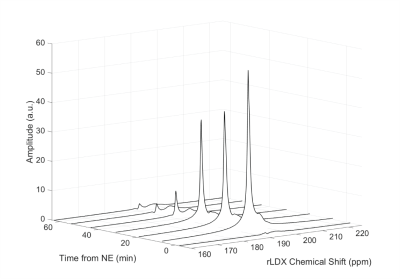 |
104 |
Absolute MR thermometry by hyperpolarized 129Xe in UCP1-/- animals reveals UCP1-independent thermogenesis
Michael Antonacci, Christian McHugh, Michele Kelley, Andrew McCallister, Tamara Branca
Direct and non-invasive temperature measurements of BAT in UCP1-/- mice could help to settle the debate on whether BAT in UCP1-/- mice is thermogenically competent and whether there exist other UCP1-independent mechanisms for non-shivering thermogenesis. Here we use hyperpolarized xenon MR thermometry for direct absolute temperature measurements of brown adipose tissue in UCP1-/- mice. Hyperpolarized xenon MR thermometry indicates a small but detectable temperature increase in the BAT of UCP1-/- mice after adrenergic stimulation of this tissue.
|
|
4131.
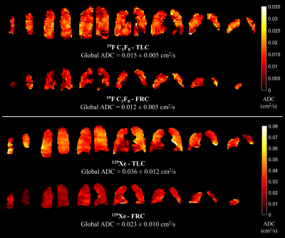 |
105 |
Comparison of apparent diffusion coefficient mapping in the lungs of healthy volunteers with 19F C3F8 and hyperpolarized 129Xe gas MRI
Ho-Fung Chan, Adam Maunder, Jim Wild
In this work we derive in-vivo 19F C3F8 and 129Xe apparent diffusion coefficient (ADC) maps from healthy volunteers’ lungs at functional residual capacity (FRC) and total lung capacity (TLC) to evaluate the sensitivity of C3F8 ADC to changes in alveolar dimensions. Each volunteer’s C3F8 ADC values at FRC was significantly smaller than the respective TLC value (P=0.008), and both FRC and TLC values were smaller than the free diffusion coefficient of C3F8. Furthermore, a significant correlation between 129Xe and C3F8 ADC was observed (r=0.69, P=0.029); demonstrating the clinical potential of in-vivo C3F8 ADC mapping.
|
|
4132.
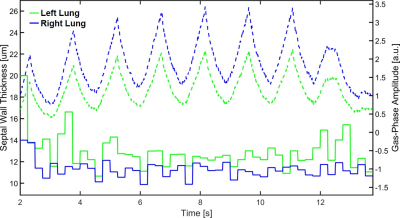 |
106 |
Measuring Septal Wall Thickness Using Rapid 1D Hyperpolarized Xenon-129 CSSR Acquisitions
Kai Ruppert, Yi Xin, Faraz Amzajerdian, Hooman Hamedani, Luis Loza, Tahmina Achekzai, Ryan Baron, Ian Duncan, Harrilla Profka, Sarmad Siddiqui, Mehrdad Pourfathi, Federico Sertic, Maurizio Cereda, Rahim Rizi
Septal wall thickness measurements using hyperpolarized xenon-129 MRI are sensitive to inflammatory or fibrotic pathologies but currently require multi-second breath-holds. We investigated the feasibility of sampling the recovery of the xenon dissolved-phase magnetization in the lung parenchyma in real time using a rapid 1D acquisition technique combined with periodic saturation of the xenon dissolved-phase magnetization, allowing us to extract the septal wall thickness during short breath holds or even in free-breathing rabbits. Our measurements indicate that the correlation between the derived septal wall thickness and the respiratory cycle are low enough to obtain consistent wall-thickness measurements in free-breathing, non-cooperative subjects.
|
|
4133.
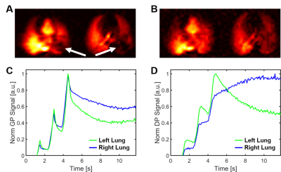 |
107 |
Monitoring of Pulmonary Function in Real-time with 1D Hyperpolarized Xenon-129 MRI
Kai Ruppert, Yi Xin, Faraz Amzajerdian, Hooman Hamedani, Luis Loza, Tahmina Achekzai, Ryan Baron, Ian Duncan, Harrilla Profka, Sarmad Siddiqui, Mehrdad Pourfathi, Federico Sertic, Maurizio Cereda, Rahim Rizi
Data sampling for pulmonary function measurements using hyperpolarized gas MRI typically lasts for several seconds due to the time requirements for spatial signal encoding. Consequently, highly-dynamic processes are largely invisible to existing lung-imaging techniques. In an initial attempt to observe pulmonary function in real time, we traded spatial resolution for a high temporal resolution of 10 ms by limiting spatial information to 1D projection acquisitions. We tested the technique in a rabbit model by observing pulmonary signal oscillations throughout the lung and by detecting alveolar collapse during expiration at high PEEP in acid-induced acute lung injury.
|
|
4134.
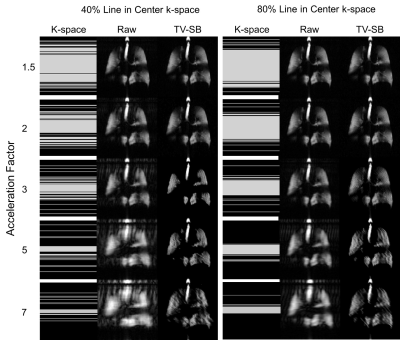 |
108 |
Compressed Sensing Hyperpolarized Noble Gas Ventilation MRI: Towards a Maximum Clinically Acceptable Acceleration Factor
Andrew Westcott, Alexei Ouriadov, Rachel Eddy, David McCormack, Miranda Kirby, Grace Parraga
Compressed sensing has been applied to hyperpolarized gas MRI to accelerate acquisition and allow for increased data or resolution. To better understand the effect of compressed sensing on biomarkers derived from static ventilation images, data were retrospectively undersampled in 10 individuals with chronic obstructive pulmonary disease and k-means clustering was performed. Raw reconstruction resulted in differences less than the ventilation defect percent minimal clinically important difference up to an acceleration factor of 3. The total variation Split-Bregman reconstruction resulted in qualitatively adequate images, however, further optimization of regularized reconstruction techniques is required to achieve consistent ventilation signal clustering.
|
|
4135.
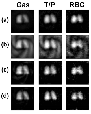 |
109 |
Accelerated Spiral-IDEAL for Gas and Dissolved Phase Hyperpolarized 129Xe in the Lungs of Healthy Human Volunteers: Effect of k-space Calibration Data on Image Quality and Gas Exchange Analysis
Brandon Zanette, Giles Santyr
Parallel imaging with multi-channel receivers provides flexible acceleration of scan duration and is an important tool in both clinical and research settings. The application of parallel imaging to hyperpolarized 129Xe MRI may be useful in improving the clinical utility of parametric gas exchange mapping by reducing breath-hold durations or allowing for the acquisition of additional temporal/spatial information. In this work, we investigate the effect of k-space calibration data on parallel imaging reconstruction of time-resolved measurements of hyperpolarized 129Xe uptake using spiral-IDEAL and quantify resultant changes in the estimation of lung physiology.
|
|
4136.
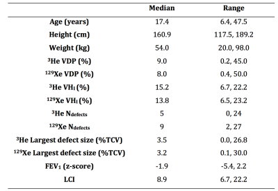 |
110 |
Comparison of hyperpolarised 3He and 129Xe ventilation MRI to assess lung disease in cystic fibrosis
Laurie Smith, Guilhem Collier, Helen Marshall, Paul Hughes, Alberto Biancardi, Graham Norquay, Jody Bray, Oliver Rodgers, Martin Wildman, Noreen West, Alex Horsley, Jim Wild
Ventilation MRI using hyperpolarised gases is highly sensitive to early cystic fibrosis lung disease. With the recent move towards 129Xe from 3He, we aimed to assess both gases in parallel in patients with CF, at baseline and follow-up, to determine whether any inherent bias was present when assessing lung disease. We found that there was no inherent bias for VDP between the two gases present although at an individual level differences were evident. Despite this, when followed up at a later date both gases similarly reflected changes in ventilation, suggesting both are capable of reflecting CF lung disease severity.
|
|
4137.
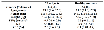 |
111 |
129Xe ventilation and 1H anatomical MRI to detect functional and structural abnormalities in sub-clinical cystic fibrosis lung disease
Laurie Smith, David Hughes, Ho-Fung Chan, Kevin Johnson, Jody Bray, Oliver Rodgers, Guilhem Collier, Graham Norquay, Alberto Biancardi, Paul Hughes, Sailesh Kotecha, Martin Wildman, Noreen West, Alex Horsley, Pierluigi Ciet, Piotr Wielopolski, Harm Tiddens, Helen Marshall, Jim Wild
Lung MRI in cystic fibrosis (CF) has the potential to measure both structure and function during the same session. Here we assessed 14 patients with CF and normal spirometry for lung abnormalities using 129Xe ventilation and 1H 3D SPGR and UTE anatomical MRI. 129Xe ventilation defects were evident in all subjects assessed, whilst gas trapping was evident on expiratory 3D SPGR in 71% of patients. Further anatomical abnormalities were evident on UTE in 57% of patients. Ventilation and anatomical abnormalities are therefore present in sub-clinical CF and highlights the potential of MRI in routine CF lung imaging.
|
|
4138.
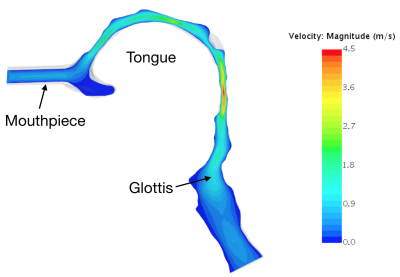 |
112 |
In vivo Validation of Upper Airway Respiratory Computational Fluid Dynamics (CFD) with Phase-Contrast MRI of Hyperpolarized 129Xe
Alister Bates, Matthew Willmering, Robert Thomen, Chamindu Gunatilaka, Md Hossain, Charles Dumoulin, Jason Woods
Computational fluid dynamics (CFD) can provide clinicians with otherwise unobtainable information such as regional airway resistance and breathing effort. However, CFD has yet to be validated in vivo and has only been compared to in vitro experiments that do not represent all aspects of airway physiology. To achieve in vivovalidation, velocities produced by CFD were compared to those measured by phase-contrast MRI of inhaled hyperpolarized 129Xe.
Voxelwise comparison of velocities between the two methods produced an R2 value of 0.75 and a slope of 0.91. Phase-contrast MRI produces a benchmark that allows validation and optimization of CFD simulations.
|
|
4139.
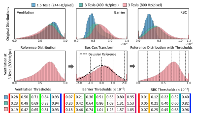 |
113 |
Quantifying Hyperpolarized 129Xe Gas Exchange MRI Across Platforms, Field Strength, and Acquisition Parameters
Ziyi Wang, Mu He, Rohan Virgincar, Elianna Bier, Sheng Luo, Bastiaan Driehuys
Quantitative analysis of 129Xe gas exchange MRI has previously employed linear binning, using thresholds derived from the mean and standard deviation of a healthy reference population. However, such distributions can be skewed from a purely Gaussian shape by differences in acquisition strategy and field strength, thereby complicating threshold determination. Here we demonstrate a generalization of previous binning methods by applying a Box-Cox transformation to derive non-linearly spaced reference thresholds. We provide new thresholds appropriate for 3T acquisitions and demonstrate the robustness of the approach by showing consistent quantification of subjects scanned longitudinally across platforms, field strengths, and acquisition parameters.
|
|
4140.
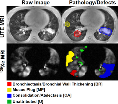 |
114 |
The Nature of Early Airway Obstruction in Cystic Fibrosis Lung Disease using UTE MRI and 129Xe MRI
Robert Thomen, Laura Walkup, Nara Higano, David Roach, Zackary Cleveland, Andrew Schapiro, Alan Brody, John Clandy, Jason Woods
Cystic Fibrosis (CF) lung disease is characterized by identifiable structural abnormalities on CT or UTE MRI, such as mucus plugs and bronchiectasis. Similarly, hyperpolarized 129Xe MRI can provide functional information related to regional pulmonary ventilation, with high sensitivity to early obstruction. Here we combined UTE and 129Xe MRI to quantify the extent to which specific structural abnormalities contribute to regional ventilation defects. Mucus plugs and bronchiectasis account for the majority of ventilation defects that can be attributed to structural abnormalities. However, structurally-unattributable ventilation defects are more common, implying higher sensitivity than structural imaging to lung dysfunction in milder disease.
|
|
4141.
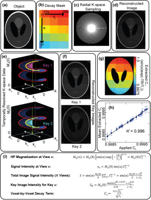 |
115 |
Mapping and Correcting Hyperpolarized Magnetization Decay Using Radial Keyhole Imaging
Peter Niedbalski, Matthew Willmering, Scott Robertson, Matthew Freeman, Wolfgang Loew, Randy Giaquinto, Christopher Ireland, Ronald Pratt, Charles Dumoulin, Jason Woods, Zackary Cleveland
Hyperpolarized (HP) media enable biomedical imaging applications otherwise unachievable using standard MRI contrast agents. However, quantitative analysis of HP images is complicated by spatially varying signal decay due to T1 relaxation and RF excitation. By imaging with center-out trajectories, HP signal intensity is collected with every acquisition, thereby encoding signal decay alongside k-space data. Using keyhole reconstruction, multiple temporally resolved images were reconstructed to generate voxel-by-voxel maps of hyperpolarized signal decay following a simple analytical model. Here these maps are applied to correct spatially varying magnetization decay in HP 129Xe, improving the quantitative accuracy of ventilation images.
|
|
4142.
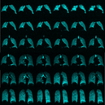 |
116 |
Feasibility of Single Breath-hold Isotropic Voxel 129Xe MRI in Patients
Andrea Barker, Andrew Westcott, Rachel Eddy, David McCormack, Grace Parraga, Alexei Ouriadov
Hyperpolarized 129Xe gas MRI has been established as an effective tool to investigate lung function in a number of pulmonary diseases. Improved 129Xe polarization techniques provide the opportunity to use a high-resolution 3D static ventilation image acquisition sequence (FGRE) in a 16-second breath-hold. Isotropic-voxel images were acquired in one healthy-volunteer and two patients with asthma; images were compared to coronal images acquired in two other patients with asthma. Isotropic images had sufficient SNR to analyze VDP. This proof-of-concept investigation establishes the feasibility of using an isotropic-voxel acquisition sequence in single breath-hold MRI of the lungs.
|
|
4143.
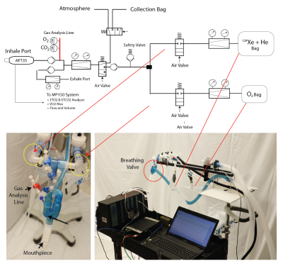 |
117 |
Multi-breath Wash-in Hyperpolarized 129Xenon MRI in Human Subjects
Hooman Hamedani, Ryan Baron, Stephen Kadlecek, Kai Ruppert, Ian Duncan, Yi Xin, Francisca Bermudez, Sarmad Siddiqui, Mehrdad Pourfathi, Faraz Amzajerdian, Luis Loza, Tahmina Achekzai, Federico Sertic, Rahim Rizi
We have shown the feasibility of multi-breath wash-in imaging of regional fractional ventilation using HP 129Xe MRI for the first time in both healthy and diseased human subjects.
|
|
4144.
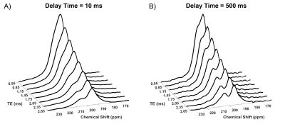 |
118 |
The Impact of Echo-time on Hyperpolarized Xenon-129 CSSR Measurements
Faraz Amzajerdian, Kai Ruppert, Yi Xin, Hooman Hamedani, Luis Loza, Tahmina Achekzai, Ryan Baron, Ian Duncan, Harrilla Profka, Sarmad Siddiqui, Mehrdad Pourfathi, Federico Sertic, Maurizio Cereda, Rahim Rizi
Chemical Shift Saturation Recovery (CSSR) MR spectroscopy and imaging are powerful techniques for deriving a number of important physiological pulmonary parameters. We investigated the impact of measurement echo time on the spectral composition of the acquisition and the subsequent numerical data analysis in rabbits. We found that lengthening the TE resulted in a vastly improved separation between the dissolved-phase resonances by suppressing short T2* signal components. As consequence of this phenomenon, the measured septal wall thickness changed with TE by 1.0 ± 0.13 μm/ms. Similarly, we hypothesize that CSSR-derived measures of pulmonary physiology might be field strength-dependent.
|
|
4145.
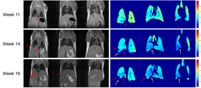 |
119 |
Measurement of Regional Fractional Ventilation in a Mouse Model of Lung Cancer under Free-Breathing
Luis Loza, Stephen Kadlecek, Hooman Hamedani, Mehrdad Pourfathi, Tahmina Achekzai, Ian Duncan, Rahim Rizi
Hyperpolarized gas MRI is a powerful modality for the assessment of lung structure and function. However, its utility as an investigative tool for animal disease models is limited by the terminal intubation procedures necessary for precise gas delivery and ventilation. Here, we present a technique for measuring regional fractional ventilation longitudinally in a mouse model of lung cancer under spontaneous respiration using hyperpolarized 129Xe MRI. Fractional ventilation maps at different stages of cancer revealed significant structural changes, a decline in regional ventilation, and an increase in ventilation heterogeneities associated with tumor progression.
|
|
4146.
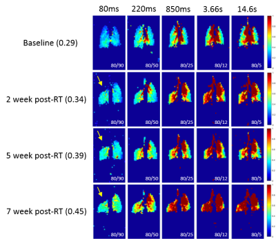 |
120 |
A Technique for Measuring Long-Term Alterations in Pulmonary Gas Uptake under Free-Breathing in a Murine Model of Radiation-Induced Lung Injury
Luis Loza, Stephen Kadlecek, Mehrdad Pourfathi, Tahmina Achekzai, Kai Ruppert, Ian Duncan, Rahim Rizi
Computed tomography (CT), pulmonary function tests (PFT) and positron-emission tomography (PET) are all currently used to assess lung structure and function clinically. However, the sensitivity and safety of these modalities limit their use in cases of progressive lung disease, where longitudinal measurements are of value for both therapeutic planning and monitoring response to treatment. Hyperpolarized 129Xe (HXe) MRI provides robust, specific information about the lungs and is suitable for longitudinal measurements. In this study, we demonstrate an HXe MRI technique for detecting and assessing alterations in pulmonary gas uptake in a mouse model of RILI under spontaneous respiration.
|
|
4147.
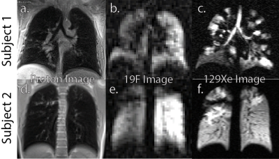 |
121 |
Initial Comparison of 19F and Hyperpolarized 129Xenon Gas Lung Imaging in Cystic Fibrosis
Andrew McCallister, Sang Chung, Michael Antonacci, Yueh Lee, Jennifer Goralski, Scott Donaldson, Rosa Branca
Gas enhanced MRI has the potential to provide regional information about lung function that pulmonary function tests alone cannot provide. Hyperpolarized xenon gas is a well-established tool for the assessment of regional lung function in humans, and 19F gas MRI has been proposed as a lower cost alternative that can provide, in addition to ventilation defects, wash in and wash out rates. Here we report the initial findings of a study aiming at comparing these two techniques for assessment of lung ventilation function in subjects with cystic fibrosis.
|
|
4148.
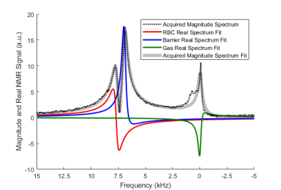 |
122 |
129Xe Gas-Exchange MRI in Pediatrics with Lung Disease
Matthew Willmering, Ziyi Wang, Hui Wang, Laura Walkup, Bastiaan Driehuys, Zackary Cleveland, Jason Woods
The solubility properties of hyperpolarized 129Xe have made it attractive in many translational pulmonary imaging studies; the large chemical-shift range can be exploited to obtain semi-quantitative ventilation, barrier-uptake, and RBC-transfer images. To date, the technique has demonstrated significant changes in gas exchange in adult lung disease, but has yet to be explored in children, where pulmonary function testing can be more challenging (or impossible). This pediatric gap caused us to pursue 129Xe gas-exchange MRI at the now-common field of 3T; here, we present the first demonstration of dissolved-phase xenon imaging in pediatrics (with cystic fibrosis and post-bone marrow transplant).
|
|
4149.
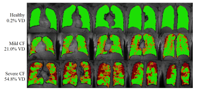 |
123 |
Xenon-129 MRI of the lung in cystic fibrosis – preliminary results from a clinical trial
Vicki Huang, Dana Albon, Lucy Gettle, Kun Qing, Nicholas Tustison, Yun Shim, John Mugler, James Patrie, Jaime Mata
Results demonstrate sensitivity to early physiologic changes in subjects with cystic fibrosis (CF) using 3D single breath-hold chemical shift imaging (3D-SBCSI). 3D-SBCSI is a technique that uses MRI and hyperpolarized Xenon-129, an inert, non-radioactive gas, to assess lung physiology by detecting Xe-129 in three compartments in the lungs: airspaces, tissue parenchyma and plasma, and red-blood-cells. Results of 3D-SBCSI were compared to pulmonary function test results, Xe-129 ventilation images, blood panels and showed early physiologic trends in mild subjects that progressed on severe CF subjects. 3D-SBCSI could be used to monitor treatment responses and disease progression before symptoms become clinically apparent.
|
|
4150.
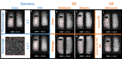 |
124 |
Multi-center Comparison of Image Quality Using a Thermally Polarized 129Xe Phantom
Elianna Bier, John Nouls, Ziyi Wang, Mu He, Ralph Hashoian, John Mugler, Bastiaan Driehuys
Multiple centers are starting to use hyperpolarized 129Xe MR imaging and spectroscopy to quantify pulmonary function. This drives the need for standardization and quality assurance (QA), which is challenging for short-lived hyperpolarized agents. To address this, we developed a high pressure thermal 129Xe phantom constructed from high-density polyethylene and an associated loader shell that mimics the human torso. The phantom was imaged at 8 institutions across North America comprising a variety of scanners, vendors, coils, and field strengths. Image SNR measured with a minute-long scan agreed within a factor of 2 across sites and enabled rapid comparison of MR configurations.
|
|
Back to Program-at-a-Glance |  Back to Top Back to Top |
| The International Society for Magnetic Resonance in Medicine is accredited by the Accreditation Council for Continuing Medical Education to provide continuing medical education for physicians. |
