Digital Poster Session
Acquisition, Reconstruction & Analysis Back to Program-at-a-Glance Back to Program-at-a-Glance
|
Thursday, 16 May 2019
Digital PosterAcquisition, Reconstruction & Analysis
4376 -4398 Using MRI to Measure Numbers Outside of the Brain
4399 -4423 Quantitative Mapping of the Brain
4424 -4448 Motion Correction: Brain
4449 -4472 Robust & Reproducible Quantitation
4473 -4497 Motion Correction: Non-Brain
4498 -4521 System Imperfections
4522 -4545 Optimization of Quantitative Mapping Techniques
4546 -4570 Quantitative Mapping: Relaxometry & Beyond
4571 -4594 Going Faster: New Sequences & Acquisition Protocols
4595 -4619 Artifacts, Implants & Corrections
4620 -4643 New RF & Gradient Strategies
4644 -4668 Machine Learning for Image Reconstruction: A New Frontier
4669 -4693 Improving Definition & Reducing Artifacts
4694 -4718 Machine Learning for Image Reconstruction: Breakthroughs |
| |
Using MRI to Measure Numbers Outside of the Brain
Digital Poster
Acquisition, Reconstruction & Analysis
Thursday, 16 May 2019
| Exhibition Hall |
08:15 - 09:15 |
| |
|
Computer # |
|
4376.
 |
1 |
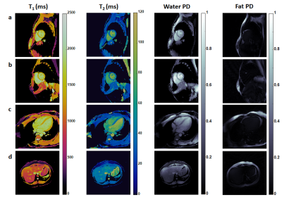
Fat/Water Separation and T1 and T2 Quantification Using MRF with a Rosette Trajectory in the Heart and Liver

Yuchi Liu, Jesse Hamilton, Mark Griswold, Nicole Seiberlich
Cardiac Magnetic Resonance Fingerprinting (cMRF) has recently been introduced for simultaneous T1 and T2 quantification in the myocardium. One important feature of the MRF framework is the potential to measure multiple tissue properties beyond T1 and T2. Here we propose an approach for simultaneous fat imaging and T1 and T2quantification based on the cMRF framework with a rosette trajectory. The accuracy in T1 and T2 measurements and the efficacy in water-fat separation were demonstrated in the ISMRM/NIST system phantom and a multi-compartment water/oil phantom, respectively. Preliminary results in the heart and liver in healthy subjects are also shown.
|
|
4377.
 |
2 |
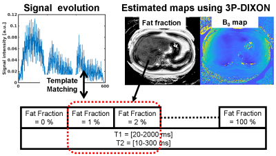
Mapping T1, T2, and proton density fat fraction of the liver using MR Fingerprinting with three-point DIXON and 6-peak fat model
Daiki Tamada, Hiroshi Onishi, Utaroh Motosugi
An MR Fingerprinting (MRF) simultaneously combining the three-point DIXON (3P-DIXON) method for the fatty liver was proposed. The MRF-FISP sequence with multi-TR/TE/flip angle was developed. The six-peak fat model was used to calculate a dictionary for the MRF. Template matching using the acquired signal evolutions and the rough fat fraction map estimated by 3P-DIXON provided quantification of T1, T2, and fat fraction. The phantom and volunteer studies demonstrated the feasibility of our study.
|
|
4378.
 |
3 |
Three-Dimensional, Free-Breathing Magnetic Resonance Fingerprinting for Whole-Liver Coverage
Kathleen Ropella-Panagis, Yong Chen, Yun Jiang, Jesse Hamilton, Wei-ching Lo, Dan Ma, Mark Griswold, Nicole Seiberlich, Vikas Gulani
In this proof-of-concept study, a 3D free-breathing abdominal MR Fingerprinting sequence is applied to the abdomen. Full-liver coverage with spatial resolution of 1.6×1.6×5 mm3 is attained in 8 minute 40 seconds.
|
|
4379.
 |
4 |
Free-Breathing Liver T1 and Fat Mapping Using a Golden-Angle-Ordered Variable Flip Angle Stack-of-Radial Sequence
Le Zhang, Tess Armstrong, Holden Wu
Measurement of T1 and proton-density fat fraction (PDFF) in the liver can provide information about fibrosis and steatosis, respectively. Existing Cartesian acquisition schemes generally require breath-holding, which limits spatial coverage and may be difficult for sick, elderly or pediatric patients. In this study, we propose a golden-angle-ordered (GA) 3D stack-of-radial variable-flip-angle (VFA) sequence that can map T1 and PDFF simultaneously with close to full liver coverage under five minutes during free-breathing. Pilot studies in phantom and healthy subjects demonstrate feasibility and show good measurement repeatability.
|
|
4380.
 |
5 |
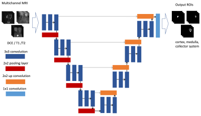
DeepKidney: Deep segmentation of MR images for automated glomerular function quantification in heterogeneous pediatric patients
Edgar Rios Piedra, Morteza Mardani, Ukash Nakarmi, Joseph Cheng, Shreyas Vasanawala
Automated segmentation of kidneys and their sub-components is a challenging problem, particularly in pediatric patients and in the presence of a pathology or some anatomical deformation. We present a segmentation framework using a multimodal U-Net that allows for the automated segmentation of the multiple kidney components as well as a functional evaluation of the glomerular filtration rate. Results achieve an average Dice similarity coefficient of 0.912, 0.853, and 0.917 for kidney cortex, medulla, and collector system, respectively.
|
|
4381.
 |
6 |
Field Map Estimation from Magnitude-Based Water-Fat Separation
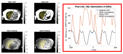
Alexandre Triay Bagur, Chloe Hutton, Benjamin Irving, Michael L. Gyngell, Matthew D. Robson, Michael Brady
Complex-based MRI chemical-shift encoded water-fat separation depends on accurate field map convergence, which is often mitigated with spatial regularization. This is prone to error propagation and over-smoothing of fat-fraction maps. Magnitude-based separation circumvents field mapping but is reportedly limited in fat-fraction range (0-50%). We have recently presented MAGO, a magnitude-based method that resolves this water-fat ambiguity. In this study, we compare MAGO to state-of-the-art fat-fraction quantification on N=150 volunteers, and we expand the method for field map calculation using previously estimated water and fat images. MAGO is comparable to regularized hybrid-based decomposition and shows promise in higher field inhomogeneity regimes.
|
|
4382.
 |
7 |
Phase Correction for Abdominal Quantitative Susceptibility Mapping with Bipolar Readout Gradients Sequence
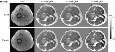
Rui Tong, Jianqi Li, Xu Yan, Yi Wang
Bipolar acquisition in abdominal multi-echo quantitative susceptibility mapping (QSM) could reduce echo-spacing and total scan time. However, the bipolar acquisition introduces phase error between odd and even echoes. A phase correction method in image domain was proposed to address this problem. We demonstrated the feasibility of generating a quantitative susceptibility map in human abdomen using bipolar multi-echo GRE sequence. Quantification analysis showed an excellent agreement between bipolar and unipolar methods.
|
|
4383.
 |
8 |
Quantifying liver function using artificial neural networks to estimate gadoxetic-acid uptake rate in temporally sparse gadoxetic-acid enhanced MRI
Josiah Simeth, Yue Cao
Though methods exist for quantifying regional liver function from dynamic gadoxetic-acid enhanced (DGE) MRI, errors are introduced when using the clinically typical temporally sparse acquisition scheme (6 volumes over 20 minutes) relative to a temporally dense dynamic acquisition (volumes every 5-10 sec over a similar period). This motivates a data driven approach. An artificial neural network (ANN) was trained to reproduce the results of the fully characterized analysis using only the restricted dataset. Across the patients evaluated the ANN solution resulted in lower mean and median WMAPE, as well as a reduction in MSE in most cases.
|
|
4384.
 |
9 |
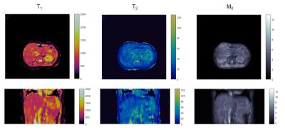
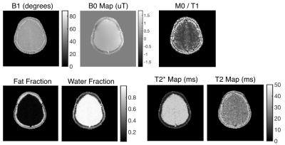
multiMap: A Gradient Spoiled Sequence for Quantitation of B1, B0, T1, T2, T2*, and Fat Fraction
Nicholas Dwork, Adam Kerr, Ethan Johnson, John Pauly
multiMap is a single sequence that combines standard techniques for measuring several quantities of interest: B0, B1, T1, T2, T2*, and Fat Fraction.
|
|
4385.
 |
10 |
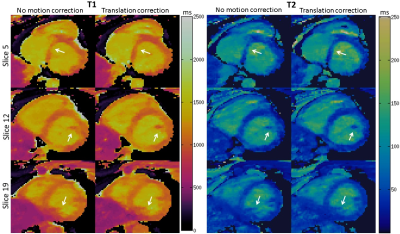
Toward 3D Free-breathing Cardiac Magnetic Resonance Fingerprinting
Gastao Cruz, Olivier Jaubert, Torben Schneider, Aurelien Bustin, René Botnar, Claudia Prieto
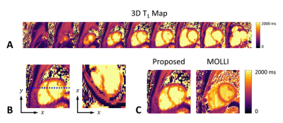
Magnetic Resonance Fingerprinting (MRF) has been introduced to simultaneously estimate multiple quantitative parameters but mainly applied to static organs. Recently the feasibility of 2D triggered cardiac MRF (cMRF) under breath-hold has been demonstrated and provides single slice simultaneous T1 and T2 maps. However, 2D cMRF provides insufficient coverage of the heart. Here we sought to develop a free-breathing 3D triggered cMRF sequence. Respiratory bellows drive an autofocus algorithm that is used to perform translation correction of respiratory motion followed by a low rank MRF reconstruction. The proposed 3D cMRF approach was evaluated in three healthy subjects, demonstrating considerable improvements in parametric maps when compared to no motion correction.
|
|
4386.
 |
11 |
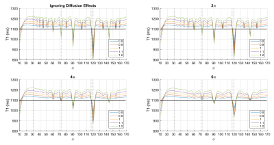
Confounding Factors in Breast Magnetic Resonance Fingerprinting: B1+, Slice Profile and Diffusion Effects
Teresa Nolte, Mariya Doneva, Thomas Amthor, Peter Koken, Nicolas Gross-Weege, Tianyu Han, Hannah Scholten, Volkmar Schulz
In this study, we evaluate the effect of three potentially confounding factors (B1+ inhomogeneity, slice profile, diffusion) on the outcome of 2D Magnetic Resonance Fingerprinting measurements in the female breast for six healthy volunteers. Each of these factors was included into an MRF dictionary and matching results were compared to a reference dictionary that excluded the correction. For the given MRF sequence, both B1+ inhomogeneity and slice profile correction affected the quantitative relaxation times in the female breast, whereas this was not the case for diffusion.
|
|
4387.
 |
12 |
A Protocol for Comprehensive Quantitative 3D Ultrashort Echo Time (UTE) Cones MR Imaging of the Knee Joint with Motion Correction
Mei Wu, Wei Zhao, Jonathan Lee, Lidi Wan, Saeed Jerban, Eric Chang, Jiang Du, Yajun Ma
We propose a protocol for comprehensive quantitative 3D UTE-Cones imaging of the knee joint with motion correction. The protocol includes 3D UTE-Cones actual flip angle imaging (UTE-Cones-AFI) for T1 measurement, UTE-Cones with variable TEs for T2* measurement, UTE-Cones with adiabatic T1ρ preparation for AdiabT1ρmeasurement, and UTE-Cones-MT for measuring MTR and modeling of macromolecular fraction (f) for various knee joint tissues including the cartilage, menisci, ligaments, tendons and muscle. An elastix motion registration method was used for motion correction. In our study, three knee specimens and 15 volunteers were evaluated. Mean and standard deviation of the measurements for various knee joint tissues are reported.
|
|
4388.
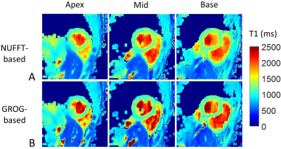 |
13 |
Free-running 3D Radial Myocardial T1 Mapping using Self-Calibrating GRAPPA Operator Gridding for Accelerated Iterative Reconstruction
Haikun Qi, Aurelien Bustin, Olivier Jaubert, René Botnar, Claudia Prieto
Free-running 3D radial (kooshball) sampling is suitable for fast and self-navigated whole-heart cardiovascular imaging. However, iterative undersampled 3D radial reconstruction requires computational demanding gridding/regridding steps in each iteration, which leads to long reconstruction time and may limit the applications of this imaging strategy. In this work, we investigate the feasibility of accelerating iterative reconstruction for a free-running 3D myocardial T1 mapping sequence using GRAPPA Operator Gridding (GROG)-based pre-reconstruction interpolation. Image quality and T1 estimation accuracy of the accelerated GROG-based reconstruction were compared with conventional non-uniform FFT (NUFFT)-based reconstruction in a standardized phantom and five healthy subjects.
|
|
4389.
 |
14 |
Efficient Quantitative Susceptibility Mapping of Popliteal Artery Wall
Yan Wen, Thanh Nguyen, Ajay Gupta, Yi Wang
The objective of this study was to develop and optimize the pulse sequence and post-processing for an efficient and high quality QSM of the popliteal artery wall. We showed that high quality QSM could be achieved in 4 minutes without the need for cardiac gating.
|
|
4390.
 |
15 |
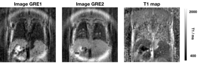
Development of a 3D UTE MP2RAGE sequence for mouse pulmonary T1 mapping at 7T
Thibaut Faller, Colleen Cardiet, Wilfried Souleyreau, Lin Cooley, Andreas Bikfalvi, Baudouin Denis de Senneville, Sylvain Miraux, Emeline Ribot
The 3D Magnetization Prepared 2 Gradient Echo (MP2RAGE) sequence is very useful to obtain high contrasts between brain tissues and between metastases and the surrounding healthy brain at high clinical magnetic fields (≥3T). In order to apply this sequence for the detection and T1 mapping of lung metastases in mice at 7T, major modifications were done. We developed an ultra-short echo time (UTE) MP2RAGE sequence by replacing the Cartesian encoding by a radial one. This encoding enables (i) to shorten echo time to less than 0.1ms and consequently obtain lung T1 maps; and (ii) to track respiration motion through a self-gating strategy to evaluate the displacements of the metastases due to breathing.
|
|
4391.
 |
16 |
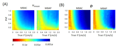
Model Based Analysis of Complex Difference Images for Measuring Diameters and Velocities of Penetrating Arteries
Xiaopeng Zong, Weili Lin
Pathological changes of penetrating arteries (PAs) may be an important contributing factor of cerebral small vessel disease (SVD). Measurement of PA flow velocity and diameter with phase contrast (PC) MRI remains challenging due to the presence of strong partial volume effects. Here we propose model-based analysis of complex difference (MBAC) images to quantify diameter and velocity of PAs. We demonstrated the accuracy of the MBAC method with simulation and phantom studies. In vivo PA diameter and velocity were obtained for the first time. The MBAC method may serve as a useful tool for understanding the etiopathogenesis of SVD.
|
|
4392.
 |
17 |
MR Relaxivity Mapping using multi-dimensional integrated (MDI) complex signal ratio
Yongquan Ye, Jingyuan Lyu
A novel relaxivity mapping method for MR transverse relaxivity mapping (e.g. T2*) is proposed and demonstrated. By extracting an overall complex signal ratio by means of multi-dimensional integration (MDI) , our method offers significantly improved SNR and homogeneous parametric mappings. With MDI, no explicit multi-channel combination operation is required, and calculation efficiency is extremely high for inline calculation.
|
|
4393.
 |
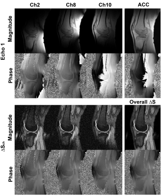
18 |
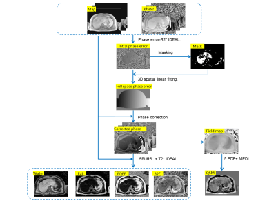
Spine Quantitative Susceptibility Mapping Using In-Phase Echoes to Initialize the Nonconvex Optimization Problem of Fat-Water Separation (R2*-IDEAL)
Yihao Guo, Zhe Liu, Yan Wen, Pascal Spincemaille, Honglei Zhang, Ramin Jafari, Shun Zhang, Sarah Eskreis-Winkler, Kelly M. Gillen, Yanqiu Feng, Yi Wang
This work aims to investigate the initializations of R2* and field maps in R2*-IDRAL for developing a robust quantitative susceptibility mapping (QSM) in the spine. A 3D multi-echo GRE sequence was implemented to acquire out-phase and in-phase (IP) echoes in 10 subjects. The R2* and background field maps estimated by fitting the magnitude and phase of IP echoes were used to initialize R2*-IDEAL to obtain final R2*, field, water, and fat maps. The final field map was further processed to generate QSM. The results demonstrated that IP initializations of R2* and field in R2*-IDEAL provide robust QSM of the spine.
|
|
4394.
 |
19 |
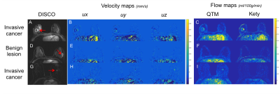
Quantitative Transport Mapping (QTM): a new AIF-free perfusion technique to distinguish malignant and benign breast lesions
Sarah Eskreis-Winkler, Natsuko Onishi, Liangdong Zhou, Pascal Spincemaille, Ramin Jafari, Meredith Sadinski, Elizabeth Sutton, Elizabeth Morris, Yi Wang
Quantitative perfusion imaging is challenging in the breast because the requisite arterial input function (AIF) is difficult to measure given the lack of large-caliber feeding arteries. To overcome this problem, we show that quantitative transport mapping (QTM), a new AIF-free perfusion model, is not only technically feasible in the breast, but has the potential to better distinguish malignant from benign breast lesions compared to conventional perfusion modeling.
|
|
4395.
 |
20 |
Texture analysis of multi-phase magnetic resonance images to discriminate expression of Ki67 in hepatocellular carcinoma
Yueming Li, Chuan Yan
Aims: This study aimed to determine whether texture analysis of preoperative magnetic resonance images could predict expression of Ki67 in hepatocellular carcinoma(HCC). Methods: 83 patients confirmed HCC were included. Texture analysis on 3.0 Tesla MR Unit included histogram, co-occurrence matrix, run-length matrix, gradient, auto-regressive model, and wavelet transform features as calculated by MaZda software.
Results: HCC with higher Ki67 label index tend to display a lower differentiation pattern. Larger tumors usually had higher Ki67 label index. Texture parameters generated from arterial phase imaging was the most frequently significant correlation.
Conclusions: Texture analysis could be used to discriminate Ki67proliferation status in HCC.
|
|
4396.
 |
21 |
Free-Breathing 3D T1 Mapping of the Whole-Heart Using Low-Rank Tensor Modeling
Paul Han, Debra Horng, Yoann Petibon, Jinsong Ouyang, Nathaniel Alpert, Georges El Fakhri, Chao Ma
T1 of the myocardium is an emerging quantitative biomarker for a variety of heart diseases. However, T1 mapping is challenging in the heart due to the cardiac and respiratory motion. To overcome this issue, T1 mapping methods utilizing ECG triggering with breath-hold or respiratory control using bellow or navigators have been developed in the past. Recently, low-rank tensor-based approach has been proposed for MR to enable image reconstruction at extremely high acceleration factors, and has demonstrated promising results. This work presents a low-rank tensor-based approach for free-breathing 3D T1 mapping of the whole-heart.
|
|
4397.
 |
22 |
Diagnostic performance of chemical shift in/opposed phase (IOP) and fat-fraction to evaluate the presence of intra-tumoral fat in HCC
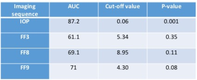
Kritisha Rajlawot, Jing Zhou, Churong Lin, Sichi Kuang, Jingbiao Chen, Yao Zhang, Hao Yang, Ying Deng, Bingjun He, Diego Hernando, Jin Wang, Scott B Reeder
Hepatocellular carcinoma (HCC) is the most common primary malignancy of the liver. Previous studies have showed that the presence of intra-tumoral fat has morefavorable prognosis of HCC. We aim to compare the diagnostic accuracy of chemical shift encoded fat-fraction at three different flip angles (FA) 3°, 8° and 9°using IDEAL-IQ, in comparison to chemical shift imaging (IOP) to evaluate intra-tumoral fat in HCC.Our results showed higher positive rates of detection of intra-tumoral fat in HCC with IOP (100%) compared to IDEAL IQ at FA 3° (44.4%), FA 8° (55.6%) and FA 9° (88.9%).
|
|
4398.
 |
23 |
DeepBLESS: learning inverse Bloch equations for rapid prediction of myocardial relaxation parameters
Jiaxin Shao, Vahid Ghodrati , Kim-Lien Nguyen, Peng Hu
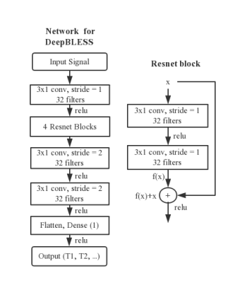
Bloch equation simulation provides accurate estimation of soft tissue relaxation parameters for many applications. To speed up using Bloch equation for relaxation parameter estimation, we propose a general approach - deep learning with Bloch equation simulations (DeepBLESS) - to learn inverse Bloch equation for rapid myocardial relaxation parameter prediction. Using the Modified Look-Locker inversion recovery (MOLLI) sequence and a self-designed simultaneous radial T1 and T2 mapping sequence as examples, we demonstrated that DeepBLESS was adaptive to heart rate variation with good estimation accuracy and precision while reducing the inline computation time compared to the conventional Bloch-equation-based approaches.
|
|
| Top |
Quantitative Mapping of the Brain
Digital Poster
Acquisition, Reconstruction & Analysis
Thursday, 16 May 2019
| Exhibition Hall |
08:15 - 09:15 |
| |
|
Computer # |
|
4399.
 |
26 |
Direct Myelin Volume Fraction Mapping with Correction for Magnetization Transfer and Diffusion Effects Using a Four-pool White Matter Model
Zhe Wu, Ilana Leppert, David Rudko
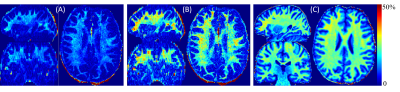
We propose an accelerated myelin water fraction (MWF) imaging technique that employs wave encoding combined with double inversion-recovery weighting (wave-CAIPI ViSTa) for rapid human brain myelin volume fraction (MVF) measurement. In addition, we investigate the contributions of both magnetization transfer and diffusion to the wave-CAIPI ViSTa MWF signal. A four-pool white matter model and extended phase graph framework are applied for magnetization transfer and diffusion modeling. The proposed MVF mapping method is a promising candidate for measuring white matter properties in human demyelinating conditions with limited dependence on model fitting parameters.
|
|
4400.
 |
27 |
Accelerating Multi-Echo GRASE with CAIPIRINHA for Fast and High-Resolution Myelin Water Imaging
Gian Franco Piredda, Tom Hilbert, Erick J. Canales-Rodríguez, Marco Pizzolato, Reto Meuli, Josef Pfeuffer, Jean-Philippe Thiran, Tobias Kober
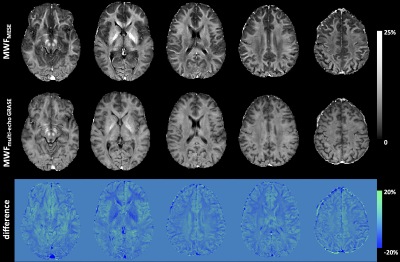
Impaired myelin plays a central role in a wide range of degenerative brain diseases. A method for non-invasive and in vivo assessment of myelin content within clinically acceptable acquisition times is thus desirable. In this work, a 3D multi-echo gradient and spin-echo (GRASE) sequence was accelerated with CAIPIRINHA to achieve high-resolution and whole-brain myelin imaging in less than ten minutes. Myelin water fraction (MWF) maps were derived from multi-echo GRASE data in a cohort of healthy subjects and values proved to be consistent with MWF maps computed from a conventional multi-echo spin-echo acquisition.
|
|
4401.
 |
28 |
A fast, joint sparsity constraint algorithm for improved myelin water fraction mapping
Martijn Nagtegaal, Burkhard Mädler, Thomas Amthor, Peter Koken, Mariya Doneva
A new method for myelin water fraction mapping from multi-echo spin echo data using a joint sparsity constraint is proposed, which is faster than previously proposed methods. This method is based on the assumption that the T2 spectrum is sparse and consists of a common small set of discrete relaxation times for all voxels. The method finds an estimation of the flip angle inhomogeneity map from the data itself, to remove the bias caused by B1 inhomogeneities. The proposed method is compared to state of the art MWF approaches in 3T brain measurements.
|
|
4402.
 |
29 |
Non-negative least squares fitting of multi-exponential T2 decay data: Are we able to accurately measure the fraction of myelin water?
Vanessa Wiggermann, Irene Vavasour, Enedino Hernandez-Torres, Gunther Helms, Alexander MacKay, Alexander Rauscher
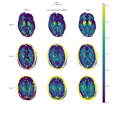
The ability to determine the myelin water fraction (MWF) in vivo is essential to assessments of neurodevelopmental myelination and myelin damage in neurodegenerative diseases. The analysis of multi-exponential T2 decay data relies on the non-negative-least-squares (NNLS) fitting, which may be sensitive to the chosen fitting parameters. We performed simulations to explore the outcomes of NNLS under different parameter selection. The lowest allowed T2 was found to have the largest effect on correctly estimating the T2 of different water pools as well as the MWF. Lower refocusing FAs led to further underestimation of the MWF.
|
|
4403.
 |
30 |
Myelin Water Fraction Estimation using Small-Tip Fast Recovery MRI
Steven Whitaker, Gopal Nataraj, Mingjie Gao, Jon-Fredrik Nielsen, Jeffrey Fessler
Myelin water fraction (MWF) is a good biomarker for myelin content. Traditional methods for acquiring MWF maps require long scan times. Recent work has estimated MWF from faster steady-state scans. In this work, we propose to acquire MWF maps from an optimized set of small-tip fast recovery (STFR) scans that can exploit resonance frequency differences between myelin water and the slow-relaxing water compartment.
|
|
4404.
 |
31 |
Myelin Water Imaging Profiles Along White Matter Tracts
Tobias Baumeister, Shannon Kolind, Alex MacKay, Martin McKeown
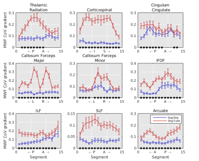
Myelin water fraction (MWF) maps are spatially noisy. Here we investigated a possible inherent spatial structure of MWF values along diffusion tensor imaging (DTI)-derived white matter (WM) tracts in 41 healthy subjects. Sixteen major fibre bundles were extracted and MWF was computed in sub-segments along each fibre tract and compared to surrounding voxels. MWF values were more spatially coherent along fibre bundles than elsewhere. The profile along the trajectory of fibre bundles estimated subjects’ age more accurately than tract-averaged MWF. We conclude that the spatial MWF distribution in WM consistently follows a distinct pattern along underlying fibre bundles across subjects.
|
|
4405.
 |
32 |
Single-point macromolecular proton fraction mapping at 7T in healthy and demyelinated mouse brain
Lucas Soustelle, Maria-Cristina Antal, Paulo Loureiro de Sousa, Laura Harsan
Assessment of myelin content in the brain is essential for monitoring pathologies such as multiple sclerosis. Quantitative MRI methods including quantitative magnetization transfer imaging (qMTI) have been employed in animal and human studies to assess demyelination processes. Animal studies have reported high correlations between myelin content and the macromolecular proton fraction (MPF), a metric derived from qMTI. The single-point MPF mapping method requires the acquisition of a single MT-weighted image, hence reducing protocol scan duration. In this work, we propose the adaptation of this method at 7T in a study involving healthy and demyelinated mice.
|
|
4406.
 |
33 |
Development of quantitative water content mapping in human brain at high magnetic field
Hidehiro Watanabe, Nobuhiro Takaya, Fumiyuki Mitsumori
The method for quantitative water content mapping of a human brain at high magnetic field was proposed. We demonstrated that B1- is proportional to B1+ in a uniform area even on a nonuniform image measured at 4.7T. B1-s of the reference phantom and a human brain can be compared by measurable B1+s in uniform areas and water content of human brain can be computed from that comparison. Our method was validated in the experiments of the mixture phantom of H2O and D2O. Quantitative water content maps of human brains were obtained by our method.
|
|
4407.
 |
34 |
Global relaxometry and volumetry of the brain using synthetic MR: possible implications for the neurobiology of human brain ageing in healthy adults
Lu Yu, Chunmei Li, Bing Wu, Jianxun Qu, Yuwei Jiang, Min Chen
Synthetic MR is an emerging technique capable of providing quantitative relaxation maps and conventional contrast weighted images simultaneously. This study aims to study the relaxation and volumetric characteristics in the ageing process with synthetic MRI. We found volume is a primary metrics for assessing brain ageing and relaxometry may provide additional quantitative biomarkers and possible implications for studying brain ageing.
|
|
4408.
 |
35 |
Fast multiparametric imaging in the brain using a stationary balanced steady state cartesian approach
Christian Guenthner, Sebastian Kozerke, Mathieu Sarracanie
We propose a multiparametric balanced steady-state 3D Cartesian sequence that exploits model based and pattern matching reconstruction strategies for a series of 20 flip angles and repetition times, allowing for the simultaneous quantification of B0, B1+, T1, T2, and proton density. Time-varying signal patterns at the steady state are reached that allow for the acquisition of unique signal patterns in each image voxel for any acquisition scheme. We show the feasibility of our technique in-vivo in the human brain in 11 minutes, here with Cartesian acquisition and no acceleration strategies.
|
|
4409.
 |
36 |
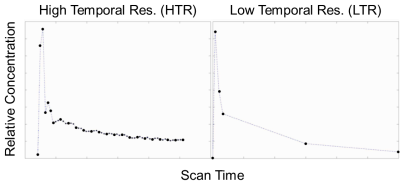
Improving quantification accuracy in whole brain high spatial resolution 3D kinetic mapping: Development of a novel dual temporal resolution DCE-MRI technique
Ka-Loh Li, Daniel Lewis, Alan jackson, Sha Zhao, Xiaoping Zhu
Mapping microvascular parameters from DCE MRI traditionally requires a compromise between temporal resolution, spatial resolution, and volume coverage. This study developed a dual-temporal-resolution-based analysis method which concatenates acquired high temporal (HT) and high spatial (HS) tissue contrast agent concentration curves into a unified HTHS merged volume and then pixel-by-pixel reconstructed the HT first pass concentration curve to a HS resolution before undertaking kinetic analysis. In vivo assessment of this method was undertaken in 12 patients with neurofibromatosis type II, and demonstrated the potential of the new method to provide high spatial resolution kinetic map with HT comparable accuracy and quality.
|
|
4410.
 |
37 |
High-resolution 3D T1 and T2 Mapping in the Brain Using Compressed Sensing and Dictionary Fitting
Emilie Mussard, Tom Hilbert, Christoph Forman, Reto Meuli, Jean-Philippe Thiran, Tobias Kober
Quantitative magnetic resonance imaging (qMRI) aims at directly measuring physical tissue properties to be more independent from technical influences. However, parameter mapping is often long and 2D-based. In this work, we propose a protocol for 3D brain T1 and T2 mapping accelerated by compressed sensing. To improve T2 accuracy, we also implemented a T1-informed T2 dictionary fitting technique. Preliminary results showed the ability of the protocol to provide T1 and T2 maps at a 1x1x1.2mm3 resolution in 14:05min as well as the accuracy of the mapping. Establishing a fast 3D protocol will enable generating high-resolution atlases as a next step.
|
|
4411.
 |
38 |
Effects of acquisition and compressed sensing reconstruction parameters on 3D-QALAS multi-parameter quantitation and synthetic imaging of the brain
Ken-Pin Hwang, Marcel Warntjes, Naoyuki Takei, Suchandrima Banerjee, Drew Mitchell, R. Stafford, Linda Chi, David Fuentes
3D QALAS is a promising new technique that simultaneously maps T1, T2, and PD in a single 3D acquisition. We investigate the robustness of this technique to several acquisition and compressed sensing reconstruction parameters in phantom and brain images. Parameter maps were shown to be robust to B1 through the center portion of the slab, while compressed sensing did not demonstrate any effects on parameters in phantom or cause additional artifacts on parameter maps in human brain. 3D QALAS thus presents an attractive quantification method for therapy planning and tissue volume measurement applications.
|
|
4412.
 |
39 |
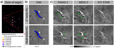
Influence of SWI Sequences and QSM Reconstruction Methods on Measured Magnetic Susceptibility in Cerebral Veins
Ronja Berg, Jakob Meineke, Andreas Hock, Claus Zimmer, Christine Preibisch
Quantitative Susceptibility Mapping (QSM) has recently been used for assessing the cerebral oxygen metabolism. However, a systematic investigation on the most suitable imaging parameters and reconstruction algorithms for determining the venous susceptibility values is missing. Therefore, we investigated both, the impact of flow compensation and accelerated acquisition as well as different reconstruction methods on measured venous susceptibility. Our results suggest that the choice of reconstruction technique can significantly influence the venous susceptibility values while the investigated imaging parameters did not considerably affect its accuracy. Thus, the applied QSM reconstruction technique has to be considered carefully when quantifying the venous oxygenation.
|
|
4413.
 |
40 |
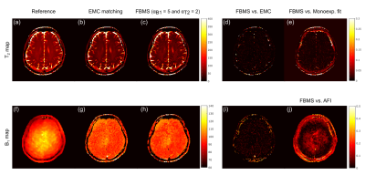
Improving T2 and B1 parametric estimation in the brain with multi spin-echo MR and fusion bootstrap moves solver (FBMS)
Andreia Freitas, Inês Sousa, Andreia Gaspar, Rui Teixeira, Joseph Hajnal, Rita Nunes
Multi spin-echo (MSE) sequences have been prescribed for efficient T2 mapping. This can be further improved by matching to pre-computed echo-modulation curves (EMC). Previous use of this method to estimate T2 and B1 resulted in bias in the latter. We investigated the possibility to improve B1 by taking advantage of its spatial smoothness, using a fusion bootstrap moves solver (FBMS). The two methodologies were compared using a numerical phantom and in-vivo brain data. While T2 estimation was accurate and equivalent, B1 accuracy was improved using the FBMS. Future work is required to tune the regularization parameters of the FBMS algorithm.
|
|
4414.
 |
41 |
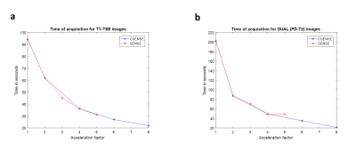
To Evaluate Effect of SENSE and CSENSE on Quantitative T1 and T2 mapping of Human Brain
Dinil Sasi S, Anup Singh, Rupsa Bhattacharjee, Ayan Debnath, Snekha Sehrawat, Rakesh K Gupta, Indrajit Saha, Marc Van Cauteren
Parallel-imaging and compressed-sensing based approaches are playing crucial role in accelerating MRI data acquisition. Objective of the study was to accelerate the data acquisition of T1, T2 and PD-weighted TSE images and to evaluate the accuracy of T1 and T2 mapping in the human brain. Data was acquired using SENSE parallel-imaging and Compressed-SENSE technique for different factors as well as without any acceleration. T1 and T2 values obtained using data with SENSE (upto factor of 3) and CSENSE (upto factor of 6) were comparable to those acquired without any acceleration. Errors in T1 and T2 increased with increase in acceleration factor.
|
|
4415.
 |
42 |
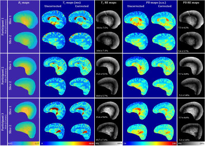
Four angle method for accurate and rapid clinical high-resolution whole-brain mapping of longitudinal relaxation time and proton density with B1 inhomogeneity correction
Abinand Rejimon, Luis Cortina, Richard Spencer, Mustapha Bouhrara
Changes in longitudinal relaxation time (T1) and proton density (PD) are sensitive markers of microstructural damage associated with different neurological conditions including myelin degradation, axonal loss, inflammation, and edema. In this study, we propose an accurate and rapid approach to mapping T1 and PD with B1inhomogeneity correction. This four angle method (FAM) is based on the use of four images acquired with different flip angles and short repetition times using the spoiled-gradient recalled-echo sequence available on all preclinical and clinical MRI machines. The accuracy and ease of implementation of the FAM renders it of great potential for clinical investigations.
|
|
4416.
 |
43 |
Towards in-vivo voxel-wise parcellation of human brain cortex
Shahrzad Moeiniyan Bagheri, Viktor Vegh, David Reutens
The research aims to establish the feasibility of developing an automated method for in vivo voxel-wise parcellation of the human brain cortex. We combined our previously proposed residual analysis Magnetic Resonance Fingerprinting (MRF) approach with supervised classification. We show that extraction of a feature vector from a patch of voxels about a voxel of interest improves prediction accuracy by about 10%, as measured using the Area Under the Curve (AUC) metric. Our approach leads to an increase in the prediction accuracy rate for areas of distinct microstructural heterogeneity, such as the primary motor cortex.
|
|
4417.
 |
44 |
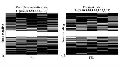
Variable Rates Undersampling Scheme for Fast brain T1? mapping
Yuanyuan Liu, Yanjie Zhu, Jing Cheng, Xin Liu, Dong Liang
T1ρ mapping requires several T1ρ-weighted images with different spin lock times to obtain the T1ρ maps, resulting in a long scan time.Compressed sensing has shown good performance in fast quantitative T1ρ mapping. In this work, we developed a variable acceleration rates undersampling strategy to reduce the scan time. A signal compensation with low-rank plus sparse model was used to reconstruct the T1ρ-weighted images. Specifically, a feature descriptor was used to pick up useful features from the residual images. Preliminary results show that the proposed method achieves a 5.76-fold acceleration and obtain more accurate T1ρ maps than the existing methods.
|
|
4418.
 |
45 |
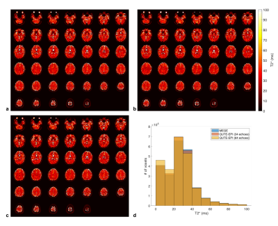
Fast and whole-brain T2* mapping using QUTE-EPI at 7T
Seonyeong Shin, Seong Dae Yun, N. Jon Shah
Quantification of T2* relaxation time is of great interest as knowledge of it can be used for clinical diagnosis or optimisation of MR imaging parameters. A typical approach to quantify T2* is to acquire multi-echo data. Although this approach is effective, it still requires a substantial acquisition time for whole-brain coverage. This work aims to employ quantitative echo-planar imaging (QUTE-EPI) at 7T for fast and whole-brain T2* mapping. The performance of QUTE-EPI was directly compared to that of a conventional multi-echo gradient-echo sequence (MEGE). The estimated T2* values were quantitatively analysed for the regions of grey matter (GM) and white matter (WM).
|
|
4419.
 |
46 |
Joint T1 and T2 Mapping with Tiny Dictionaries and Subspace-Constrained Reconstruction
Volkert Roeloffs, Martin Uecker, Jens Frahm
Dictionaries as used in multi-parametric mapping are typically very large in size, take long to compute, and scale exponentially with the number of parameters. Here, we break the bond between dictionary size and representation accuracy by two modifications: First, we approximate the Bloch-response manifold by piece-wise linear functions, and second, we allow the sampling grid to be refined adaptively depending on the precision needed. Phantom and in vivo studies demonstrate efficient multi-parametric mapping with tiny dictionaries and subspace-constrained reconstruction. The presented method preserves accuracy and precision with dictionaries reduced in size by a factor of 10 and beyond.
|
|
4420.
 |
47 |
T1 Mapping at 7T Using a Novel Inversion-Recovery Look-Locker 3D-EPI Sequence
Rüdiger Stirnberg, Yiming Dong, Jonas Bause, Philipp Ehses, Tony Stöcker
We propose a novel Inversion-Recovery Look-Locker 3D-EPI sequence for rapid T1 mapping. The inherent SNR benefit of a 3D acquisition, segmentation along both phase encode directions and a turbofactor introduced to reduce the number of required inversions can be traded freely for acquisition speed, SNR, resolution and geometric distortions. Aside from quantitative validation, two high-resolution T1 mapping applications are demonstrated at 7T: whole-brain with minimal distortions, and reduced field-of-view with geometric distortions matched to corresponding fMRI data. The results show high T1 accuracy for several turbofactor and flip angle combinations compared to a single-slice inversion-recovery 2D-EPI reference.
|
|
4421.
 |
48 |
Fast quantitative susceptibility reconstruction via total field inversion with L0 norm approximation
Shuhui Cai, Li Zhang, Congbo Cai, Zhong Chen
Quantitative susceptibility mapping (QSM) is a meaningful MRI technique owing to its unique relation to actual physical tissue magnetic properties. The reconstruction of QSM is usually decomposed into three sub-problems which are solved independently. Here, we propose a fast reconstruction method named as fast TFI based on total field inversion. It accelerates the total field inversion by using specially selected preconditioner and the advanced solution of weighted L0 regularization. Results from gadolinium phantom and in vivo data verified that the new method has good performance.
|
|
4422.
 |
49 |
Six-direction diffusion tensor MRI using a convolutional neural network
Qiyuan Tian, Berkin Bilgic, Qiuyun Fan, Chanon Ngamsombat, Congyu Liao, Yuxin Hu, Thomas Witzel, Kawin Setsompop, Jonathan Polimeni, Susie Huang
Diffusion tensor imaging (DTI) is widely used for clinical neuroimaging and neuroscientific research but has traditionally suffered from relatively length acquisition. Here, we propose a new approach to obtain both scalar and orientational DTI metrics from six diffusion-weighted images with optimal directional encoding. Through the careful choice of diffusion directions, we compute initial tensor results that are then denoised using a convolutional neural network. Our results provide comparable scalar and orientational DTI metric maps to those acquired with 90 directions.
|
|
4423.
 |
50 |
Silent 3D Parameter Mapping using Variable Flip Angle Looping Star
Florian Wiesinger, Nikou Damestani, David Lythgoe, Emil Ljungberg, Tobias Wood , Mark Symms, Fernando Zelaya, Gareth Barker, Steven Williams, Ana Beatriz Solana
This abstract presents a new method for silent and 3D parameter mapping, including proton density (PD) , T1, T2*, and quantitative susceptibility mapping (QSM), by combining Looping Star 3D silent T2*-weighted imaging with the concept of variable flip angle (VFA) PD and T1 mapping.
|
|
| Top |
Motion Correction: Brain
Digital Poster
Acquisition, Reconstruction & Analysis
Thursday, 16 May 2019
| Exhibition Hall |
08:15 - 09:15 |
| |
|
Computer # |
|
4424.
 |
51 |
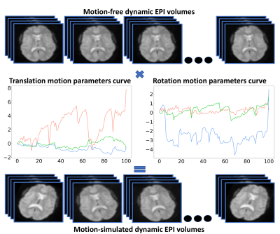
Three-dimensional motion correction in Magnetic Resonance Fingerprinting (MRF)
Jan Kurzawski, Matteo Cencini, Pedro Gómez, Rolf Schulte, Giada Fallo, Alessandra Retico, Michela Tosetti, Mauro Costagli, Guido Buonincontri
Two-dimensional MRF is considered to be less sensitive to in-plane motion than conventional imaging techniques. However, in scanning populations prone to rapid and extensive motion, challenges remain. Here, we suggest a two-step 3D MRF procedure that includes the correction of subject motion during the reconstruction. In the first step, we reconstruct the data in small segments consisting of images with equal contrast and calculate the between-segment motion. In the second step, we perform motion correction and use corrected images for matching with dictionary. This results in higher quality of reconstructed images and better precision of quantitative maps.
|
|
4425.
 |
52 |
Motion-corrected and high-resolution anatomically-assisted (MOCHA) reconstruction of arterial spin labelling MRI
Abolfazl Mehranian, Andrew Reader, Enrico De Vita
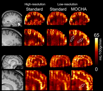
A MOtion-Corrected and High-resolution Anatomically-assisted (MOCHA) reconstruction framework is proposed for ASL MRI. The method simultaneously accounts and corrects for rigid motion and partial volume effects (PVE), and reduces noise by guided high-resolution anatomical MR images without any need for segmentation. The proposed method was compared with standard methods and a 3D linear regression (3DLR) correction method using realistic simulations and in-vivo data. Results show that MOCHA outperforms 3DLR not only in preservation of structural and local details, including simulated lesions, but also in PVE correction of deep grey matter structures, often subject to segmentation errors in conventional methods.
|
|
4426.
 |
53 |
Deep Learning based motion artifact correction improves the quality of cortical reconstructions
Ben Duffy, Lu Zhao, Arthur Toga, Hosung Kim
Cortical reconstruction is prone to failure without high quality structural imaging data. Here, motion simulation was performed on good quality structural MRI images and used to train a regression convolutional neural network to predict the motion-free images as the output. We show that performing retrospective motion correction using a convolutional neural network is able to significantly reduce the number of cortical surface reconstruction quality control failures.
|
|
4427.
 |
54 |
Improved motion correction of submillimetre 7T fMRI time series with boundary-based registration (BBR)
Pei Huang, Johan Carlin, Richard Henson, Marta Correia
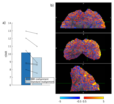
Here, we present a novel approach of utilizing Boundary-Based Registration for realigning submillimetre 7T fMRI time series. We collected fMRI data from 6 human participants and processed the data using either standard rigid body realignment using SPM or our BBR realignment method. We compared the two pre-processed datasets with multiple metrics (tSNR, fCNR and percentage of variance explained by the model) and show that realigning using BBR consistently outperforms conventional methods.
|
|
4428.
 |
55 |
A fast approach for simultaneous measurement of head motion and induced magnetic field changes using FID navigators
Tess Wallace, Onur Afacan, Tobias Kober, Simon Warfield
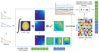
Incorrect spatial encoding due to subject motion is a dominant source of artifacts in MRI. Even if changes in head pose are measured and corrected, motion-induced perturbations in the local magnetic field are a further source of image degradation, particularly for imaging at longer echo times and higher field strengths. We propose a fast approach for simultaneously measuring head motion and spatiotemporal B0 changes using FID navigators (FIDnavs) and simulation of the acquisition physics. Rigid-body motion and first-order field coefficients estimated from FIDnavs exhibit a high degree of agreement with ground-truth values in both phantom and volunteer experiments.
|
|
4429.
 |
56 |
Robust retrospective correction of 3D golden-ratio radial MRI using electromagnetic tracking
Tess Wallace, Simon Warfield, Onur Afacan
Radial MRI is intrinsically more robust to motion than Cartesian sampling; however, if large rotational motion occurs, the uniform sampling of conventional 3D radial acquisitions is disrupted and is difficult to recover retrospectively. The golden angle ratio has been used to generate a quasi-isotropic distribution of spokes over time in 2D, but is limited to fully correct for motion, which occurs in three dimensions. Extending the flexibility of golden-ratio spoke ordering to 3D radial sampling, combined with rigid-body motion tracking using electromagnetic sensors, enables robust retrospective correction by maintaining relatively uniform sampling, even in the presence of large-amplitude rotational motion.
|
|
4430.
 |
57 |
Correction of Out-of-FOV Motion Artifacts using Convolutional Neural Network Derived Prior Image
Chengyan Wang, Yuan Wu, Yucheng Liang, Danni Yang, Siwei Zhao, Yiping P. Du
This study presented a new motion correction algorithm with the incorporation of convolutional neural network (CNN) derived prior image to solve the out-of-FOV motion problem. A modified U-net network was developed by introducing motion parameters into the loss function. We assessed the performance of the proposed CNN-based algorithm on 1113 MPRAGE images with simulated oscillating and sudden motion trajectories. Results show that the proposed algorithm outperforms conventional TV-based algorithm with lower NMSE and higher SSIM. Besides, robust reconstruction was achieved with even 20% data missed due to the out-of-FOV motion.
|
|
4431.
 |
58 |
Non-contact measurement of head movements inside a 7 T Scanner using a 16-channel field camera
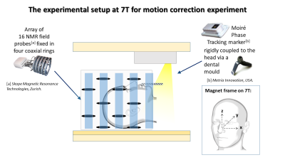
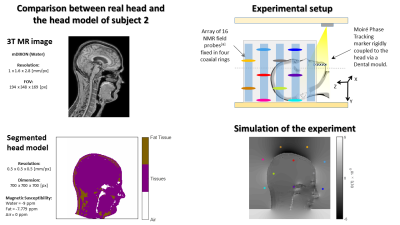
Laura Bortolotti, James Smith, Penny Gowland, Richard Bowtell
The extra-cranial magnetic field changes due to changes in head position have been measured in a 7T scanner using a 16-channel field camera and used to estimate the head movements. A partial least squares regression was used to identify the relationship between field changes and head position data that was simultaneously measured using an optical camera. By applying spherical harmonic spatial filtering to the field measurements it was possible to reduce the unwanted effect of chest movement in respiration, and to then predict head position changes with good accuracy. This provides a step forward towards a non-contact motion monitoring technique.
|
|
4432.
 |
59 |
Simulation of external magnetic field changes due to head motion during 7 Tesla MRI scan
Laura Bortolotti, James Smith, Penny Gowland, Richard Bowtell
One potential method for monitoring the effects of head movement in the scanner consists of using a fixed array of field probes to measure field changes produced outside the head by small changes in head position and angulation. This method has the advantage of requiring neither attachment of markers or probes to the head, nor modification of the imaging sequence. Here, we use realistic head models to simulate the external field changes produced by typical head movements in a 7T scanner and use the results to explore the relationship between the magnetic field perturbation and changes in head position.
|
|
4433.
 |
60 |
Markerless real-time motion correction for 2D RARE: reducing artefacts in clinical T2 and FLAIR MRI
Robert Frost, F. Karahanoglu, Camilo Jaimes, Paul Wighton, Richard Robertson, P. Grant, M. Tisdall, André van der Kouwe
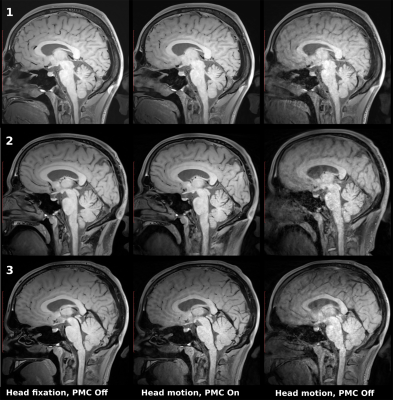
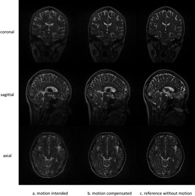
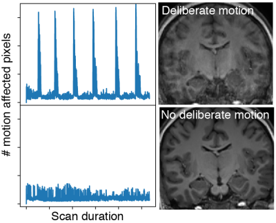
This study investigates high-frequency prospective motion correction (PMC) using markerless face tracking for artefact reduction in clinical T2 and FLAIR MRI. The FOV pose was corrected before the acquisition of each slice in PMC sequences and five subjects were scanned with 1:16 min T2, 4:55 min T2, and 1:50 min FLAIR protocols. The multi-slice segmented RARE sequences showed high sensitivity to changes in head position but use of PMC scans consistently recovered good image quality with higher image sharpness as measured by the Tenengrad metric.
|
|
4434.
 |
61 |
Robust motion regression of resting-state data using a convolutional neural network model
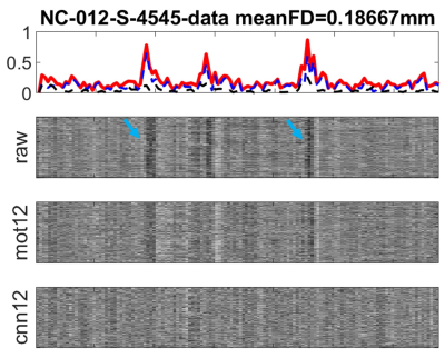
Zhengshi Yang, Xiaowei Zhuang, Karthik Sreenivasan, Virendra Mishra, Dietmar Cordes
The fluctuation introduced by head motion considerably confounds the interpretation of resting-state fMRI data. Specifying motion regressors without taking fMRI data itself into consideration may not be sufficient to model the impact of head motion. We proposed a robust and automated deep neural network (DNN) to derive motion regressors with both fMRI data and estimated realignment parameters considered. The results show that DNN-derived regressors outperform traditional regressors based on several quality control measurements.
|
|
4435
 |
62 |
High resolution 3D GRASE BLADE Arterial Spin Labelling sequence: evaluation of the performance with various level of motion: simulations and validation in volunteers and patients
Video Permission Withheld
Manjunathan NANJAPPA, Thomas Troalen, Matthias Günther, Magalie Viallon, Huber Jörn
In MRI, longitudinal acquisition protocols such as arterial spin labeling are susceptible to patient motion; this work focused on implementing 3D GRASE with BLADE readout trajectory as an alternative to Cartesian readout to increase robustness of sequence with regards to motion. Virtual data simulation and involuntary patient motion data were used to evaluate the performance of this approach with different levels of patient motion. Image reconstruction embedded with self-referenced custom rigid motion correction algorithm was developed and tested on both simulated and patient data. Results confirming superiority of SNR and motion correction capabilities offered by Blade strategy over Cartesian.
|
|
4436.
 |
63 |
A novel, coil-integrated camera for prospective optical motion correction of brain imaging at 7T
Phillip DiGiacomo, Elizabeth Tong, Julian Maclaren, Murat Aksoy, Roland Bammer, Brian Rutt, Michael Zeineh
The advancements in signal to noise ratio (SNR), contrast, and resolution enabled by high-field MR systems may visualize more nuanced brain anatomy and pathology. In order to translate these advancements to the discovery and clinical implementation of novel neuroimaging biomarkers, motion artifact resulting from requisite long scan times must be addressed. Here, we demonstrate a novel prospective optical motion tracking and correction system using a camera seamlessly integrated into the 7T Tx/Rx head coil. The integrated camera allows tracking of head motion by visualizing an optical marker on the forehead of human subjects in a 7T MR system.
|
|
4437.
 |
64 |
Prospective motion correction for 2D slice-selective FISP-MRF in the brain using an in-bore camera system
Gregor Körzdörfer, Mario Bacher, Thomas Kluge, Randall Kroeker, Dominik Paul, Josef Pfeuffer, Bernhard Hensel, Mathias Nittka
In contrast to motion artifacts in conventional MRI, which can often be identified by visual inspection, the effect can be more subtle in quantitative MRI (qMRI) methods such as Magnetic Resonance Fingerprinting (MRF). Subject motion during qMRI scans can lead to altered parameter maps without affecting their morphologic appearance which limits the user’s possibility to assess the scan quality. One way to mitigate motion artifacts is to track the subject’s movement and prospectively correct for the motion. Here, we present results of applying prospective motion correction using an in-bore camera system for MRF.
|
|
4438.
 |
65 |
Towards motion-robust MRI – Autonomous motion timing and correction during MR scanning using multi-coil data and a deep-learning neural network
Rafi Brada, Michael Rotman, Ron Wein, Sangtae Ahn, Itzik Malkiel, Christopher Hardy
We propose a method for timing and correcting for rigid-body in-plane patient motion during an MRI scan. The motion is detected using differences between coil-intensity-corrected images from different coils in the receiver array together with the scan-order information. The method allows for the detection and timing of multiple movements during the scan. For each scan where motion was detected, k-space data are divided into different motion states, which are used as input to a deep neural network whose output is a motion-corrected image. The system shows promising results on MR data containing simulated and real motion.
|
|
4439.
 |
66 |
Investigation of the impact of receive field sensitivity on motion corruption in 3D-EPI for fMRI
Nadine Graedel, Nadege Corbin, Yael Balbastre, Oliver Josephs, Martina Callaghan
High temporal signal-to-noise ratio (tSNR) is crucial in fMRI to maximise functional sensitivity. The use of high-density receiver arrays can greatly improve tSNR and enables parallel imaging, a requirement for imaging with high spatial resolution while maintaining reasonable scan times. The 3D-EPI approach enables through plane acceleration but at the cost of increased motion sensitivity. Here we explore the impact of rapidly varying sensitivity fields on the degradation of tSNR in the presence of motion in the context of 3D-EPI.
|
|
4440.
 |
67 |
Direct comparison of fat navigators and Moiré phase tracking for retrospective brain motion correction at 7T
Frederic Gretsch, Hendrik Mattern, Daniel Gallichan, Oliver Speck
Retrospective rigid body motion correction based on FatNavs or MPT motion information are directly compared. Both modalities significantly improve image quality of very high resolution anatomical images, but both suffer from drawbacks: rigid marker fixation during long scans for MPT and low temporal resolution for FatNavs. Quantitative analysis confirms these visual observations.
|
|
4441.
 |
68 |
Prospective motion correction for compressed sensing 3D TSE sequence
Patrick Hucker, Esther Raithel, Maxim Zaitsev, Axel Krafft
A compressed sensing 3D TSE Sequence prototype (CS-SPACE) was enhanced by prospective motion correction (PMC). For T1-weighted imaging this sequence uses a center-out trajectory along each echo train and sparser sampling with increasing distance from the center. Motion during such echo trains can result in unexpected image artifact behavior. In this work, we investigate whether for a particular echo train structure, a center-out trajectory and compressed sensing PMC can correct for motion artifacts.
|
|
4442.
 |
69 |
Translational Motion Compensation for 3D FSE Parallel Imaging using Autocalibration Signals
Chaoping Zhang, Stefan Klein, Alexandra Cristobal-Huerta, Juan Antonio Hernandez-Tamames, Dirk Poot
Motion during scanning deteriorates MR image quality, especially in 3D fast spin echo (FSE) acquisitions which typically require long acquisition time, even with parallel imaging. Instead of prospective motion compensation which is often difficult to perform, we propose a retrospective translational motion compensation method using autocalibration signals. The proposed method estimates the motion by minimizing the GRAPPA prediction error of the motion corrected signal in the autocalibration signal region. In-vivo experimental results demonstrate the effectiveness of our method.
|
|
4443.
 |
70 |
Video-based head motion assessment for improved quantitative neuroanatomy studies
Heath Pardoe, Allan George, Samantha Martin, Pablo Velasco, Orrin Devinsky
In-scanner head motion systematically varies with age and diagnosis, and this motion causes bias in morphometric estimates derived from neuroanatomical MRI. There are currently no widely available methods for directly assessing head motion during acquisition of neuroanatomical sequences. In this project we developed a method for measuring head motion via analysis of video obtained from an in-scanner eye tracker. Data obtained from 5 healthy controls demonstrates the feasibility of the technique. The system has minimal set up requirements for subjects or MR technicians, which suggests the technique may be well suited to the young, elderly, or impaired populations in which participant compliance may be a problem.
|
|
4444.
 |
71 |
Super-resolution reconstruction applied to neonatal MRI: multi-orientation vs through-plane slice shift MRI acquisition and segmentation
Nurten Ceren Askin, Laura Gui Levy, Joana SaDeAlmeida, Michel Kocher, Petra Huppi, Francois Lazeyras
In this study, the super-resolution (SR) method is used to reconstruct high-resolution MRI volumes from multi-orientation and through-plane shift low-resolution neonatal MRI. Multi-orientation low-resolution images yield higher quality SR results than through-plane shift low-resolution images. SR reconstructed volumes and high-resolution volumes from the scanner are segmented with a morphology-based segmentation algorithm. Segmentation quality is similar between the SR reconstructed volume and the high-resolution volume. Since low-resolution acquisitions are faster, they are less prone to motion artifacts, and thus the reconstructed SR volumes are an alternative to lengthy high-resolution acquisitions.
|
|
4445.
 |
72 |
Motion correction in Brain MR imaging using a Structure Light based Optical MOtion Tracking system (SLOMO)
Chunyao Wang, Chen Zhang, Yu Wang, Haikun Qi, Tianqi Huang, Jin Liu, Chun Yuan, Hongen Liao, Huijun Chen
Motion artifact is an important challenge in MR imaging. Optical tracking based motion correction technique has been verified effective with the advantages of perfect accuracy, real-time performance and no effect on sequence and scan time. However, most traditional system need an additional Reflective Marker to trace and quantify the motion parameters, which complicated the scan procedure. Recently, our group proposed a markerless optical tracking solution(NORMS) and validated its ability in non-rigid motion detection and correction for carotid artery imaging. In this study, we aim to develop a parallel line Structure Light based Optical Motion Tracking system (SLOMO) to accurately correct rigid motion by acquiring the whole 3D surface. The results demonstrated the feasibility of SLOMO system in motion correction for brain imaging.
|
|
4446.
 |
73 |
Improvement of Glutamate Chemical Exchange Saturation Transfer (GluCEST) Imaging in a Rat Model of Epileptic Seizure Using Retrospective Motion Correction
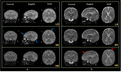
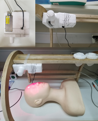
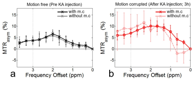
Dong-Hoon Lee, Do-Wan Lee, Jae-Im kwon, Chul-Woong Woo, Sang-Tae Kim, Jin Seong Lee, Choong Gon Choi, Kyung Won Kim, Jeong Kon Kim, Dong-Cheol Woo
GluCEST is a novel molecular MR imaging technique to detect glutamate in the brain parenchyma by measuring the exchange of glutamate amine protons with bulk water. However, a disadvantage of CEST imaging is the relatively long scan time required to collect the data while varying the resonance frequency around the water. In this abstract, we describe the application of a retrospective motion correction approach using a gradient-based motion correction (GradMC) algorithm to CEST data for investigating the feasibility of motion correction, using an epileptic seizure rat model with head motion. Our results clearly show that the GradMC can be used in CEST imaging to efficiently correct for motion.
|
|
4447.
 |
74 |
Deep learning motion compensation for Cartesian and spiral trajectories
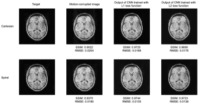
Quan Dou, Xue Feng, Zhixing Wang, Daniel Weller, Craig Meyer
Movement of the subject during MRI acquisition causes image quality degradation. In this study we adopted a deep CNN to correct motion-corrupted brain images. To get paired training datasets, synthetic motion artifacts were added by simulating k-space data along different sampling trajectories. Quantitative evaluation showed that the CNN significantly improved the image quality. The spiral trajectory performed better than the Cartesian trajectory both before and after the motion deblurring. A network trained with an L1 loss function achieved better RMSE and SSIM than one trained with an L2 loss function after convergence. Overall, deep learning yields rapid and flexible motion compensation.
|
|
4448.
 |
75 |
Deep learning based motion estimation from highly under-sampled EPI volumetric navigators
Mykhailo Hasiuk, Kamlesh Pawar, Shenjun Zhong, Richard McIntyre, Zhaolin Chen, Gary Egan
Dynamic EPI volumetric navigators are widely used to track head motion in MRI, and accurate motion estimation requires EPI volumes to be inserted in every several seconds or even less. However, the use of dynamic EPI volumes to track motion significantly degrades the overall data acquisition efficiency. To address this issue, in this work we introduce a deep learning based motion estimation method from highly under-sampled (i.e. acceleration factor of 16) EPI volumetric navigators. The method directly estimates motion parameters from the under-sampled data, and does not require reconstruction of images.
|
|
| Top |
Robust & Reproducible Quantitation
Digital Poster
Acquisition, Reconstruction & Analysis
Thursday, 16 May 2019
| Exhibition Hall |
08:15 - 09:15 |
| |
|
Computer # |
|
4449.
 |
76 |
Fast Multi-Parametric Mapping Competition: MR Fingerprinting vs. Triple-Echo Steady State
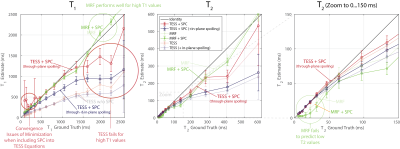
Christian Guenthner, Thomas Amthor, Sebastian Kozerke, Mariya Doneva
Magnetic Resonance Fingerprinting (MRF) and triple-echo steady-state (TESS) are two sequences that both allow for the simultaneous quantification of T1 and T2. While MRF relies on the transient response of tissue and noise-like under-sampling artifacts, TESS acquires the two lowest order SSFP-FIDs and the lowest order SSFP-Echo in the steady-state of a rapid, spoiled SSFP sequence. In this work, we compare the performance of the two sequences in a phantom study, where imaging parameters and total acquisition duration between the two scan techniques were matched. In addition, a slice-profile correction for TESS is proposed and included in the comparsion.
|
|
4450.
 |
77 |
Experimental Validation of Augmented Fractional MR Fingerprinting
Lixian Zou, Haifeng Wang, Huihui Ye, Shi Su, Xin Liu, Dong Liang
Magnetic resonance fingerprinting is a time-efficient acquisition and reconstruction framework to provide simultaneous measurements of multiple parameters including the T1 and T2 maps. The accuracy of the mapping dictionary of MRF is very important for its clinical applications. In this work, we validated the dictionary performance of the augmented fractional order Bloch equations on MRF in the experimental phantom study. Representative results of experimental phantom demonstrate that the utilization of the augmented fractional model is able to improve the accuracy of the T1 and T2 values.
|
|
4451.
 |
78 |
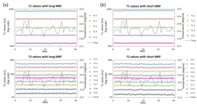
The impact of shorter acquisition time in MRF: Long term repeatability and reproducibility study on ISMRM/NIST phantom and volunteers.
Yutaka Kato, Kazushige Ichikawa, Toshiaki Taoka, Hirokazu Kawaguchi, Katsutoshi Murata, Katsuya Maruyama, Gregor Koerzdoerfer, Josef Pfeuffer, Mathias Nittka, Shinji Naganawa
This study focused on the stability of MRF in a phantom and volunteers, and explored the feasibility of MRF with a shorter acquisition time. Phantom scans on 40 days and volunteer scans on 5 days over 3 months showed comparable repeatability and reproducibility of T1 and T2 values between MRF with acquisition times of 41 sec and 20 sec. Shorter acquisition time has the potential to expand the clinical usage of MRF.
|
|
4452.
 |
79 |
Cross-system reliability for rapid quantitative MRI
Catharina Petersen, Peter Johansson, Marcel Warntjes
Absolute quantification of R1 and R2 relaxation rates and proton density PD has been gaining considerable attention in recent years. It is of utmost importance that these measurements entirely reflect patient properties and no influence is detectable on which specific MRI scanner system the quantitative maps were obtained. The SyMRI software was verified on Philips, GE and Siemens scanners at both 1.5T and 3T showing cross-system reliability.
|
|
4453.
 |
80 |
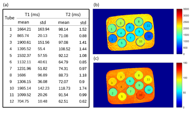
Repeatability of T2 Relaxation Measurements over a Four-Year Period
Xing Wang, Cheryl McCreary, Marina Salluzzi, Richard Frayne
The reliability of a T2 relaxation quantification technique was assessed by repeatedly scanning four subjects (total of 12 scans at 4 time points over 4 years). Both total, biological and scanner variability were assessed across the whole brain and in the frontal, occipital, parietal temporal lobes. Total variability (coefficient-of-variation CoV < 10.3%) was dominated by biological variation (CoV < 10.3%). Scanner variability was low (CoV < 1.6%) despite scanner software and hardware upgrades during this interval. These results suggest that quantitative T2 estimates are reproducible over 4 years and robust to scanner upgrades.
|
|
4454
 |
81 |
The traveling heads 2.0: Reproducibility of quantitative imaging methods at 7 Tesla
Video Permission Withheld
Maximilian Voelker, Oliver Kraff, Steffen Goerke, Frederik Laun, Kerrin Pine, Philipp Ehses, Moritz Zaiss, Andrzej Liebert, Sina Straub, Korbinian Eckstein, Simon Robinson, Armin Nagel, Oliver Speck, Mark Ladd, Harald Quick
The “traveling heads” is a study to assess the comparability and reproducibility of multicenter human brain imaging at 7T. In previous experiments, we compared typical UHF sequences for structural brain imaging. In this study, we focus on the reproducibility of quantitative imaging and compare methods for volumetry, relaxometry, QSM and CEST between different sites. In addition, three generations of 7T MR systems are compared, i.e. the older installed base consisting of passively and actively shielded magnets of the first and second generation, respectively, as well as the most recent generation which has been approved as a medical device.
|
|
4455
 |
82 |
Knee T2 relaxometry using quantitative DESS: reproducibility across imaging vendors
Video Permission Withheld
Quin Lu, Brian Hargreaves, Dave Hitt, Akshay Chaudhari
T2 is a promising MR-based biomarkers for early diagnosis of osteoarthritis (OA). Studies have shown that quantitative DESS (qDESS) is capable of performing simultaneous knee morphometry and T2 relaxometry. In this study, we investigate the cross-vendor reproducibility of knee T2 relaxometry using qDESS. By comparing measured cartilage and meniscus T2 values in volunteers scanned on both Philips 3T and GE 3T scanners, we show that qDESS has good intra-vendor scan-rescan repeatability (CCC = 99.2% and 98.8% ) and cross-vendor reproducibility (CCC=96.3%). With continued effort, we hope to show that qDESS T2 relaxometry can serve as a reliable clinical biomarker for early OA diagnosis.
|
|
4456.
 |
83 |
Inter-site Reproducibility of Cardiac Magnetic Resonance Fingerprinting T1 and T2 Quantification in the ISMRM/NIST MRI System Phantom and Human Heart
Yuchi Liu, Luuk Hopman, Jesse Hamilton, Elizabeth Hillier, Matthias Friedrich, Nicole Seiberlich
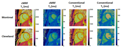
Cardiac Magnetic Resonance Fingerprinting (cMRF) is a novel technique for simultaneous T1 and T2 quantification in the myocardium. Because cMRF has the potential to take heart rate variations and any variable system properties into account, it is hypothesized that cMRF will enable more reproducible measurements of T1 and T2. The purpose of this study is to evaluate the inter-site reproducibility of cMRF. Excellent agreement of cMRF measurements between two sites (University Hospitals Cleveland Medical Center, Cleveland, US and McGill University Health Center, Montreal, CA) was achieved in the ISMRM/NIST phantom and in the hearts of healthy subjects.
|
|
4457.
 |
84 |
Exploring the sensitivity of Magnetic Resonance Fingerprinting to k-space trajectory uncertainties
Alessandro Palombit, Alessandra Bertoldo, Zidan Yu, Riccardo Lattanzi, Martijn Cloos
In this work, we experimentally explore the sensitivity of Magnetic Resonance Fingerprinting (MRF) to k-space trajectory uncertainties typically encountered in non-cartesian imaging. We demonstrate that T1 and T2* quantification can be affected by minor gradient delays observed in stack-of-stars 3D MRF implementations, particularly resulting in severely disrupted T2* measures. As a first approximation, we modeled these imperfections as constant readout sampling shifts of a few integer k-space steps along every trajectory direction. We show that by simply shifting back the nominal sampling locations before the reconstruction can restore reliable MRF parametric estimates.
|
|
4458.
 |
85 |
Three-dimensional, fast parameter mapping at 7.0T with SSFP MR Fingerprinting: comparison of radial and spiral projections k-space trajectories
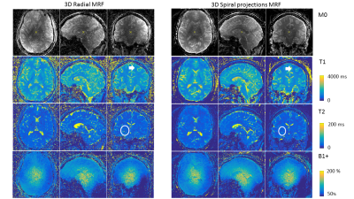
Guido Buonincontri, Pedro Gómez, Matteo Cencini, Mauro Costagli, Graziella Donatelli, Rolf Schulte, Michela Tosetti
When using ultra-high field MRI scanners (UHF, B0>= 7T), quantitative imaging is challenging due to B0 and B1+ non-uniformities. Magnetic resonance fingerprinting (MRF) represents a great opportunity for quantitative imaging at UHF as it can estimate these effects at the same time of the parameters of interest. Here, we compare two novel 3D SSFP MRF approaches, one based on a three-dimensional spiral projection acquisition and one using a radial acquisition in vivo at 7.0T. We estimate M0, T1, T2 and B1+ simultaneously at high resolution (1mm isotropic) within 6.5 minutes acquisition time.
|
|
4459.
 |
86 |
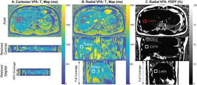
T1 mapping with golden-angle radial sampling: A comparison of direct and indirect reconstruction
Nikolaos Kallistis, Ian Rowe, Steven Sourbron
The purpose of the study is to compare a direct model-based reconstruction with an indirect compress sensing reconstruction for the estimation of T1-map, from simulated radial sampled datasets. Comparisons are performed for the binning strategy that is optimal in each case as measured by T1-errors. The direct reconstruction solves the nonlinear-least-squares optimization problem with a gradient-based L-BFGS algorithm without regularization, while for the indirect method the images are reconstructed using the iGRASP technique.
The accuracy for both methods is similar, however the computational time of the model-based reconstruction is a limiting factor for clinical applications.
|
|
4460.
 |
87 |
Information Quantification of Subsequent Acquisitions for Minimizing Synthetic MRI Reconstruction Uncertainty
Drew Mitchell, David Fuentes, Jason Stafford, James Bankson, Ken-Pin Hwang
A mutual information-based mathematical framework is developed to quantify the information content of various acquisition parameters and subsampling approaches. A recursive conditional formulation quantifies information content given previous acquisitions. This framework is applied to 3D QALAS. Mutual information between reconstructed M0, T1, and T2 uncertainty and measurement noise is calculated for an in silico phantom and the results applied to measurements on a System Standard Model 130 phantom. Reconstructions from these measurements demonstrate the potential use of information theory in guiding pulse sequence design to maximize reconstruction quality.
|
|
4461.
 |
88 |
The in vivo impact of diffusion spoiling on the estimate of T1 using spoiled gradient echoes with variable flip angles
Nadège Corbin, Shaihan Malik, Martina Callaghan
Incomplete spoiling of the transverse MRI signal causes errors in the T1 time estimated from variable flip angle measurements acquired with spoiled gradient-echo images. Diffusion spoiling is thought to lessen these effects. However, these conclusions are based on phantom experiments, using very long T2 times, or from in vivo simulation using infeasibly strong diffusion spoiling. Here we perform simulation and in vivo experiments to characterise the impact of diffusion spoiling in the short T2, low spoiling regime. We show that even under these conditions, diffusion spoiling reduces the dependence of the estimated T1 on both the phase-increment and the transmit field.
|
|
4462.
 |
89 |
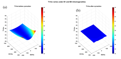
Robust quantitative T1rho imaging in the presence of B1 RF and B0 field inhomogeneities
Huimin Zhang, Baiyan Jiang, Queenie Chan, Weitian Chen
T1rho is a valuable biomarker to probe macromolecular environment of tissue. However, T1rho imaging suffers from B1 RF and B0 field inhomogeneities. In this work, we present an approach to address this problem. The performance of our proposed method was demonstrated by simulations, phantom and in vivo experiments.
|
|
4463.
 |
90 |
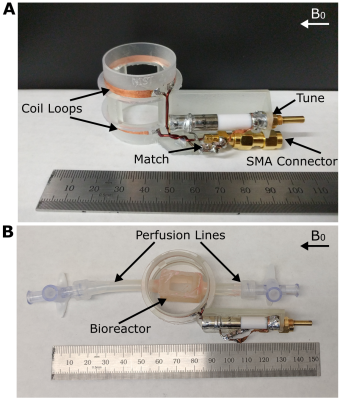
MR imaging of a 3D-printed bioreactor with a dedicated radiofrequency coil for cellular level validation of quantitative MR metrics
Megan Poorman, Slavka Carnicka, Jeanne Barthold, Michele Martin, Karl Stupic, Corey Neu, Kathryn Keenan
Quantitative MRI methods have the potential to push the clinical standard of care towards quantitative diagnosis. However, controlled systems are needed to study the effects of underlying cellular properties on the MRI signal to validate quantitative MRI measures. To meet this need, our group previously developed an MR-compatible bioreactor to monitor cell behavior using MRI validated with optical microscopy. The present work develops a dedicated RF coil for improved MR imaging of the bioreactor and uses it to explore the effects of cell culture on T1 and T2 in our system.
|
|
4464
 |
91 |
Assessment and optimisation of bias field correction using N4ITK for PD mapping
Video Permission Withheld
Sara Lorio, David Carmichael
Proton density (PD) maps measure the amount of free water molecules in the tissue and can be used in a range of neurological disorders. We previously developed a new approach for PD mapping based on a multi-contrast acquisition protocol, and a data-driven estimation method for inhomogeneity correction and map scaling. Here we evaluate the robustness of the inhomogeneity correction method and its effect on the PD value estimation using data acquired with different receiver coils. This allowed us to assess the impact of the spatial variability of the receiver coil profile on the PD map.
|
|
4465.
 |
92 |
Performance comparison of channel combination methods for multi-echo chemical shift-encoded MRI
Nathan Roberts, Timothy Colgan, Kang Wang, Diego Hernando
Many coil combination methods have been developed, including methods that require pre-calibrated coil sensitivity maps, as well as methods that do not require additional sensitivity maps. Several of these methods have been adapted to CSE-MRI, where accurate signal combination is particularly critical as it needs to preserve consistent phase and magnitude information across echoes; however, their relative performance remains unknown. Therefore, the purpose of this work is to compare theoretically, in simulation, and experimentally the bias and noise performance of quantitative parameter maps resulting from five commonly used coil multi-echo coil combination techniques.
|
|
4466.
 |
93 |
Correlating the measured fast/slow decay components of tissue sodium to the intra-/extra-cellular sodium concentrations
Presentation Not Submitted
Jinhu Xiong, William Kearney, Mathews Jacobs, Rolf Schulte, Baolian Yang, Vincent Magnotta
We have developed two models (anisotropic-anisotropic (AAS) vs. anisotropic-isotropic (AIS) models) to correlate the measured fast/slow decay components of tissue sodium to the intra-/extra-cellular sodium concentrations. The models were evaluated based on theoretical and experimental results. Our results indicate that AAS model fits experimental data much better than AIS model does.
|
|
4467.
 |
94 |
In-vivo cardiac DTI using motion compensated optimized diffusion encoding (MODE): A reproducibility study
Prateek Kalra, Waqas Majeed, Mohammad Maddah, Xiaokui Mo, Richard White, Arunark Kolipaka
Diffusion-weighted imaging (DWI) is used to identify heterogeneous infarcted region by calculating ADC(apparent diffusion coefficient) and FA(fractional anisotropy). However, performing DWI in heart is very challenging because of heart motion. Earlier method used convex optimized diffusion encoding (CODE) to optimize diffusion encoding gradients (DEG) waveform. However, due to limitations of CODE waveforms, earlier we proposed motion compensated diffusion encoding (MODE) to achieve higher b-value for a given DEG duration. The aim of this study is to validate and assess the reproducibility of MODE technique in computing ADC and FA maps in healthy subjects. Preliminary results demonstrated good reproducibility using MODE.
|
|
4468.
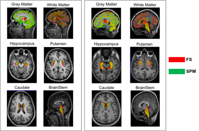 |
95 |
Repeatability and Reproducibility of brain volume measurements with SPM and Freesurfer and their impact on subtle between-group differences
Letizia Palumbo, Paolo Bosco, Elisa Ferrari, Piernicola Oliva, Giovanna Spera, Alessandra Retico
The main aim of the study is to investigate whether the adoption of a processing method has a relevant influence on the results of a neuroimaging research. We evaluated the intra-method repeatability and the inter-method reproducibility of two widely-used automatic segmentation methods for brain MRI: FreeSurfer (FS) and Statistical Parametric Mapping (SPM) software packages. We segmented the gray matter, the white matter and the subcortical structures in test-retest MRI data of healthy volunteers from two publicly available datasets. High intra-method repeatability was found for both SPM and FS, but SPM was more consistent than FS in measuring ROIs volumes.
|
|
4469.
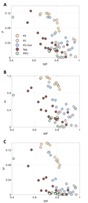 |
96 |
The Effect of Membrane Lipids on qMT Exchange Constants
Oshrat Shtangel, Aviv Mezer
Quantitative Magnetization Transfer(qMT) is a proposed method for deeper characterization of brain tissue. Yet, a connection between qMT parameters and the components of cellular tissue is required. Myelin is composed of various types of lipids, which their amount and composition are changed between brain areas, disease states and across the lifespan. In this work, we formulated liposomes to model the environment of abounded lipids in the human brain and systematically estimated their effect on qMT parameters. We found qMT technique useful to identify differences between lipids. This result can pave the way to future research the molecular environments of human tissue in-vivo.
|
|
4470.
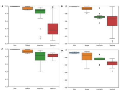 |
97 |
Repeatability of radiomics features in double baseline MR imaging of glioblastoma
Katharina Hoebel, Andrew Beers, James Brown, Ken Chang, Jay Patel, Marco Pinho, Bruce Rosen, Tracy Batchelor, Elizabeth Gerstner, Jayashree Kalpathy-Cramer
Extraction of radiomic features has to be repeatable in order to be clinically useful. We investigated the repeatability of radiomic feature extraction on a unique dataset consisting of a double baseline MRI scans in 48 patients diagnosed with glioblastoma. Size and shape features which are mostly governed by tumor segmentation showed on average higher repeatability than intensity and texture-based features which are more dependent on image acquisition and preprocessing. More research on the influence of image acquisition and preprocessing on the repeatability and reliability of radiomic features has to be undertaken to make radiomics a safe image-analysis tool.
|
|
4471.
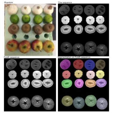 |
98 |
Influence of image processing on the robustness of radiomic features derived from magnetic resonance imaging - a phantom study
Barbara Wichtmann, Ulrike Attenberger, Felix Harder, Stefan Schönberg, David Maintz, Kilian Weiss, Daniel Pinto dos Santos, Bettina Baeßler
The emerging field of radiomics aims at analyzing quantitative features derived from medical images to characterize tissue and support clinical decision-making. However, an extensive knowledge about the robustness and reproducibility of radiomic features has to precede translation into routine clinical practice. Very little is known regarding the robustness of radiomic features derived from magnetic resonance imaging. We want to assess the influence of image processing parameters on the robustness of radiomic features. Our results showed that variation of image processing parameters has considerable impact on the robustness as well as on absolute values of radiomicfeatures extracted from MRI data.
|
|
4472.
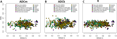 |
99 |
Repeatability of radiomic features for prostate cancer diffusion weighted imaging obtained using b-values up to 2000 s/mm2
Harri Merisaari, Rakesh Shiradkar, Jussi Toivonen, Amogh Hiremath, Mohammadhadi Khorrami, Ileana Montoya Perez, Tapio Pahikkala, Pekka Taimen, Janne Verho, Peter Boström, Hannu Aronen, Anant Madabhushi, Ivan Jambor
We evaluated repeatability and diagnostic performance of commonly used radiomic features for prostate cancer (PCa) DWI obtained using b values up to 2000 s/mm2. Forty-eight men with diagnosed PCa under two repeated 3T MRI examinations performed on the same day. Whole mounts prostatectomy sections were manually matched with in-vivo MRI data. Fourteen of the evaluated 575 features demonstrated high repeatability with ICC(3,1)>0.9 and AUC(Gleason score 3+3 vs >3+3 PCa)>0.6. Many of the conventional radiomics feature demonstrate high AUC but low repeatability (low ICC(3,1) values)stressing the fact that high classification potential using single acquisition does not necessarily mean good overall performance.
|
|
| Top |
Motion Correction: Non-Brain
Digital Poster
Acquisition, Reconstruction & Analysis
Thursday, 16 May 2019
| Exhibition Hall |
08:15 - 09:15 |
| |
|
Computer # |
 |
4473
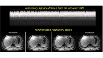 |
101 |
Comparison of free-breathing motion-resolved radial imaging with standard breath-hold imaging on liver MRI: a feasibility study
Video Permission Withheld
Kate Harrington, Feng Li, Arifa Chowdhury, Kang Wang , Ty Cashen, Ali Ersoz , Maggie Fung, Ersin Bayram , Kinh Gian Do, Ricardo Otazo
Respiratory motion remains a major challenge in clinical abdominal MRI. Recent technical advances using continuous radial imaging during free-breathing and motion-resolved compressed sensing-based image reconstruction have demonstrated improvements in motion robustness over conventional motion-gated or motion-corrected techniques, but they were not validated for liver imaging. This work implemented extra-dimensional (XD) reconstruction for free-breathing RadialLAVA acquisitions and compared it against conventional breath-held CartesianLAVA. We demonstrate that the XD technique matches that, and in some instances, is superior to that of standard breath-hold technique in terms of overall image quality in the evaluation of post-contrast liver images.
|
|
4474.
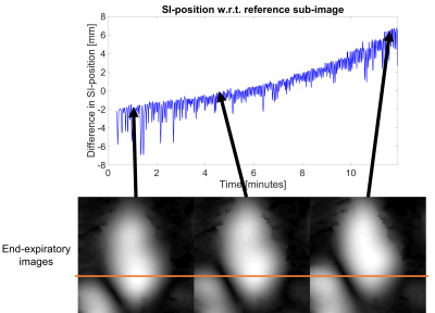 |
102 |
Respiratory Motion Signals Extracted from 3D Image-Based Navigation and from PCA of SI-Projections: Initial Findings in Whole-Heart Imaging using a Free-Running Framework
John Heerfordt, Nemanja Masala, Lorenzo Di Sopra, Christopher Roy, Bastien Milani, Jérôme Yerly, Jessica Bastiaansen, Davide Piccini, Matthias Stuber
In continuously acquired whole-heart coronary MRA, it is not fully understood how unitless respiratory self-gating signals relate to actual respiratory displacement and drift. Therefore, self-gating signals extracted from principal component analyses of 1D projections oriented in the superior-inferior (SI) direction were compared to image-based navigators. Whole-heart data from continuous uninterrupted 3D radial bSSFP acquisitions were used to reconstruct time series of 3D sub-images with a temporal resolution of 0.6 seconds. Preliminary findings suggest that the SI-directed motion obtained from these sub-images is better described by respiratory self-gating signals created from three principal components rather than from one principal component alone.
|
|
4475.
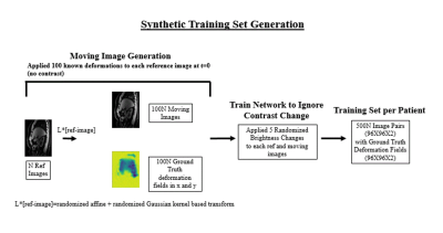 |
103 |
DCE-Abdominal MR Image Registration using Convolutional Neural Networks
Zachary Miller, Kevin Johnson
Convolutional neural networks (CNNs) have had incredible success solving image segmentation problems. We explore whether CNNs could have a similar level of success on difficult image registration problems. To this end, we developed a modified U-net to remove respiratory motion, but preserve contrast changes in abdominal free breathing dynamic contrast enhanced (DCE)-MRI. We then compared this network to a state of the art iterative registration algorithm. We demonstrate that our modified U-net outperforms iterative methods both in terms of registration quality and speed (600 registrations in <1 sec vs. Elastix in 2 hours)
|
|
4476.
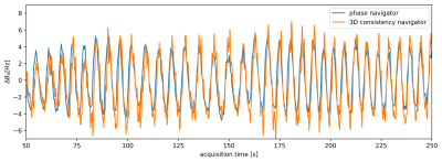 |
104 |
Data Consistency Driven Correction of B0-Fluctuations in 2D and 3D Gradient-Echo MRI of the Spine
Jakob Meineke, Tim Nielsen
We demonstrate the data-consistency driven determination and correction of B0-fluctuations induced by respiratory motion in 2D and 3D gradient-echo images of the cervical spine. By promoting data-consistency in the multi-channel raw data, it is possible to estimate the instantaneous off-resonance. Furthermore, we demonstrate a marked improvement in image quality by correcting the k-space data using the measured B0-fluctuations.
|
|
4477.
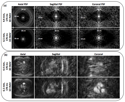 |
105 |
Unraveling the effect of spatial resolution and scan acceleration on 3D image-based navigators for respiratory motion tracking
Srivathsan Koundinyan, Mario Malavé, Dwight Nishimura
Beat-to-beat 3D image-based navigators (3D iNAVs) enable nonrigid respiratory motion tracking of the heart. In this work, we study the accuracy of motion information extracted from 3D iNAVs with different choices of two parameters: spatial resolution and scan acceleration factor. We demonstrate that high spatial resolution coupled with aggressive scan acceleration results in residual blurring and aliasing following iterative reconstruction, which corrupts the derived motion estimates. Through simulations, we identify the optimal combination of spatial resolution and scan acceleration for acquiring 3D iNAVs. In vivo studies presenting sharp motion correction outcomes demonstrate a capability for monitoring motion with high fidelity.
|
|
4478.
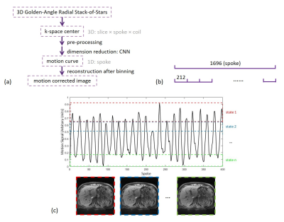 |
106 |
Self-navigation Liver Respiratory Motion Correction Based on Deep Learning
Yu Wang, Haikun Qi, Guanhua Wang, Yuze Li, Huijun Chen
Correction of respiratory motion with 100% acquisition efficiency is of great significance for clinical abdominal imaging. In this study, we propose a novel self-navigation liver respiratory motion correction method for 3D radial sampling. This new approach is based on the fact that radial acquisition enables oversampled k-space center to extract motion-state signal and neural network can be used for data dimensionality reduction. Both regular and irregular hepatic breathing experiments were conducted and the proposed method has shown similar reconstruction image quality with bellow.
|
|
4479.
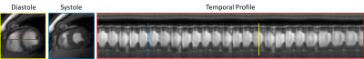 |
107 |
Free breathing & Ungated Multi-Slice cardiac cine MRI using spiral-SToRM
Abdul Haseeb Ahmed, Sunrita Poddar, Stanley Kruger, Prashant Nagpal, Rolf Schulte, Mathews Jacob
The advantages of cardiac cine MRI are often limited by its long acquisition and breath-held requirement. To overcome these limitations, we have introduced a navigator based spiral SToRM to acquire free breathing and ungated cardiac cine MRI in a short acquisition time. Our algorithm is fully automated and does not depend on explicit binning. It gives improved image quality compared to the existing self-gated methods. Post-reconstructions, the time series can be processed to extract cardiac cycles at different respiratory phases, facilitating the estimation of anatomical and functional evaluation of the heart.
|
|
4480.
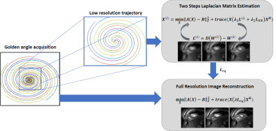 |
108 |
Navigator-less Spiral SToRM for Free breathing and Ungated Cardiac CINE MRI
Abdul Haseeb Ahmed, Ruixi Zhou, Yang Yang, Michael Salerno, Mathews Jacob
This study introduces an iterative kernel low-rank algorithm to recover images in a free breathing and ungated cardiac MRI dataset. The approach relies on the manifold structure of dynamic data to recover it from highly undersampled measurements. The data is acquired using variable density spiral acquisition. An iterative kernel low-rank algorithm is introduced to estimate the manifold structure of the images, or equivalently the manifold Laplacian matrix, from central k-space regions. Unlike previous manifold regularization implementations, the iterative algorithm, coupled with the non-Cartesian acquisitions, eliminates the need for dedicated navigators to estimate the manifold Laplacian, thus improving sampling efficiency.The iterative kernel low-rank algorithm facilitates the extension of manifold regularization to navigatorless spiral acquisitions, thus improving sampling efficiency. This algorithm provides improved reconstruction compared to the state of the art methods.
|
|
4481
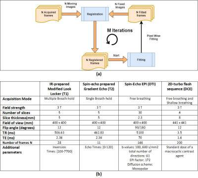 |
109 |
Motion Correction with a Model Target (MoCoMo): A universal approach for quantitative MRI?
Video Permission Withheld
Fotios Tagkalakis, Kanishka Sharma, Susmita Basak, Christopher Kelly, David Shelley, Irvin Teh, Jehill Parikh, Peter Thelwall, Neil Sheerin, Steven Sourbron
Motion correction with a model-target (MoCoMo) has been used in DCE-MRI to overcome the problem of changes in image contrast, but the method applies in principle to any other quantitative MRI method. The aim of this study is to demonstrate this hypothesis by applying the algorithm to renal DCE, DTI, T1 and T2-mapping in human subjects. The results show that MoCoMo is effective in removing even major motion effects in all 4 modalities and does not affect data where no motion is present. We conclude that MoCoMo is a suitable candidate for universal motion correction across all functional MRI modalities.
|
|
4482.
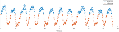 |
110 |
Free Breathing Radial Magnetic Resonance Elastography
Joseph Holtrop, Stephan Kannengiesser, Ralf Loeffler, Ruitian Song, Claudia Hillenbrand
Liver magnetic resonance elastography (MRE) to this point has clinically relied on using breath holds to produce reliable artifact free images. Here we present initial work adapting recent advances in motion compensated abdominal imaging for use in MRE. Specifically, we take advantage of a golden angle radial sampling scheme combined with a self-navigation approach for motion correction to perform free breathing MRE of the liver. Resulting images show enhanced detail compared to the standard breath hold technique while producing comparable image stiffness values.
|
|
4483.
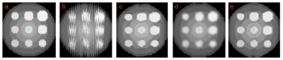 |
111 |
Deformable slice-to-volume registration for respiratory motion correction in abdominal and in-utero MRI
Alena Uus, Tong Zhang, Laurence Jackson, Mary Rutherford, Joseph Hajnal, Maria Deprez
This work introduces deformable slice-to-volume registration (DSVR) integrated into super-resolution reconstruction framework for correction of respiratory motion MRI. Using the initial estimation of respiratory motion as an input this method allows reconstruction of high-resolution volumes for specific respiratory positions using all slices. Based on diffeomorphic free-form deformation model, DSVR provides robust registration of deformable objects as well as out-of-plane motion correction. The feasibility of the method was successfully evaluated on a ‘motion-corrupted’ phantom and a free-breathing in-utero MRI scan. The results also indicated that the accuracy of spatial features in reconstructed volume is directly defined by the initial motion estimation.
|
|
4484.
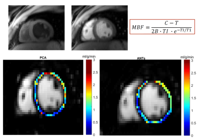 |
112 |
Groupwise Non Rigid Registration For Temporal Myocardial Arterial Spin Labeling Images
Veronica Aramendía-Vidaurreta, Pedro Macías-Gordaliza, Marta Vidorreta, Rebeca Echeverria-Chasco, Gorka Bastarrika, Arrate Muñoz-Barrutia, María Fernández-Seara
Arterial Spin Labeling (ASL) enables quantitative measurement of myocardial blood flow (MBF) by averaging over multiple ASL pairs providing a voxelwise map in units of milliliters of blood per gram of tissue per minute (ml/g/min). However, its estimation accuracy in free breathing acquisitions depends critically on the quality of the image registration algorithm. In this work, a groupwise non-rigid registration method with a similarity measure based on Principal Component Analysis (PCA) was applied to ASL images of the heart acquired during free breathing. The method was compared against a pair-wise registration algorithm provided by the advanced normalization tools software (ANTs). Results demonstrate the feasibility of using PCA-groupwise for temporal ASL image registration.
|
|
4485.
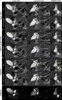 |
113 |
Reconstruction-based Super-Resolution for High-Resolution Abdominal MRI: A Preliminary Study
Michael Ebner, Premal Patel, David Atkinson, Lucy Caselton, Stuart Taylor, Alan Bainbridge, Sebastien Ourselin, Manil Chouhan, Tom Vercauteren
Magnetic resonance (MR) cholangio-pancreatography (MRCP) is an established specialist method for imaging the upper abdomen and biliary/pancreatic ducts. Due to limitations of either MR image contrast or low through-plane resolution, patients may require further evaluation with contrast-enhanced computed tomography (CT) images. However, CT fails to offer the high tissue-ductal-vessel contrast-noise ratio available on T2-weighted MRI. MR Super-Resolution Reconstruction (SRR) frameworks can provide high-resolution visualizations from multiple low through-plane resolution single-shot T2-weighted (SST2W) images as currently used during MRCP studies. Here, we investigate the clinical potential of using additional SST2W acquisitions in multiple directions with SRR for higher diagnostic yield.
|
|
4486.
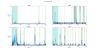 |
114 |
Self-navigated bulk motion detection for feed and wrap renal dynamic radial VIBE DCE-MRI
Jaume Coll-Font, Onur Afacan, Alto Stemmer, Richard Lee, Jeanne Chow, Simon Warfield, Sila Kurugol
Dynamic Radial VIBE (DRV) DCE-MRI allows to image with sufficient spatio-temporal resolution for functional imaging of kidneys. However, fast movements of babies during the scan corrupt individual lines in k-space and severely compromise the quality of the reconstructed images and limits the clinical utility of non-sedated imaging. In this work, we evaluate a self-navigated bulk motion detection approach to identify these corrupted lines. We applied this approach on non-sedated infants undergoing feed-and-wrap DCE-MRI with DRV. Our results show that this approach correctly identifies the bulk motion and allows for post-processing correction of the DCE absorption curves.
|
|
4487.
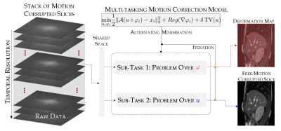 |
115 |
Motion Correction Resolved for MRI via Multi-Tasking: A Simultaneous Reconstruction and Registration Approach
Veronica Corona, Noémie Debroux, Angelica Aviles-Rivero, Guy Williams, Martin Graves, Carole Le Guyader, Carola-Bibiane Schoenlieb
The prolonged time required to form an MR image continues to impose different challenges at both theoretical and clinical levels. With this motivation in mind, this work addresses a central topic in MRI, which is how to correct the motion problem, through a new multitask optimisation framework. The significance is that by tackling the reconstruction and registration tasks −− simultaneously
and jointly −− one
can exploit their strong correlation reducing error propagations and resulting in a significant motion correction. The clinical potentials of our approach are reflected in having higher image quality with fewer artefacts whilst keeping fine details. We evaluate our approach through a set of quantitative and qualitative experimental results.
|
|
4488.
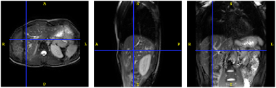 |
116 |
Free-breathing MRI of the upper abdomen assisted by motion modelling
Robert Johnstone, David Atkinson, Manil Chouhan, Ricky Sharma, Jamie McClelland
This study demonstrates the use of motion modelling and super resolution reconstruction (SRR) to produce an isotropic 3D image of the upper abdomen during free breathing.
Sagittal and coronal 6 mm 2D slices are acquired throughout the volume of interest. The slices are repeated with sub-voxel offsets to facilitate SRR. An interleaved navigator slice is also acquired.
The navigator slice is processed with non-rigid registration and principal component analysis, to give two motion surrogate signals. These signals are used to control the motion model. The motion model and the SRR are jointly optimised using an iterative scheme.
|
|
4489.
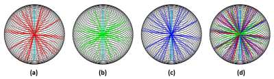 |
117 |
A segmented ultra-short echo (UTE) sequence equipped with robustness to respiratory motion
Cihat Eldeniz, Uday Krishnamurthy, Hongyu An
Synopsis
|
|
4490.
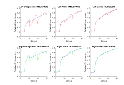 |
118 |
Effects of Image Registration in Dynamic Contrast-Enhanced MRI of the TMJ
Lea Starck, Erling Andersen, Ondrej Macícek, Oskar Angenete, Thomas Augdal, Erlend Hodneland, Radovan Jirík, Karen Rosendahl, Renate Grüner
The effect of elastic and affine motion correction in dynamic contrast enhanced MRI ofthe temporomandibular joints in children is investigated. Imaging in children is particularly difficultdue to motion. This hampers DCE-MRI and pharmacokinetic estimations for their potentialdiagnostic value in these children with Juvenile Idiopathic Arthritis with possible TMJ involvement.The relative enhancement curves obtained with different motion correction approaches arecompared with the curves calculated with the Gamma Capillary Transit Time model. It is found thatwhen image registration is applied, a greater number of participants can be analysed. The elasticmotion correction approach outperforms the affine approach.
|
|
4491.
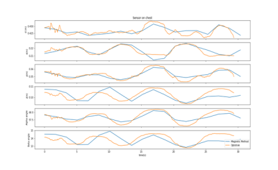 |
119 |
Magnetic tracking of ECG sensors for respiratory motion correction
Benjamin Roussel, Joris Pascal, Jacques Felblinger, Julien Oster
Monitoring the respiration motion is a crucial step for motion correction. We propose a magnetic tracking system, using a magnetic sensor and a Helmholtz coil as the magnetic field source. By comparing the sensed magnetic fields with theoretical values under the dipole approximation, we were able to locate sensors placed on a subject’s chest and track their motion during breathing. With a sub-centimeter resolution and the current sources of imprecision being identified, we are confident this method can be a viable solution for accurate motion monitoring in MRI, especially by using the magnetic fields generated by the gradient coils.
|
|
4492.
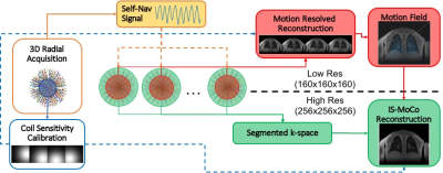 |
120 |
Iterative Static Motion Compensated(IS-MoCo) Reconstruction: application to high resolution lung imaging
Xucheng Zhu, Kevin Johnson, Michael Lustig, Peder Larson
High resolution 3D MRI thoracic and abdominal MRI is always challenging, due to long acquisition time and susceptibility to subject motion. We proposed a novel reconstruction method, named Iterative Static Motion Compensated(IS-MoCo) reconstruction, to compensate motion affects during the reconstruction instead of gating. The proposed method is applied to high resolution free breathing lung imaging, outperforms widely used motion correction strategies with higher SNR and less residual motion artifacts.
|
|
4493
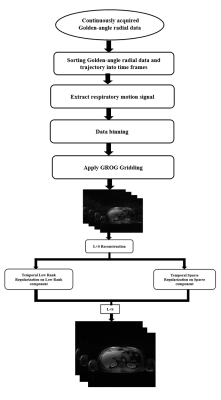 |
121 |
Respiratory Motion Corrected GROG based L+S Reconstruction for Free Breathing Golden-Angle Radial MRI
Video Permission Withheld
Ahmad Hussain, Faisal Najeeb, Ibtisam Aslam, Hammad Omer, Mujahid Nisar
Respiratory motion during MRI scan causes inconsistencies in the acquired k-space data providing strong blurring artifacts in the reconstructed images. In this work, a new method ( respiratory motion corrected GROG followed by L+S reconstruction for free breathing Golden-Angle Radial DCE-MRI) is presented.The proposed method is tested on 3-T free-breathing Golden angle radial DCE liver MRI data. The proposed method is compared with the conventional L+S reconstruction model. The proposed method provides 90% improvement in Artefact Power and 42% in RMSE as compared to conventional L+S reconstruction at acceleration factor 8.
|
|
4494.
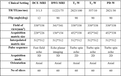 |
122 |
Impact of registration on multi-parametric breast MRI data and parameters: Qualitative and Quantitative Assessment
Snekha Thakran, Subhajit Chatterjee, Dinil Sasi, Ayan Debnath, Rupsa Bhattacharjee, Rakesh Gupta, Anup Singh
Multi-parametric(mp)-MRI data such as conventional MRI, DCE-MRI, DWI, etc. are routinely acquired for breast cancer patients. Any motion during mp-MRI data acquisition can affect qualitative as well as quantitative mp-MRI results. In this study, impact of registration on mp-MRI data as well as on quantitative parameters was evaluated qualitatively and quantitatively. Study included mp-MRI data of 40 patients with breast cancer. B-spline based registration performed better than Affine and SyN. It showed highest dice-coefficient, correlation coefficient. It also provided better histograms of quantitative maps and provided lowest sum-of-squared error in signal-intensity curves from ROI at edge and center of lesion.
|
|
4495.
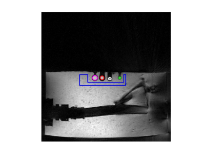 |
123 |
MR-assisted PET motion correction improves tumor-to-background and contrast-to-noise ratios in a phantom study with ground truth reference
Sihao Chen, Cihat Eldeniz, Richard Laforest, Hongyu An
Respiratory motion leads to signal blurring and reduced tumor-to-background (TBR) and contrast to noise (CNR) ratios. As a result, it can severely affect the detectability of lesions in PET imaging.1,2 Simultaneous PET/MR imaging uniquely allows for MR assisted motion correction in PET imaging.3 In this study, we have demonstrated that the MR assisted PET motion correction significantly improves both tumor-to-background and contrast-to-noise ratios, leading to better lesion detection.
|
|
4496
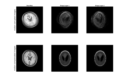 |
124 |
Blind Sparsity Based Motion Estimation and Correction Model for Arbitrary MRI Sampling Trajectories
Video Permission Withheld
Anita Möller, Marco Maass, Tim Parbs, Alfred Mertins
A blind retrospective MRI motion estimation and compensation algorithm is designed for arbitrary sampling trajectories. Using the idea of natural images being sparsely representable, the algorithm is based on motion estimation between a motion corrupted image and it’s sparse representative. Therefore, rigid motion models are designed and used in gradient descent methods for image quality optimization. As the motion estimation and compensation work on arbitrary real valued sampling coordinates, the algorithm is capable for all trajectories. Image reconstruction is performed by computationally efficient gridding. The exact motion estimation results are shown for PROPELLER and radial trajectory simulation.
|
|
4497.
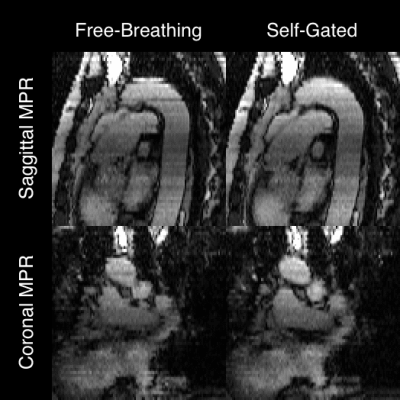 |
125 |
Self-Gated Pulmonary Embolism Imaging with Multi-Slice Golden-Angle Radial bSSFP
Alexander Fyrdahl, Roberto Vargas-Paris, Koshiar Medson, Sven Nyrén, Martin Ugander, Peter Lindholm, Andreas Sigfridsson
Free-breathing, non-contrast bSSFP MRI has shown great potential for imaging of pulmonary embolisms in patients with contraindication for contrast-enhanced computed tomography angiography. While the free-breathing approach is convenient, it limits the possibility for multiplanar reformatting which otherwise could aid in visualizing the pulmonary vasculature. In this work, we propose a methodology for deriving a motion signal from the free-breathing data and we incorporate this signal in the reconstruction pipeline to obtain a slice-aligned image stack from which multiplanar reformatting can be performed.
|
|
| Top |
System Imperfections
Digital Poster
Acquisition, Reconstruction & Analysis
Thursday, 16 May 2019
| Exhibition Hall |
08:15 - 09:15 |
| |
|
Computer # |
|
4498.
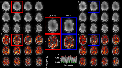 |
126 |
Minimal Linear Networks for MR Image Reconstruction
Gilad Liberman, Benedikt Poser
We propose minimal linear networks (MLN) for MR image reconstruction that employ complex-valued, axis-dependent and fully- and neighborhood-connected layers with shared and independent weights, Their topology is restricted to the minimum required by the MR-physics, without nonlinear activation layers. The suggested MLN perform well in reconstructing imaging data acquired under challenging real-world imaging conditions, specifically an Arterial Spin Labeling perfusion experiment with spiral sampling at 7 Tesla. Despite the strong B0 field inhomogeneities at 7T, artifact-free images are obtained that are capable of resolving the minute perfusion signal changes. The results show that even without nonlinear activation and higher-order image manifold description as used by others, deep-learning algorithms and framework, and learning from large realistic datasets, can play a significant role in the success of image reconstruction.
|
|
4499.
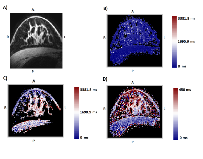 |
127 |
Correcting breast MRI with a generic B-1(+) template for T-1 map calculation
Lieke van den Wildenberg, Erwin Krikken, Jannie Wijnen, Josien Pluim, Dennis Klomp, Michael van Rijssel
In 7T breast MRI, the use of local transmit coils causes an inhomogeneous B1+ field, decaying towards the pectoral muscle. This leads to differences in image contrast throughout the breasts and in dynamic contrast enhanced (DCE) MR images it has a direct influence on the enhancement kinetic curves. Therefore a correction is necessary. We used B1+ simulations to generate a template to correct the images, because the dynamic range of measured B1+ maps is often insufficient. We validated the template on eleven volunteers. T1-maps were calculated using the generic template as a first step of correcting the DCE images.
|
|
4500.
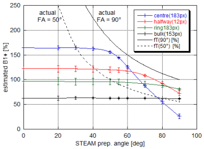 |
128 |
Removing bias and increasing dynamic range in DREAM flip angle mapping at 7T
Gunther Helms, Hampus Olsson, Mads Andersen
DREAM is an ultra-fast multi-slice B1+-mapping technique based on the single-shot STEAM sequence. To study noise and bias related to slice-profiles, DREAM B1+-maps at 3.75mm resolution were acquired at 7T in phantoms and in human brain with nominal flip angles (FA) between 20° and 90° of the two STEAM preparation pulses. B1+ was decreasing at actual FAs above 50°; noise became apparent at actual FAs below 20° reducing dynamic range. By varying the preparation FA, this reliable range (20°<FA<50°) is shifted over a B1+ range from 20% to 250%. The FA map is constructed from overlapping B1+ maps after thresholding.
|
|
4501.
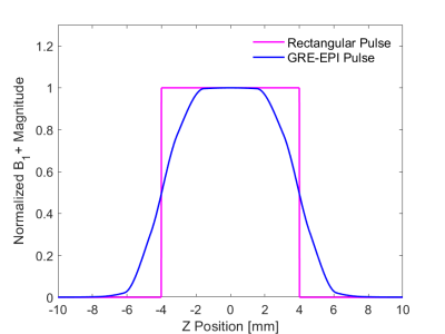 |
129 |
Optimal Flip Angle Range for B1+ Mapping at 3T with Slice Profile Correction Using a Dual Angle EPI Sequence
Gabriela Belsley, Damian Tyler, Matthew Robson, Elizabeth Tunnicliffe
Mapping B1+ inhomogeneity, using commonly available pulse sequences, is essential for widespread, accurate determination of T1 using variable flip angle methods. We investigated the accuracy of B1+ mapping with different flip angles (FA) using the double angle method with a 2D multi-slice GRE-EPI sequence. At lower FAs, we found that B1+ accuracy is affected by SNR, whereas the extent of B1+ inhomogeneities imposes an upper limit on the FAs that can be employed. For a B1+ inhomogeneity of ±40% and a SNR of 29 at 30°, the optimal FA pairs were found to lie between 43°/86° and 74°/148°.
|
|
4502.
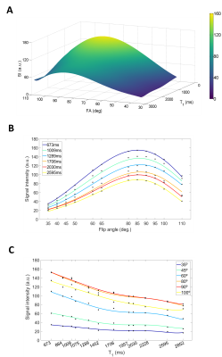 |
130 |
Retrospective transmission field (B1+) and sensitivity profile (B1-) correction for transceive surface RF coils: an empirical solution for RARE
Paula Ramos Delgado, Andre Kuehne, Ludger Starke, Jason M. Millward, Joao Periquito, Thoralf Niendorf, Sonia Waiczies, Andreas Pohlmann
Improving the low signal-to-noise ratio (SNR) inherent to emerging MRI methods such as fluorine MRI is challenging. To enhance sensitivity, SNR-efficient pulse sequences such as RARE and cryogenically-cooled surface RF coils (CRP) are used. Transceive surface RF coils show variation in the excitation field (B1+), impairing quantification. To compensate, previous studies have used an analytical signal intensity equation to perform a retrospective B1+-correction. However, this is unfeasible for RARE due to the absence of such an equation. To overcome this challenge, we propose and validate a numerical method using experimental data acquired with a volume resonator (reference) and a 1H-CRP.
|
|
4503.
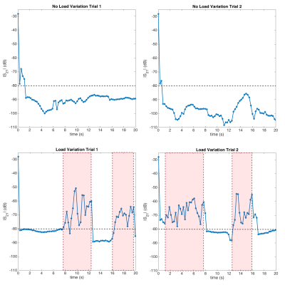 |
131 |
Dynamic Decoupling for Simultaneous Transmission and Acquisition in MRI
Bilal Tasdelen, Alireza Sadeghi-Tarakameh, Ugur Yilmaz, Ergin Atalar
In order to use simultaneous transmission and acquisition in clinical MRI for living subjects, robustness to load and environmental changes has to be established, especially for uncooperative subjects. High isolation can be achieved with active cancellation methods, but maintaining it over a long time is a challenge. A look-up table based method is proposed with a smart search algorithm that enables fast dynamic decoupling of transmit/receive coils using an active decoupling circuit. Experiments with a birdcage coil used as a transceiver show that maintaining >80 dB isolation is possible even under the presence of load variation.
|
|
4504.
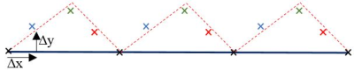 |
132 |
Simple and effective trajectory estimation for image reconstruction of accelerated k-space acquisition on non-rectangular periodic trajectories
Kazi Rafiqul Islam, Jingxin Zhang
Precise coordinates of trajectories are essential for image reconstruction of k-space data acquired from non-rectangular trajectories, and measurement of the trajectories often requires prescan calibration that complicates the process. This abstract presents a simple and effective method to estimate the coordinates of non-rectangular periodic trajectories from normal scan data and demonstrates its efficacy in image reconstruction of in vivo scan data acquired from ZIGZAG trajectory.
|
|
4505.
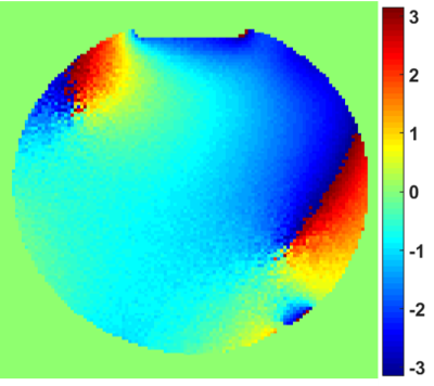 |
133 |
Absolute Phase of Radio-frequency Transmit Field B1+ for a Dual Transmit Coil System
Jinghua Wang, Yu Ding, Christopher Sica, Qing Yang
The spatial absolute phase information is important in various stages of MRI scanning, such as parallel image reconstruction, the combination of MR image or MR spectroscopy from each element of multiple receivers and exploration of new contrast and biomarker. Currently, the absolute phase of a transmit field can only be roughly estimated as half of the transceiver phase. This method is not only inaccurate but also not applicable for a transceiver coil. Thus, the accurate estimation of the absolute phase for an arbitrary RF coil system is an unsolved problem and an unmet need of the MR society. Here we propose a new approach to solve this old problem.
|
|
4506.
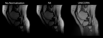 |
134 |
Evaluation of the Uniform Combined Reconstruction (UNICORN) Algorithm for Improving 7T Knee MRI Uniformity
Venkata Veerendranadh Chebrolu, Peter Kollasch, Vibhas Deshpande, John Grinstead, Benjamin Howe, Matthew Frick, Andrew Fagan, Thomas Benner , Robin Heidemann , Joel Felmlee , Kimberly Amrami
MR image intensity non-uniformity is often observed at 7T. A novel algorithm termed ‘Uniform Combined Reconstruction’ (UNICORN) was developed recently to correct for intensity non-uniformity in MR images without the use of a calibration/reference scan. In this work, 3 fellowship trained musculoskeletal radiologists with cumulative experience of 42 years evaluated the efficacy of UNICORN in 33 7T musculoskeletal MRI volumes. The uniformity, contrast, signal-to-noise-ratio and overall image quality were evaluated. Without the use of a reference scan, UNICORN was rated to provide better image uniformity, contrast and overall image quality than the N4 bias-field correction algorithm at 7T.
|
|
4507.
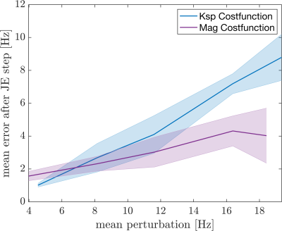 |
135 |
Investigation of the Cost Function for Joint Estimation of Object and B0
Franz Patzig, Bertram Wilm, Klaas Pruessmann
Due to their short read-out time single-shot techniques are frequently used for several imaging modalities but they are prone to static B0 off-resonance artifacts. To avoid separately acquired field maps joint estimation of the object and the B0 map has been proposed as a potential solution alternating between updating an object and a field map guess. A measure to compare cost functions is introduced and two different joint estimation cost functions are investigated whereby a new cost function in image space is suggested. It shows its potential if only a less reliable B0 map guess is given.
|
|
4508.
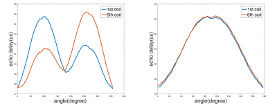 |
136 |
Coil-induced phase removal during gradient delay estimation
Presentation Not Submitted
Chao Wang, Gaojie Zhu, Hai Luo, Bei Lv, Xiang Zhou, Ziyue Wu
Gradient delay can lead to severe artifacts in radial imaging. While several methods have been proposed to correct the linear phase caused by gradient delay, no publications have mentioned the impact of coil sensitivity phase during the estimation of gradient delay to our knowledge. This work reports the impact of this factor and presents a simple method to remove the coil-induced phase during the gradient delay estimation. Both phantom and in-vivo test results are provided to demonstrate the effectiveness of this method.
|
|
4509.
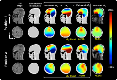 |
137 |
Magnetic field estimation with ultrashort echo time (UTE) imaging.
Jiazheng Zhou, Ali Aghaeifer, Jonas Bause, Alexander Loktyushin, Gisela Hagberg, Klaus Scheffler
We have used UTE sequence to obtain the subject-specific susceptibility distribution, which was then used to simulate motion-induced B0 change at two head positions. A Fourier-based dipole-approximation method was used to map susceptibility to B0. We have evaluated the simulation results against the measured B0 at the same positions and observed a good agreement between the simulated and real data.
|
|
4510.
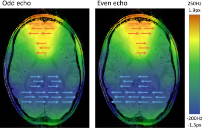 |
138 |
Correction for Geometric Distortion in Bipolar Gradient Echo Images from B0B0 Field
Variations
Korbinian Eckstein, Siegfried Trattnig, Simon Robinson
In bipolar multi-echo gradient echo imaging, signal is acquired during positive and negative readout gradients, giving an efficiency advantage over monopolar imaging in which no signal is acquired during “fly-back”/rewind periods. This increased acquisition efficiency allows higher resolution, shorter echo spacing or increased SNR. In bipolar acquisitions, however, B0B0-related
distortion along the readout axis occurs in opposite directions for odd and even echoes, leading to blurring when images from echoes are combined. We show that a simple unwarping scheme, based on B0B0 field
maps derived from the multi-echo data themselves, is effective in correcting this effect in multi-echo SWI.
|
|
4511.
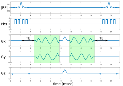 |
139 |
Routine B0 eddy current measurements with TOPPE for more robust spiral imaging
Jon-Fredrik Nielsen, Gopal Nataraj, Jeffrey Fessler, Douglas Noll
Spiral imaging is an SNR- and time-efficient alternative to conventional cartesian MRI, but is relatively sensitive to gradient system imperfections. Unfortunately, measuring the k-space trajectory and B0 eddy currents for a particular spiral readout is cumbersome and not routinely performed. We propose to leverage the TOPPE development environment for rapid pulse sequence prototyping to easily measure both k-space trajectory and B0 eddy currents using “pencil-beam” acquisitions. To demonstrate this setup, we obtained k-space and B0 measurements of a pair of spiral-in and spiral-out readouts. We show that compensating for B0 eddy currents can improve image quality, and that TOPPE provides a convenient platform for these types of measurements.
|
|
4512.
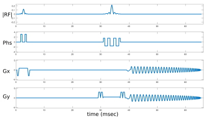 |
140 |
Cross-vendor spiral gradient calibration using TOPPE and Pulseq
Jon-Fredrik Nielsen, Douglas Noll, Sairam Geethanath
Spiral imaging is fast and SNR-efficient, but is relatively sensitive to gradient system imperfections. Unfortunately, these imperfections are generally not known, and can furthermore be expected to vary across different scanner vendor platforms. This complicates multi-site, multi-vendor studies that can benefit from rapid spiral imaging, e.g., those involving fMRI. Here we demonstrate that it is in fact possible to characterize and directly compare spiral gradient performance across two major vendors (GE and Siemens), using the TOPPE and Pulseq frameworks for rapid pulse sequence prototyping. Our observations indicate that B0 eddy currents are substantial on both vendor platforms, and underscore the need for measuring and correcting for B0 effects in spiral imaging.
|
|
4513.
 |
141 |
Multiparametric evaluation of geometric distortions in stereotactic MR imaging at 1.5 and 3 Tesla with a plexiglass phantom: towards practical recommendations for clinical imaging protocols
Gizem Temiz, Fernando Pérez-García, Catherine Jenny, Stéphane Lehéricy, Marguerite Cuttat , Didier Dormont, Damien Galanaud, Chales Valery, Carine Karachi, Romain Valabregue , Sara Fernandez-Vidal, Nadya Pyatigorskaya, Eric Bardinet
Accurate MRI-based targeting is a critical issue for stereotactic surgery. Therefore, geometric distortions need to be evaluated for any pre-operative MR imaging protocol. In this study, we investigated MRI protocols used in Deep Brain Stimulation and Gamma Knife radiosurgery, and focused on the influence of 5 factors on the geometric distortions, at 1.5T and 3T, for 3D T1-weighted and 3D FLAIR images. We found that in order to minimize geometric distortions in stereotactic imaging operator training, careful centering in the MR scanner and systematic activation of constructor’s distortion correction filter are essentials.
|
|
4514.
 |
142 |
MRI quality data assessment in the Italian IRCCS advanced neuroimaging network using ACR phantoms
Fulvia Palesi, Anna Nigri, Domenico Aquino, Ruben Gianeri, Alice Pirastru, Marcella Laganà, Laura Biagi, Michela Tosetti, Maria Grazia Bruzzone, Claudia Gandini Wheeler-Kingshott, The Italian IRCCS advanced neuroimaging network
Generating big-data is becoming imperative with the advent of machine learning. Neuroimaging networks respond to this need. Italian Research Neurological Institutes have formed an advanced neuroimaging network to develop protocols for multisite studies. The present work reports on ACR phantom data across sites and evaluates accuracy and longitudinal reproducibility of: uniformity and ghosting, geometric accuracy, slice thickness, high-contrast and low-contrast object detectability. Our findings show that uniformity, geometric accuracy, low-contrast object detectability are measures that failed at some sites. We intervened to correct these issues improving protocol quality and scanner stability, establishing levels of precision relevant for future multicentre studies in quantitative imaging.
|
|
4515.
 |
143 |
Retrospective 3D spiral trajectory correction: Exponential decay model vs. GIRF
Tobias Speidel, Anja Schwarz, Kilian Stumpf, Volker Rasche
Uncorrected gradient imperfections lead to degrading image artifacts, especially in the case of demanding 3D trajectories. Different post-processing methods have been introduced to compensate for the real-time behaviour of the gradient system. This work shows that gradient waveform deviations can be vastly and nearly equally corrected using an exponential decay model as well as the gradient impulse response function. Both approaches were applied to a pure 3D spiral-like trajectory (Seiffert's spiral), achieving a comparable enhancement in image quality.
|
|
4516.
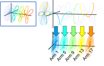 |
144 |
Retrospective gradient delay correction in multi-shot multi-echo rosette acquisition
Alexey Dimov, Nanyque Boyd, Keigo Kawaji, Timothy Carroll
Rosette k-space sampling is an attractive tool for a variety of applications such QSM, DCE, etc. However, as many non-Cartesian acquisition schemes, rosette is highly susceptible to the system-specific gradient delays. We present a robust technique utilizing intrinsic symmetries of multi-shot rosettes, which allows to reconstruct images with minimal artifacts due to misalignment of the k-space points.
|
|
4517.
 |
145 |
Imaging Beyond the Homogeneous Radius in Clinical Magnets
Nadine Dispenza, Yanitza Rodriguez, Robert Constable, Gigi Galiana
At the edge of the bore, both B0 and gradient fields have nonlinear distortions which typically make imaging impossible beyond a certain FOV. In this work, we show that undistorted imaging without significant loss of SNR can be achieved outside the typical 50 cm imaging volume using nonlinear spatial encoding techniques. Spatial distortions are corrected by incorporating the nonlinearities of the gradients and B0 field in the encoding equation, while intravoxel dephasing information counteracts spurious intensity modulations and blurring in the reconstructed images.
|
|
4518.
 |
146 |
Accuracy, repeatability, and reproducibility of longitudinal relaxation rate in twelve small-animal MR systems.
John Waterton, Catherine Hines, Paul Hockings, Iina Laitinen, Sabina Ziemian, Simon Campbell, Michael Gottschalk, Claudia Green, Michael Haase, Katja Hoffmann, Hans-Paul Juretschke, Sascha Koehler, William Lloyd, Yanping Luo, Irma Mahmutovic Persson, James O'Connor, Lars Olsson, Geoffrey Parker, Kashmira Pindoria, Juergen Schneider, Steven Sourbron, Denise Steinmann, Klaus Strobel, Sirisha Tadimalla, Irvin Teh, Andor Veltien, Xiaomeng Zhang, Gunnar Schütz
Many translational MR biomarkers derive from measurements of R1, but evidence for between-site reproducibility of R1 in small-animal MRI is lacking. Here R1 was measured by saturation-recovery in 2% agarose phantoms with five NiCl2 concentrations in 12 magnets at 5 field strengths in 11 centres on two different occasions within 1-13 days. R1 was analysed in three different regions of interest, giving 360 measurements in total. Root-mean-square repeatability and reproducibility coefficients of variation were calculated. Day-to-day repeatability was 2.3%. Between-centre reproducibility was 1.4%. Ni2+ relaxivity in 2% agarose was 0.66s-1mM-1 at 3T and 0.94s-1mM-1 at 11.7T.
|
|
4519.
 |
147 |
On the impact of slice profile and thickness definition across vendors in 2D bSSFP on SNR and T1-mapping in cardiac MRI
Jouke Smink, Guillaume Gilbert, Marc Kouwenhoven, Johan van den Brink
The actual slice thickness and slice profile in 2D imaging are often not taken into account when comparing SNR from different platforms. It can also have an impact in quantitative imaging such as T1-mapping. Inspired by an earlier study, we compared two definitions of slice thickness in 2D bSSFP(the workhorse in CMR): full width at 50% (FW50) and full width at 70% of maximum (FW70). The FW70 pulse definition leads to 30% thicker slices, 9-30% more SNR and it is more vulnerable to partial volume effects. These effects needs to be taken into account when comparing scans from different platforms in multi-center trials.
|
|
4520.
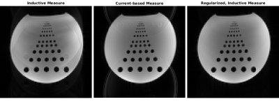 |
148 |
Regularization of Digitally Integrated, Inductive k-Space Trajectory Measures
Jan Pedersen, Christian Hanson, Rong Xue, Lars Hanson
Determining k-space trajectories inductively is conceptually simple, but rely on integration of the induced signal. Performing this integration digitally allow for higher degree of flexibility than analog integration, which is necessary to account for, e.g., refocusing RF pulses. Digital integration, however, require high bandwidth sampling of the induced signal as digitization error accumulate, making the overall approach less attractive. We show that the necessary bandwidth can be reduced by performing regularization using a gradient coil current measure.
|
|
4521.
 |
149 |
Restoring Rotation Invariance of Diffusion MRI Estimators in the Presence of Missing or Corrupted Measurements
Hans Knutsson, Evren Özarslan, Filip Szczepankiewicz, Carl-Fredrik Westin
A natural requirement of estimated tissue microstructure features is that they are rotation invariant. So far, the strategy to attain rotation invariance has been to measure in as many uniformly distributed directions as can be afforded and simply compute the projections on an appropriate set of angular basis functions. However, in the presence of missing samples, this approach is sub-optimal. We show that attaching carefully chosen weights to each measurement can achieve a significantly improved rotation invariance, even in the presence of corruptions that break the isotropic sampling symmetry.
|
|
| Top |
Optimization of Quantitative Mapping Techniques
Digital Poster
Acquisition, Reconstruction & Analysis
Thursday, 16 May 2019
| Exhibition Hall |
08:15 - 09:15 |
| |
|
Computer # |
|
4522.
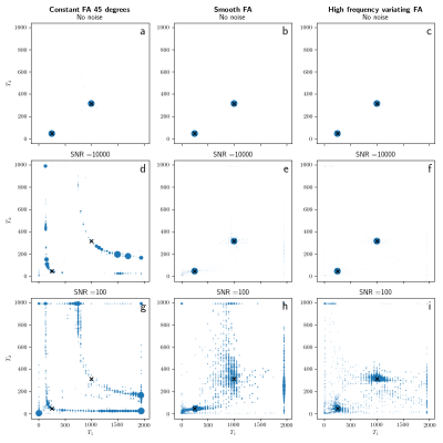 |
151 |
The need of a varying flip angle in multi-component analysis with IR-bSSFP sequences.
Martijn Nagtegaal, Thomas Amthor, Peter Koken, Mariya Doneva
A comparative analysis between IR-bSSFP and MR Fingerprinting was performed in numerical simulations for single and multi-component parameter mapping. The single component matching works for both methods, although the accuracy for T2 is better for MR Fingerprinting. The multi component matching for a constant flip angle IR-bSSFP sequence can only match to the T1* values and cannot distinguish between the underlying T1/T2 values. Using the MR Fingerprinting sequence with a varying flip angle it is possible to match to the T1/T2 components.
|
|
4523.
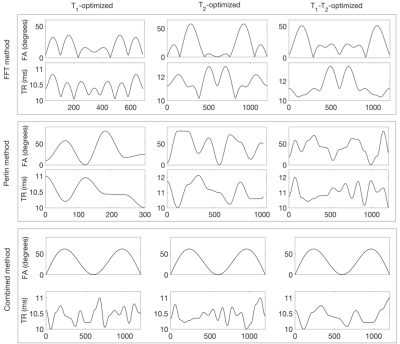 |
152 |
Schedule design for parameter quantification in the transient state using Bayesian optimisation
Giada Fallo, Matteo Cencini, Pedro Gómez, Davide Bacciu, Antonio Cisternino, Michela Tosetti, Guido Buonincontri
Magnetic resonance fingerprinting (MRF) is a useful tool for simultaneously obtaining multiple tissue-specific parameters in an efficient imaging experiment. This technique uses transient state acquisitions with pseudo-random acquisition parameters. However, specific schedules may be better suited for certain parameter ranges or sampling patterns. This work aims to introduce a framework for pulse sequence optimization, including aliasing and noise in our estimates, individually or jointly optimizing for T1 and T2 relaxation times. We demonstrated the schedules created by our algorithm using MRI acquisitions on a healthy volunteer. The design framework could improve the efficiency and accuracy of T1 and T2 acquisitions.
|
|
4524.
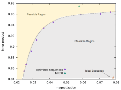 |
153 |
Optimization of MR Fingerprinting Sequence Using a Quantum Inspired Algorithm
Dan Ma, Stephen Jordan, Rasim Boyacioglu, Michael Beverland, Yun Jiang, Darryl Jacob, Sherry Huang, Helmut Katzgraber, Julie Love, Mark Griswold, Matthias Troyer, Debra McGivney
MR Fingerprinting (MRF) is a fast quantitative MR imaging technique that simultaneously quantifies multiple tissue properties. We propose to use quantum-inspired optimization to characterize the optimization landscape by using an appropriate cost function to account for signal features and create an optimization frontier. The simulation results from the optimized MRF sequences showed reduced bias and variance as compared to those from the original empirical design. The in vivo maps from the optimized sequences showed improved image quality as well.
|
|
4525.
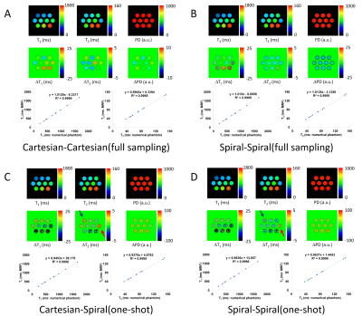 |
154 |
An accurate dictionary generation method for MR fingerprinting using a fast Bloch image simulator
Katsumi Kose, Ryoichi Kose
This study proposes a simple and accurate dictionary creation method for MR fingerprinting using a fast Bloch image simulator. A typical MR fingerprinting sequence based on a FISP sequence and a numerical phantom were used for dictionary generation. Cartesian and spiral readout gradients were used for the Bloch image simulation of the numerical phantoms. MR fingerprinting parameter maps obtained by pattern matching with the dictionaries generated by the proposed method demonstrated validity and usefulness of the method. The proposed method is simple and useful for creation of accurate dictionaries in MR fingerprinting.
|
|
4526.
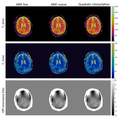 |
155 |
Towards Continuous Dictionary Resolution in MR Fingerprinting using a Quadratic Inner Product Model
Debra McGivney, Rasim Boyacioglu, Yun Jiang, Charlie Wang, Dan Ma, Mark Griswold
Magnetic resonance fingerprinting is a framework for creating quantitative tissue property maps from a single acquisition. The accuracy and precision of these maps depend upon a precomputed dictionary of simulated signal evolutions, to which acquired signals are matched using the inner product to determine the tissue property values. We propose to approximate the inner product as a quadratic function of the tissue properties in a neighborhood around the correct match in order to reduce the effect of tissue property step size in the dictionary. Results from data acquired with different MRF sequences demonstrate the value of the proposed approach.
|
|
4527.
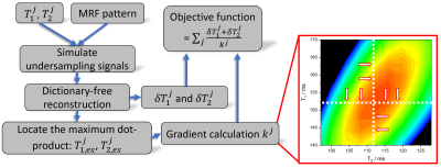 |
156 |
Dictionary-free Reconstruction Based Magnetic Resonance Fingerprinting Optimization
Tianyu Han, Teresa Nolte, Nicolas Gross-Weege, Volkmar Schulz
To make the MRF technique most suitable for clinical needs, efforts are still to be made to accelerate MRF acquisitions while maintaining the accuracy in parameter determination. However, the dictionary calculation is a heavy computational burden for each trial MRF measurement within the optimization process. In this work, we present a numerical study on the optimization of MRF-FISP sequences by using a parallel tempering algorithm. Specifically, an optimization framework tailored for MRF with severe k-space undersampling was developed based on the previously proposed dictionary-free reconstruction (DFR). In vivo measurements were carried out to evaluate the performance of the optimized sequence.
|
|
4528.
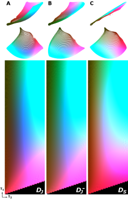 |
157 |
t-Distributed Stochastic Neighbor Embedding (t-SNE) as a Tool for Visualizing the Encoding Capability of Magnetic Resonance Fingerprinting (MRF) Dictionaries
Kirsten Koolstra, Peter Börnert, Boudewijn Lelieveldt, Andrew Webb, Oleh Dzyubachyk
In Magnetic Resonance Fingerprinting (MRF), the quality of the parameter maps depends on the encoding capability of the variable flip angle train. In this work we show how the dimensionality reduction technique t-Distributed Stochastic Neighbor Embedding (t-SNE) can be used to obtain insight into the encoding capability of different MRF sequences by embedding high-dimensional MRF dictionaries into a lower-dimensional space and visualizing them as colormaps. Experiments on example dictionaries perform comparison between different sequences and assess the effect of B1+ variations on the encoding capability.
|
|
4529.
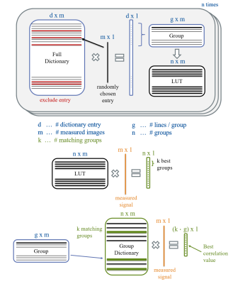 |
158 |
Optimized fast dictionary matching for magnetic resonance fingerprinting based on echo-planar imaging for enhanced clinical workflow
Ingo Hermann, Benedikt Rieger, Jascha Zapp, Sebastian Weingärtner, Lothar Schad
In this work, an optimized fast group matching reconstruction for magnetic resonance fingerprinting based on echo-planar imaging was evaluted to enhance clinical usability. This scanner based 'on the fly' reconstruction reduced the reconstruction time by an acceleration factor of 10 shortening the reconstruction to 10 seconds. The fast group matching algorithm was tested in-vivo and compared with full dictionary matching and resulted in virtually no deviation in T1 and T2* maps facilitating the use of MRF in clinical routine.
|
|
4530.
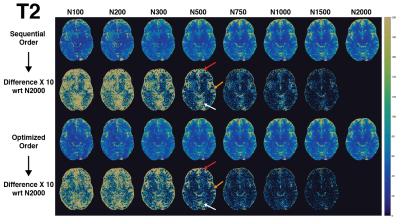 |
159 |
Magnetic Resonance Fingerprinting Optimization With Variance Based Spiral Arm Ordering
Rasim Boyacioglu, Debra McGivney, Dan Ma, Yun Jiang, Mark Griswold
Magnetic Resonance Fingerprinting (MRF) maps various tissue properties and system parameters simultaneously. MRF time series, which are matched to a precalculated dictionary, are often obtained with fast acquisition of low resolution images with undersampled spiral trajectories using a regular sampling pattern. In this work, we propose to order a set of spiral trajectories based on dictionary variance instead of the standard sequential or golden-angle ordering. Phantom and in vivo results show that the variance based optimized order converges faster to expected true values. The optimized order does not limit other MRF optimization approaches and can be applied to any MRF sequence.
|
|
4531.
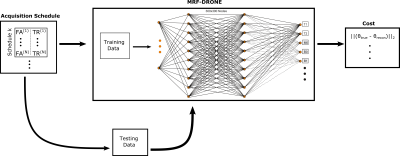 |
160 |
MR Fingerprinting SChedule Optimization NEtwork (MRF-SCONE)
Ouri Cohen
MR Fingerprinting schedule optimization can reduce scan times and improve accuracy but typically relies on minimization of indirect metrics rather than the actual reconstruction error due to the computational challenges involved in calculating the reconstruction error at each iteration of the optimization. Here we introduce a Deep Learning framework that can overcome these challenges and allow direct minimization of the reconstruction error. The proof-of-principle is demonstrated using simulations on a numerical brain phantom.
|
|
4532.
 |
161 |
MRF Dictionary Calculation and Visualization using GPU Compute Shaders
Andrew Dupuis, Dan Ma, Mark Griswold
Dictionary generation for Magnetic Resonance Fingerprinting (MRF) can be a computationally intensive procedure, especially as complexity and density increase. Conveniently, the majority of operations required for calculating dictionary entries are already enumerated in conventional computer graphics shader packages. Here, we leverage the decades of research and hardware development spent to improve computer graphics optimization to remove the need for CUDA parallelization and instead directly render MRF dictionaries into compressible video files in virtually real time.
|
|
4533.
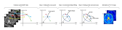 |
162 |
Constrained Ellipse Fitting for Efficient T1-T2 Mapping in Phase-cycled bSSFP Imaging
Kübra Keskin, Tolga Çukur
There is growing interest in use of balanced steady-state free precession (bSSFP) imaging for simultaneous mapping of T1T1, T2T2 and
off-resonance. An elegant ellipse fitting approach in the complex plane was recently proposed for parameter estimation from multiple phase-cycled acquisitions. Since this approach requires at least six phase-cycles, it can limit scan efficiency. Here, we propose a new technique that integrates a geometric solution with constrained ellipse fitting to enable mapping with only four phase-cycled acquisitions. The proposed method yields accurate T1T1, T2T2 and
off-resonance maps while significantly improving scan efficiency.
|
|
4534.
 |
163 |
Extracting Gold Standard Relaxation Times and Field Map Estimates from the Balanced SSFP Frequency Profile by Neural Network Fitting
Rahel Heule, Klaus Scheffler
It has been observed that the balanced steady-state free precession (bSSFP) frequency profile exhibits asymmetries if the intra-voxel frequency content is inhomogeneous and asymmetric. Recent attempts to calculate T1 and T2 values of human brain tissues from the measured bSSFP profile fail to account for anisotropies in the tissue microenvironment and are thus subject to a considerable bias, in particular for white matter. To eliminate this bias, a feedforward neural network is trained with the bSSFP profile as input and a multi-parametric output (i.e., T1, T2, B1, ?B0) using gold standard relaxation times and reference field maps as ground truth.
|
|
4535.
 |
164 |
Noise Reduction and Uncertainty Estimation for the Variable Flip Angle T1 Method with Automatic Selection of Regularization Parameters
Anders Garpebring, Max Hellström, Mikael Bylund, Tommy Löfstedt
The purpose of this work was to develop a method that simultaneously reduces and estimates the uncertainty in the T1 maps obtained with the VFA method while also avoiding the need for any manual tuning of regularization parameters. A Markov Chain Monte Carlo-based algorithm was implemented and evaluated on real and synthetic data. The results show that the method can be used to reduce both noise and noise-induced bias and simultaneously give information about the uncertainty in the estimates.
|
|
4536.
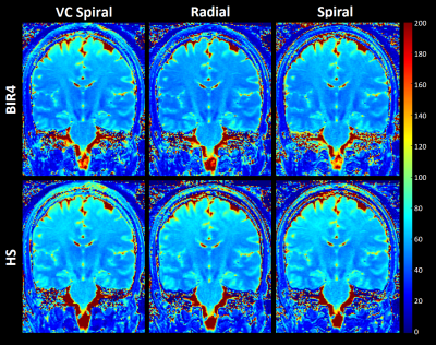 |
165 |
Optimised T2 Preparation for Brain Imaging: Application to Compressed Sensing 3D T2 Mapping
Emilie Mussard, Tom Hilbert, Christoph Forman, Ruud B. van Heeswijk, Reto Meuli, Jean-Philippe Thiran, Tobias Kober
T2-mapping is becoming an important tool to detect pathological tissue; however, achieving high isotropic resolution is challenging. This work optimises a T2-prepared 3D compressed-sensing acquisition. Two T2-preparation modules (modified-BIR4, hyperbolic secant) and three Cartesian sampling trajectories (spiral, radial, VC-spiral) are explored. The NIST-ISMRM phantom and three in vivo subjects were scanned to test T2 accuracy and homogeneity. Results show more homogeneous and accurate T2 values with BIR4, due to a decreased sensitivity to B1. In vivo data showed more homogeneous T2 in WM using a radial trajectory. Based on these results, we propose an optimised 3D T2-mapping protocol of 9:48min.
|
|
4537.
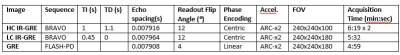 |
166 |
Practical Considerations for Mapping R1 in the Cerebral Cortex Across Sites
Laagi Yoganathan, Kim Desmond, Manpreet Sehmbi, Benicio Frey, Nicholas Bock
We map R1 in the cortex across two sites, using IR-GRE and GRE images to calculate R1 values based on the ratio of the images (IR-GRE/GRE)) using signal equations. We collect B1+ maps to analytically correct R1 inhomogeneities that might cause site-dependent variation. We tested our R1 mapping method with two different input ratio images: one formed using an IR-GRE sequence with typical neuroanatomical contrast, and one using an IR-GRE sequence optimized to produce strong intracortical contrast. We found the ratio image with the higher intracortical contrast produced more consistent R1 maps across sites, which were less sensitive to B1+.
|
|
4538.
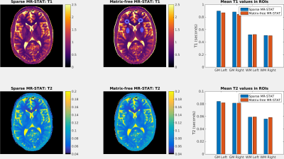 |
167 |
Sparse MR-STAT: Order of magnitude acceleration in reconstruction times
Oscar van der Heide, Alessandro Sbrizzi, Peter Luijten, Cornelis van den Berg
MR-STAT is a framework for obtaining multi-parametric quantitative MR maps using data from single short scans. A large-scale optimization problem is solved in which spatial localisation of signal and estimation of tissue parameters are performed simultaneously by directly fitting a Bloch-based volumetric signal model to the time domain data. In the current work, we exploit sparsity that is inherently present in the problem when using Cartesian sampling strategies to achieve an order of magnitude acceleration in reconstruction times. The new method is tested on synthetically generated data and on in-vivo brain data.
|
|
4539.
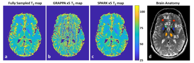 |
168 |
Accelerated T2 mapping based on Bloch signal-model with fixed rank and sparsity constraints
Daniel Grzeda, Meirav Galun, Noam Omer, Tamar Blumenfeld-Katzir, Dvir Radunsky, Ricardo Otazo, Noam Ben-Eliezer
Quantification of T2 values is valuable for a wide range of research applications and clinical pathologies. Multi-echo spin echo (MESE) protocols offer significantly shorter scan-times, at the cost of strong contamination from stimulated and indirect echoes. The echo-modulation-curve (EMC) algorithm, can efficiently overcome these limitations to produce accurate T2 values. In this work we propose a new reconstruction algorithm based on Sparsity and Fixed Rank constraints, denoted as SPARK. We compare our method against GRAPPA and show its superiority in the quantitative evaluation of T2 values from highly undersampled data.
|
|
4540.
|
169 |
Study of key properties behind a good undersampling pattern for quantitative estimation of tissue parameters
Riwaj Byanju, Stefan Klein, Alexandra Huerta, Juan Tamames, Dirk Poot
Quantitative MR (qMRI) at present is clinically unfeasible due to long scan time. Jointly performing image reconstruction and parameters estimation is expected to allow increased acceleration. In this work, we investigate properties of undersampling patterns that are most relevant for parameter estimation using a Cramer-Rao-Lower-Bound (CRLB) based metric for such an approach. We compare key properties of undersampling patterns and conclude that one of these properties, namely low discrepancy, is most relevant for achieving time-efficient qMRI.
|
|
4541.
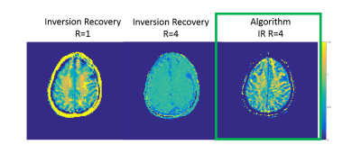 |
170 |
Accelerated R1 or R2 Mapping with Geometric Relationship Constrained Reconstruction Method
Nadine Dispenza, Gigi Galiana, Dana Peters, Robert Constable, Hemant Tagare
In this work we present a constrained reconstruction method that can produce either an R2- or R1- weighted image series, in tandem with the parameter map, from undersampled data. The method has been demonstrated in vivo for radial TSE, and radial TSE augmented with nonlinear encoding (O-space), and inversion recovery (IR) datasets. The algorithm iteratively calculates the entire series of T2 or T1 weighted images while enforcing the exponential decay posed as a geometric relationship between the images. Experimental brain images generated with these maps are in excellent agreement with the fully sampled images and show less undersampling artifact than images reconstructed from individual undersampled datasets.
|
|
4542.
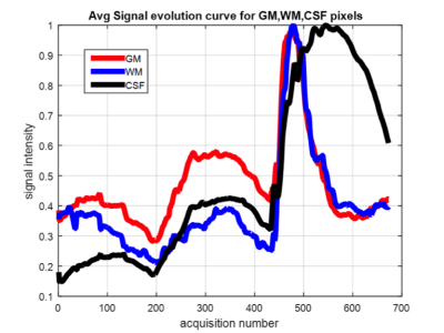 |
171 |
Reconstruction of Tailored Magnetic Resonance Fingerprinting Using Random Forest Approach
Presentation Not Submitted
Shivaprasad Chikop, Amaresh Konar, Vineet Bhombore, Fabian Balsiger, Rajagopalan Sundareshan, shaik Imam, Mauricio Antonio Reyes Aguirre, Ramesh venkatesan, Sairam Geethanath
Magnetic Resonance Fingerprinting is a new acquisition/reconstruction technique to obtain multi-parametric map. Tailored MRF has demonstrated the quantification of longer T2 components contrary to classical MRF. The supervised learning based approach model in the study does not require construction of the dictionary. Leave out one approach has been utilized as the approach for modeling the random forest approach. The dictionary approach is heavy on the computation that limits the MRF to get into the clinic.
|
|
4543.
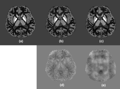 |
172 |
Dynamic streaking artifact regularization for QSM
Yongquan Ye, Xueping Li, Qinglei Zhang, Fei Zhou, Ming Li, Zhao Qing, Bing Zhang, Shuheng Zhang, Yanling Chen, Jinguang Zong
We propose dynamically estimate, formulate and update the field components that are responsible for causing streaking artifact, as an additional regularization term for solving the QSM optimization problem. As a result, streaking artifacts arising from regions with highly disrupted local fields can be well suppressed, preventing them from spatially extending and affecting other regions of interest. The proposed method can maintain the accuracy of QSM results, and has the potential to be integrated into most QSM optimization algorithms.
|
|
4544.
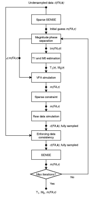 |
173 |
Sparse Pre-Contrast T1 Mapping for DCE-MRI Calibration
Zhibo Zhu, R. Marc Lebel, Yannick Bliesener, Krishna Nayak
Quantitative DCE-MRI requires fast pre-contrast T1 mapping (scan time <3 min) with matching resolution and coverage. Recent advances in imaging have substantially improved resolution and coverage of DCE-MRI but without matched improvements in the pre-contrast T1 data. Here, we demonstrate a sparse T1 mapping method and characterize a tradeoff between data acquisition and T1 statistics, using a variable flip angle (VFA) approach and sparse Cartesian spiral sampling pattern, with image domain wavelet sparsity constraint. This method provides the necessary high-resolution whole-brain T1/M0 maps for DCE-MRI tracer kinetic analysis.
|
|
4545.
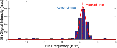 |
174 |
A fast method for field map calculation in multispectral imaging near metal implants
Yuan Zheng, Yu Ding, Yongquan Ye, Weiguo Zhang
Multispectral acquisition is an important technique for MRI near metal. It is critical to estimate the field map and correct for displacements among bin images before bin combination in order to eliminate blurring. However, current field-estimation methods are either susceptible to noise or are computationally intensive, limiting their clinical applications. We propose a robust and efficient algorithm for calculating the field map from multispectral datasets based on a previous matched-filter field estimation technique. The proposed technique was tested on a digital phantom and generated accurate field maps and high quality images with a very short calculation time.
|
|
| Top |
Quantitative Mapping: Relaxometry & Beyond
Digital Poster
Acquisition, Reconstruction & Analysis
Thursday, 16 May 2019
| Exhibition Hall |
09:15 - 10:15 |
| |
|
Computer # |
|
4546.
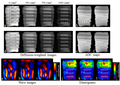 |
1 |
A robust pulse sequence for simultaneous diffusion MRI and MR elastography (diffusion-MRE)
Daiki Ito, Tomokazu Numano, Kazuyuki Mizuhara, Toshikatsu Washio, Masaki Misawa, Naotaka Nitta, Tetsushi Habe
Diffusion-magnetic resonance elastography (dMRE) can acquire diffusion and mechanical properties simultaneously. However, intravoxel phase dispersion (IVPD) interferes with the calculation of the apparent diffusion coefficient (ADC). This study presents an approach to dMRE that reduces the influence of IVPD by introducing a new pulse sequence. The ADC and stiffness, obtained using the existing and proposed dMRE techniques, were compared with spin-echo (SE)-diffusion and SE-MRE, for a phantom. In existing dMRE technique, the ADC was changed by IVPD but that of proposed dMRE technique was unchanged. The results demonstrate that our dMRE technique is a robust method for addressing the IVPD.
|
|
4547.
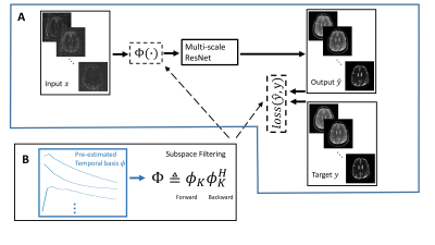 |
2 |
Deep parameter mapping with relaxation signal model driven constraints
Zhiyang Fu, Sagar Mandava, Zhitao Li, Diego Martin, Maria Altbach, Ali Bilgin
Conventional MR parameter mapping suffers from long acquisition times limiting their clinical utility. Model based iterative methods have been proposed to allow reconstructions from highly accelerated data, but these suffer from high computational costs. Deep learning based methods that can reduce reconstruction times significantly while yielding reconstruction quality comparable to the model based methods have emerged recently. In this work, we evaluate the use of signal model driven constraints in deep learning based MR parameter mapping.
|
|
4548.
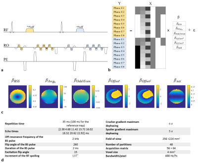 |
3 |
Robust 3D Bloch-Siegert based B1+ mapping using Multi-Echo General Linear Modelling
Nadège Corbin, Julio Acosta-Cabronero, Shaihan Malik, Martina Callaghan
Robust quantification of the longitudinal relaxation rate (R1)—a widely used proxy marker of myelin content—requires highly accurate and precise estimation of the RF transmit field (B1+). The Bloch-Siegert shift (BSS) is a B1+-mapping method that allows calibration data to be acquired with the same spoiled gradient-echo readout used for variable flip angle R1 mapping. Here we show that systematic differences in steady state phase, caused by the interleaved nature typically adopted, lead to bias or loss of precision, but that these effects can be corrected for using a multi-echo approach and GLM fitting to isolate the BSS phase.
|
|
4549.
 |
4 |
3-Dimensional Strain Mapping of the Eyeball during Adduction, Abduction Tasks
David Ouellette, Kenneth Wengler, Patrick Sibony, Xiang He, Tim Duong
This study employed high-resolution 3D-MRI to map the strain of the eyeball during adduction and abduction tasks. The strain map is highly heterogeneous with high strain toward the anterior region. Adduction induced higher strain than abduction, as expected due to more stretching of the optic nerve in the adduction position. This is the first MRI measurement of strain of the eyeball. This approach could have clinical applications in eye movement disorders and eye diseases.
|
|
4550.
 |
5 |
High resolution 3D magnetic resonance fingerprinting with hybrid radial cartesian-EPI acquisition
Dongyeob Han, Taehwa Hong, Dong-Hyun Kim
A high resolution (0.5x0.5x1mm3) 3D MRF method was proposed using a hybrid radial cartesian-EPI acquisition with both segmented & interleaved EPI strategy. For the reconstruction, k-space SVD compression and CG-SENSE were applied. An in vivo brain results were presented.
|
|
4551.
 |
6 |
Assessment of Absolute pH Using and Magnetic Resonance Fingerprinting and a Single Dysprosium-Based MRI Contrast Agent
Yifan Zhang, Andre Martins, Dean Sherry, Christian Anderson, Chris Flask
In this initial in vitro study, we used Magnetic Resonance Fingerprinting (MRF)-based T1 and T2 relaxation time maps to estimate the linear relationship between pH and relaxivity (r1 and r2) for a previously-described dysprosium (Dy) MRI contrast agent. These relaxivity estimates were then used to calculate MRF-based estimates of pH for each solution for comparison with gold-standard measurements by pH electrode at 7.0T (R = 0.93, p = <1e-6) and 9.4T (R = 0.68, p = 0.004). Results show MRF can be used in combination with a pH-sensitive paramagnetic MRI contrast agent to accurately estimate pH independent of agent concentration.
|
|
4552.
 |
7 |
Magnetic Resonance Fingerprinting with Pure Quadratic RF Phase
Charlie Wang, Rasim Boyacioglu, Debra McGivney, Dan Ma, Xin Yu, Mark Griswold
Previous work has shown that Magnetic Resonance Fingerprinting with quadratic RF phase (qRF-MRF) can be used to simultaneously quantify off-resonance, T1, T2 and T2*. This method employed a mix of bSSFP and qRF pulse sequence block segments for reliable tissue property quantification. However, the incorporation of bSSFP type acquisition schemes resulted in null-band artifacts near bSSFP signal voids. Here, we present a bSSFP-free pure qRF-MRF method with elimination of null-band artifacts, and explore its potential for tissue property mapping with reduced acquisition time.
|
|
4553.
 |
8 |
Combination of ESPIRiT and back-projection reconstruction for 3D MR fingerprinting within 2.5 minutes
Xiaozhi Cao, Qing Li, Huihui Ye, Hongjian He, Jianhui Zhong
A spiral projection acquisition scheme was implemented for 3D MR fingerprinting to achieve isotropic resolution of 1x1x1 mm3 in whole brain T1 and T2 mapping within 2.5 minutes by using efficient L1SPIRiT reconstruction (ESPIRiT) and back-projection reconstruction.
|
|
4554.
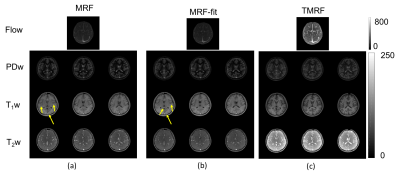 |
9 |
Rapid, simultaneous non-synthetic multi-contrast and quantitative imaging using Tailored MR Fingerprinting (TMRF)
Sairam Geethanath, Sachin Jambawalikar, Maggie Fung, Angela Lingella, John Vaughan
The goal of this work was to rapidly acquire non-synthetic multi-contrast and quantitative images simultaneously, through tailoring the MR fingerprinting acquisition schedule in contrast blocks. TMRF providing for five contrasts was designed, simulated and demonstrated on four healthy volunteer brain scans. The acquisition times for MRF and TMRF were 5:11 and 4:41 (min: sec) respectively. The spatio-temporal profiles of T1, T2, PD, water-fat and flow contrasts were reconstructed block-wise along with relaxometric maps. Comparatively, TMRF images showed higher mean to standard deviation ratios for the four volunteers over the contrast blocks for PD and T2 while maintaining similarity of relaxometric maps.
|
|
4555
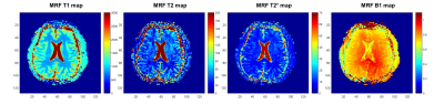 |
10 |
EPI based Dual-stage MR Fingerprinting for T1, T2, and T2* mapping
Video Permission Withheld
Young-Jung Yang, Pan Ki Kim, Dong Jin Im, Donghyun Hong, Byoung Wook Choi
We propose an improved MR fingerprinting which can generate T1, T2, and T2* maps simultaneously. This method is based on single-shot EPI and signal acquisition consists of dual-stage divided by fixed and variable echo time. Dictionary generation and pattern matching were also modified in accordance with acquisition scheme. The feasibility of proposed method was demonstrated by phantom study and the MRF results are well correlated with the conventional T1, T2, and T2* maps. In-vivo brain MRF was also performed with a healthy volunteer.
|
|
4556.
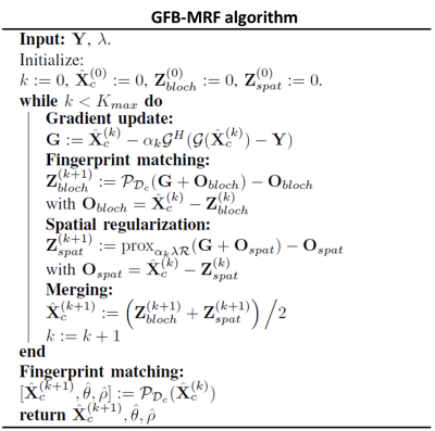 |
11 |
GFB-MRF: Parallel spatial and Bloch manifold regularized iterative reconstruction for magnetic resonance fingerprinting
Simon Arberet, Xiao Chen, Boris Mailhe, Peter Speier, Mathias Nittka, Heiko Meyer, Mariappan Nadar
We introduce a new iterative algorithm for Magnetic Resonance Fingerprinting (MRF) where spatial regularization and fingerprint matching are applied in parallel. This enables to have simultaneously a spatial regularization in addition to the time domain Bloch manifold regularization. Our proposed algorithm showed significant improvements with respect to the state of the art in particular regarding the robustness with respect to measurement noise.
|
|
4557.
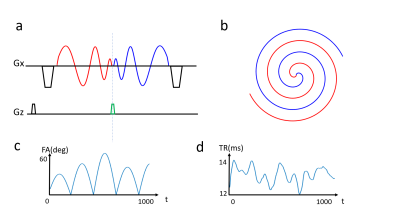 |
12 |
Accelerated Multi-band Magnetic Resonance Fingerprinting Using Spiral in-out with additional kz Encoding and Modified Sliding Window Reconstruction
Di Cui, Xiaoxi Liu, Hing-Chiu Chang, Queenie Chan, Edward Hui
Multi-band Magnetic Resonance Fingerprinting can be achieved using UNFOLD-like acquisition and dictionary matching without using parallel methods. However, the MR parametric maps after dictionary matching in one slice suffers from artifacts due to the high frequency components of other simultaneously acquired slices. In this work, a new acquisition strategy was proposed for the multi-band acquisition, where spiral-in-out trajectory was used to provide extra kz encoding. A modified sliding window reconstruction was also proposed to reduce the high frequency oscillations.
|
|
4558
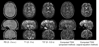 |
13 |
Computational method for T2-weighted images based on polynomial approximation using 3D MR parameter mapping with RF-spoiled gradient echo
Video Permission Withheld
Tomoki Amemiya, Suguru Yokosawa, Yo Taniguchi, Ryota Sato, Hisaaki Ochi, Toru Shirai
We propose a computational method for obtaining T2-weighted images from maps of proton density, T1, and T2* acquired by 3D RF-spoiled gradient echo. The proposed method uses a predetermined polynomial that approximates the relationship between the MR parameters and the intensity of T2WI on the basis of datasets of other subjects. Similarities between computed images and actually scanned images were improved compared with a computation method using T2* instead of T2 in the theoretical equation of the spin echo signal.
|
|
4559.
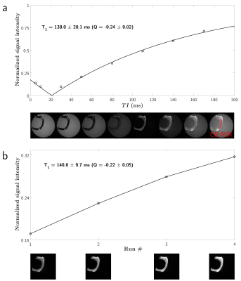 |
14 |
T1 measurement in short-T2 material with suppressed long-T2 component using an IR-UTE multishot sequence
Lucas Soustelle, Julien Lamy, François Rousseau, Jean-Paul Armspach, Paulo Loureiro de Sousa
T1 quantification of short-T2 species is challenging due to the uncommon behavior of the signal decay and magnetization tilting during excitations in conventional sequences. UTE sequences can therefore be considered with refined magnetization evolution models using the Bloch equations. In voxels comprising a mix of long and short-T2 components (e.g. myelin and water in the normal appearing white matter), an appropriate long-T2 suppression scheme is mandatory. In this work, we propose an analytical model to quantify the T1 of a short-relaxing component in an accelerated Inversion-Recovery UTE in vitro, and within long-T2 suppression condition.
|
|
4560
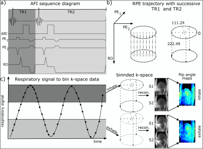 |
15 |
3D Radial Phase Encoded Flip Angle Imaging at Ultra-High Field Strength
Video Permission Withheld
Sebastian Dietrich, Christoph Kolbitsch, Tobias Schaeffter, Sebastian Schmitter
In this work a novel 3D flip angle (FA) mapping method is introduced which combines the advantages of the AFI pulse sequence (3D, low SAR) with the motion robustness of a radial phase encode acquisition scheme. Mapping the FA is needed for human body imaging at 7 tesla to improve image quality and assessment of quantitative results. Therefore, we combined an interleaved acquisition of two FIDs, S1 and S2, acquired for different repetition times TR1 and TR2 with radial phase encode trajectory. In this study we validated this new sequence with a body phantom and in two in-vivo scans. Similar results to cartesian reference scans were obtained and reasonable motion resolved abdominal FA maps were acquired.
|
|
4561.
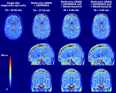 |
16 |
Combining Parallel Imaging and Model-based Reconstruction for Isotropic 3D T2 mapping with Multi-Echo GRASE
Gian Franco Piredda, Tom Hilbert, Reto Meuli, Josef Pfeuffer, Jean-Philippe Thiran, Tobias Kober
Classical T2 mapping based on 2D multi-echo spin-echo sequences achieves only limited across-slice resolutions. For clinical use, acquisitions with high isotropic resolution are however desirable, resulting in clinically prohibitive scan times. To this end, we propose a 3D multi-echo gradient and spin echo sequence with CAIPIRINHA and an additional model-based acceleration for T2 estimation. The combination of these techniques allows for whole-brain T2 mapping with 1.6mm-isotropic resolution in 3:26 min. The proposed framework was tested both in phantom and in vivo experiments.
|
|
4562.
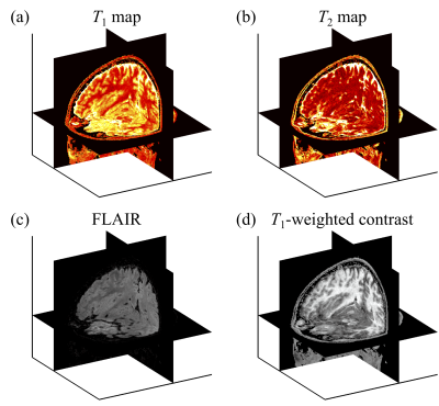 |
17 |
Quantitative and synthetic MRI using a Multi-Pathway Multi-Echo (MPME) acquisition followed by machine-learning contrast translation
Cheng-Chieh Cheng, Frank Preiswerk, Bruno Madore
Brain exams would ideally include 3D quantitative maps of several basic MR parameters, such as T1, T2, T2* and B0, along with popular qualitative contrasts such as MPRAGE and FLAIR, for example. A multi-pathway multi-echo (MPME) pulse sequence was developed that captured vast amounts of information about the imaged object relatively fast, but not necessarily with image contrasts that radiologists might be comfortable reading. A neural network was trained to act as a ‘contrast translator’, to convert information rapidly obtained from MPME scans into useful quantitative and qualitative contrasts, in effect condensing a whole exam into a single 3D scan.
|
|
4563.
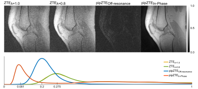 |
18 |
Variable Flip Angle T1-Mapping Using Perfect In-Phase ZTE
Mathias Engström, Axel Hartwig, Cristina Cozzini, Graeme McKinnon, Florian Wiesinger
This work details an extension of the Perfect In-Phase ZTE (pipZTE) method that allows for fast and efficient T1-mapping. By adding a variable flip angle scheme in combination with the Perfect In-Phase ZTE readout band-width modulation PD and T1 mapping can be achieved without interference from chemical shift artifacts.
|
|
4564.
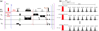 |
19 |
A Fast Multi-slice T1 mapping method based on SPatiotemporal ENcoding
Qingjia Bao, Ricardo Martinho, Lucio Frydman
A pulse sequence for T1 relaxation time mapping which enables high-resolution and multi-slice imaging in short acquisition times, is presented. The sequence combines fast, robust acquisitions based on SPatiotemporal ENcoding (SPEN), with an accelerated sampling of the T1 inversion curve via slice shuffling. The experiment allows for T1 quantifications in challenging regions subject to field inhomogeneities. In phantoms it provides reliable T1 maps that agree with standard measurements; in in vivo studies of murine brain and kidney models, it provides fewer distortions than comparable EPI-based counterparts.
|
|
4565.
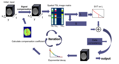 |
20 |
Accelerating Bi-exponential T1? mapping using SCOPE
Yuanyuan Liu, Yanjie Zhu, Jing Cheng, Weitian Chen, Xin Liu, Dong Liang
Mono-exponential T1ρ mapping requires 4 or 5 T1ρ-weighted images with different spin lock times (TSLs) to obtain the T1ρ maps, while bi-exponential T1ρ mapping requires a larger number of TSLs, which further prolongs the acquisition time. In this work, we develop a variable acceleration rate undersampling strategy to reduce the total scan time. A signal compensation strategy with low-rank plus sparse model was used to reconstruct the T1ρ-weighted images. We provide the reconstructed images and the estimated T1ρ maps at an acceleration factor up to 6.1 in fast bi-exponential T1ρ mapping.
|
|
4566
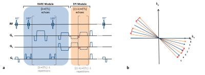 |
21 |
Dual Contrast Weighting and Simultaneous T2 and T2* Mapping with Radially Sampled RARE-EPI
Video Permission Withheld
Carl Herrmann, Katharina Paul, Till Huelnhagen, Thoralf Niendorf
MRI examinations commonly involve a series of multiple imaging contrasts and MR-metrics. Dual or even more contrast techniques offer substantial scan time reduction and eliminate the propensity to slice co-registration errors induced by bulk and physiological motion. Recognizing this opportunity this work presents a dual contrast RARE-EPI hybrid, that provides T2 (RARE module) and T2* (EPI module) contrast and facilitates simultaneous T2 and T2* mapping in a single radially (under)sampled scan (2-in-1 RARE-EPI). The applicability of 2-in-1 RARE-EPI is demonstrated in phantom and in in vivo studies and benchmarked versus conventional T2 and T2* weighted/mapping techniques.
|
|
4567.
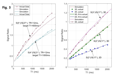 |
22 |
A novel strategy to perform the dual flip angle method for the fast and accurate T1 mapping by MRI
Steven Yee, Michael Fadell, Mark Brown, Seonghwan Yee
Fast T1 mapping can be done by utilizing dual flip angles in acquiring spoiled gradient echo signals. However, its accuracy may be questionable even when the suggested optimal flip angle pair is used. Noting that the faithful action of the prescribed flip angles is the key to the accuracy, we present here a novel dual flip angle method by which the system-specific RF-pulse fidelity of flip angles can be validated and, if necessary, calibrated to improve the T1 accuracy in a wide in vivo range. We tested this method on a few 1.5 or 3T MRI systems of major vendors.
|
|
4568
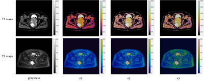 |
23 |
Evaluation of different colormaps for best visual assessment of quantitative Magnetic Resonance Fingerprinting data
Video Permission Withheld
Verena Obmann, Ananya Panda, Chaitra Badve, Jeffrey Sunshine, Vikas Gulani, Mark Griswold
Increasingly quantitative methods such as apparent diffusion coefficient, T1, T2 and T2* mapping or elastography are used in MR imaging. As quantitative data provide multidimensional characterization of pathophysiology, color provides an additional dimensionality to visualize the data. This study demonstrates the superiority of three different colormaps over grayscale display of each T1 and T2 maps for MR Fingerprinting.
|
|
4569.
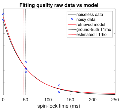 |
24 |
Relaxation parameter estimation from limited time points
Tianle Zhao, Weitian Chen, Thierry Blu
T1rho is a useful biomarker for the diagnosis of several diseases. Current imaging techniques usually use uniform sampling and require a relatively large number of samples to get reliable estimations of T1rho. We show that the intuitive uniform sampling is not optimal, and propose an optimal sampling strategy. We also propose a fast estimation algorithm, which (with the use of spatial redundancy) provides accurate estimates of the T1rho relaxation map from as few as 3 different spin-lock time samples.
|
|
4570
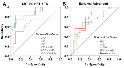 |
25 |
Predicting pathological subtypes and stages of thymic epithelial tumors using DWI: value of combining ADC and texture parameters
Video Permission Withheld
Bo Li, Xiao-cheng Wei, Yu-chuan Hu, Guang-bin Cui
To explore the value of combining apparent diffusion coefficients (ADC) and texture parameters from diffusion-weighted imaging (DWI) in predicting the pathological subtypes and stages of thymic epithelial tumors (TETs). In this study, Fifty-seven patients with TETs confirmed by pathological analysis were retrospectively enrolled. The results showed combination of ADC and DWI texture parameters improved the differentiating ability of TET grades, which could potentially be useful in clinical practice regarding the TETs evaluation before treatment.
|
|
| Top |
Going Faster: New Sequences & Acquisition Protocols
Digital Poster
Acquisition, Reconstruction & Analysis
Thursday, 16 May 2019
| Exhibition Hall |
09:15 - 10:15 |
| |
|
Computer # |
|
4571.
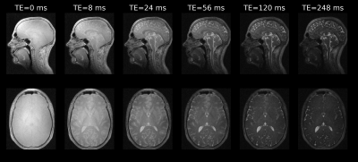 |
26 |
Rapid, multi-TE, T2-prepared RUFIS for Silent T2-weighted imaging
Emil Ljungberg, Brian Burns, Tobias Wood, Shannon Kolind, Florian Wiesinger, Gareth Barker
Zero echo time (ZTE) imaging using the RUFIS sequence allows for silent imaging with high efficiency. Without modifications, RUFIS produces proton density and/or T1-weighted images similar to a spoiled gradient echo sequence. In this work we present a novel T2-prepared RUFIS sequence with multiple echo times acquired in each shot, for efficient T2-weighted imaging. We present in vivo results acquired in 11 min with 1.5mm3 resolution, with effective echo times from 0 to 248ms.
|
|
4572.
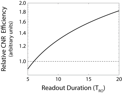 |
27 |
Rapid High Resolution T1-Weighted Hippocampus Imaging with Yarn-Ball Acquisition
Rob Stobbe, Peter Seres, Don Gross, Christian Beaulieu
For spoiled steady-state T1-weighted imaging, readout duration (TRO) and repetition time (TR) increase result in greater contrast-to-noise ratio (CNR) efficiency. Novel 3D-twisting Yarnball acquisition realizes this advantage without scan-time penalty (more of k-space sampled with increased TRO), but increased TRO results in greater point-spread-function smearing. Following TRO optimization, Yarnball is used to produce whole-brain 0.36x0.36x1.08 mm3 coronal (defined by 1/(2kmax)) images in 10 minutes (with 2 averages). Compared to 3D-MP-RAGE (same scan time and voxel volume) Yarnball images have greater resolution and grey-white CNR, facilitating sharper depiction of internal hippocampus architecture.
|
|
4573.
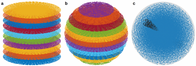 |
28 |
3D SPARKLING for accelerated ex vivo T2*-weighted MRI with compressed sensing
Carole Lazarus, Pierre Weiss, Franck Mauconduit, Alexandre Vignaud, Philippe Ciuciu
In the last decade, compressed sensing (CS) has been successfully used in MRI to reduce the acquisition time. Recently, we have proposed a new optimization-driven algorithm to design optimal non-Cartesian sampling patterns for CSMRI, called SPARKLING for Spreading Projection Algorithm for Rapid K-space sampLING. This method has a few advantages compared to standard trajectories such as radial lines or spirals: i) it allows to reproduce arbitrary densities while the other two are restricted to radial densities and ii) it is more robust to system imperfections. In this communication, we introduce an extension of the SPARKLING method for 3D imaging that allows to achieve an isotropic resolution of 600 μm in just 45 seconds for T2*-weighted ex vivo brain imaging at 7 Tesla.
|
|
4574.
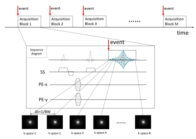 |
29 |
Capturing Time-Dependent Electric Currents Using MRI with A Sub-Millisecond Temporal Resolution
Zheng Zhong, Muge Karaman, Theodore Claiborne, Xiaohong Zhou
Neuronal current mapping using MRI has profound biomedical applications, but is hampered by limited temporal resolution. Using a technique known as Sub-Millisecond Imaging of cycLic Event or SMILE, we demonstrate that the temporal resolution of MRI can be substantially increased to the sub-millisecond scale or shorter. This allows capturing ultra-fast physical or biological processes that are cyclic. Although our experimental studies are limited to mapping time-varying currents in a phantom, the same concept can be extended to capturing more complex biological processes, including but not limited to, neuronal currents.
|
|
4575.
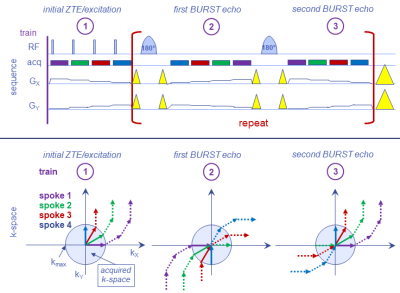 |
30 |
Spin-Echo ZTE-BURST for Quiet T2-Weighted Imaging
Rolf Schulte, Ana Beatriz Solana
ZTE was combined with single spin-echo BURST encoding for acquiring T2 weighted images in a relatively quiet manner.
|
|
4576.
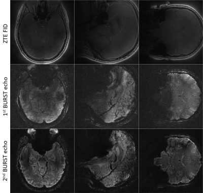 |
31 |
Silent T2* Imaging on 7T using ZTE Combined with Gradient-Echo BURST
Rolf Schulte, Mauro Costagli, Ana Beatriz Solana, Guido Buonincontri
ZTE combined with gradient-echo BURST enables silent 3D radial T2* imaging. It was implemented on 7T and T2* weighted images were acquired with isotropic resolutions of 1-3mm. From the series with different echo times, both phase and T2* maps were extracted.
|
|
4577.
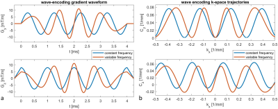 |
32 |
Variable Frequency Wave-encoded 3D Turbo Spin Echo Imaging
Zechen Zhou, Baocheng Chu, Chun Yuan, Peter Börnert
Wave encoding is an emerging approach that can take better usage of the three-dimensional (3D) spatial encoding power of multi-channel coils employed in parallel imaging (PI). In this work, a variable frequency (VF) wave encoding approach is proposed to improve the aliasing propagation property and reduce the side lobe amplitude of the transformed point spread function. This VF approach can also induce amplitude modulated wave encoding gradients to reduce eddy currents and improve the slice selection profile. The preliminary results demonstrated its improved PI performance for 3D turbo spin echo imaging over Cartesian and constant frequency wave encoding schemes.
|
|
4578.
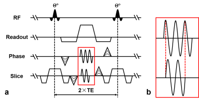 |
33 |
Accelerating Three-Dimension Balanced Steady-State Free Precession Imaging with Modified Wave-CAIPI Technique
Shi Su, Dong Liang, Zhilang Qiu, Caiyun Shi, Xin Liu, Hairong Zheng, Haifeng Wang
Balanced steady-state free precession (bSSFP) has merits such as high signal-to-noise ratio, T2/T1 contrast and rapid acquisition speed. However, bSSFP requires further acceleration in 3D imaging due to massive data collected. The acceleration of conventional parallel imaging techniques is limited. In this study, we propose wave-bSSFP by using a modified wave-CAIPI technique to highly accelerate bSSFP. Wave gradients were truncated to further reduce g-factor noise penalty with high wave amplitudes. The simulation and in vivo experiment indicate that wave-bSSFP is effective in decreasing g-factor. Here, an acceleration factor of 9 was achieved in brain scan with 0.8 mm isotropic resolution.
|
|
4579.
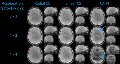 |
34 |
Highly Accelerated 3D EPI using Compressed Sensing
Patrick Liebig, Robin Heidemann, Bernhard Hensel, David Porter
In previous work, Echo-Planar Imaging (EPI) has been used in combination with a CAIPIRINHA undersampling scheme, as in SMS blipped CAIPI or 3D CAIPI EPI, for highly accelerated BOLD, perfusion and diffusion weighted imaging. In a separate development, Compressed Sensing (CS) was employed in combination with parallel imaging to significantly accelerate a range of non-EPI 3D imaging sequences. In general, this is achieved by using a variable-density randomized sampling scheme which gives aliasing artefacts a noise like appearance. This work explores the use of CS to accelerate 3D EPI acquisitions and demonstrates an improved performance compared to the CAIPIRINHA approach.
|
|
4580.
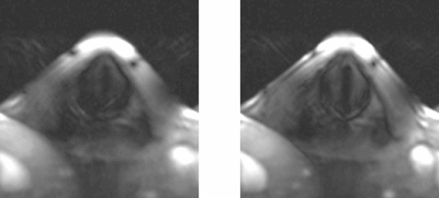 |
35 |
Highspeed Imaging of the Vocal Folds Oscillations with Image-based Motion Correction
Johannes Fischer, Ali Özen, Matthias Echternach, Bernhard Richter, Michael Bock
Highspeed imaging of the vocal folds oscillations is possible by applying a very short phase encoding gradient along the direction of motion. Due to repeated breathing cycles of the volunteer, motion and shifts are introduced that impair image quality. With the use of phase only cross correlation, we correct for this motion prior to the gated reconstruction by applying a linear phase to the k-space data. The proposed method is shown to improve reconstruction of anatomical features and SNR.
|
|
4581.
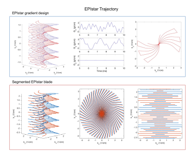 |
36 |
Twisted radial echo planar trajectory (EPIstar) for 3D self-navigated golden angle structural and functional MRI
Christoph Rettenmeier, Danilo Maziero, V. Andrew Stenger
A new 3D trajectory design for efficient, self-navigated golden angle high-resolution MRI acquisition is presented along with results in SWI and BOLD functional MRI.
|
|
4582.
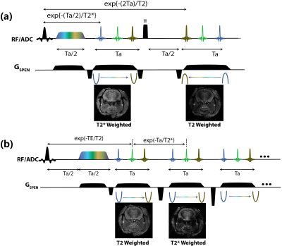 |
37 |
Simultaneous T2* and T2 weighted imaging based on ultrafast SPEN MRI
Qingjia Bao, Lucio Fydman
This work presents new sequences to acquire multislice images with different contrasts –a T2* weighted one for enhancing BOLD and a T2 weighted one for faithful location– in a single shot. The sequences rely on SPatiotemporal ENcoding (SPEN), an ultrafast MRI method with immunity to artifacts, and they utilize a “full-refocusing” mode to obtain a T2 weighted image and a “non full-refocusing” mode to obtain T2* weighted images. Two variants are proposed, differing in the order with which they collect their T2*- and the T2-weighted information, and are demonstrated with in vivo studies of brain and kidney in mice.
|
|
4583.
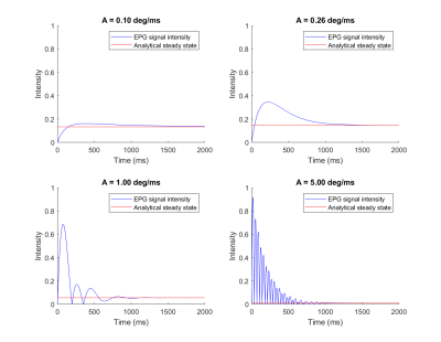 |
38 |
On the signal strength of Simultaneous Transmission and Reception (STAR) acquisition: EPG simulation and analysis
Gehua Tong, Sairam Geethanath, John Vaughan
Simultaneous Transmission And Reception (STAR) has the potential to remove the constraint of temporal separation between transmission and reception. In principle, much shorter acquisition times with significantly higher signal strength compared to pulsed sequences should be achievable. However, the signal characteristics differ from that of the conventional pulsed-RF framework. In this work, we characterize STAR characteristics with extended phase graph (EPG) simulation. We show the signal evolution from a simple STAR experiment as well as how tissue contrast could be generated in steady state.
|
|
4584.
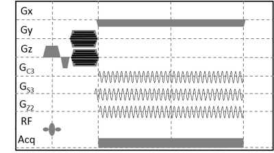 |
39 |
Accelerated Volumetric FRONSAC with WAVE and CAIPI
Nadine Dispenza, Robert Constable, Gigi Galiana
This work demonstrates the potential of FRONSAC, which adds oscillating nonlinear gradients to the Cartesian readout, for 3D accelerated imaging. In undersampled trajectories using either standard Cartesian encoding, CAIPI encoding, or WAVE-CAIPI encoding, significant further improvements are achieved when FRONSAC is applied in addition to these approaches.
|
|
4585.
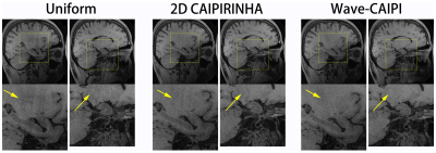 |
40 |
Wave-CAIPI accelerated whole brain structure imaging using three-dimensional T1 weighted SPACE sequence
Zhilang Qiu, Sen Jia, Haifeng Wang, Xin Liu, Hairong Zheng, Dong Liang
Three-dimensional (3D) SPACE (sampling perfection with application optimized contrast using different flip angle evolutions) sequences are the workhorse for volume imaging with isotropic spatial resolution. However, spatial resolution is often scarified to achieve clinically acceptable scan time. Conventional one- and two-dimensional parallel imaging techniques could help reducing the scan time but would lead to deteriorated signal-to-noise (SNR) performance at submillimeter spatial resolutions. In this study, three-dimensional parallel imaging technique-Wave-CAIPI is utilized to improve the SNR performance for whole brain SPACE imaging with isotropic 0.6 mm resolution. In vivo results demonstrated that Wave-CAIPI could improve the SNR at 5x acceleration.
|
|
4586.
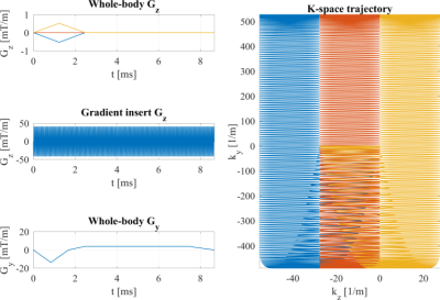 |
41 |
Supersonic imaging with a silent gradient axis driven at 20 kHz
Edwin Versteeg, Dennis Klomp, Jeroen Hendrikse, Jeroen Siero
Gradient inserts allow for faster switching and higher gradient strengths than conventional whole-body gradient coils. However, the higher gradient performance is accompanied by an increase in acoustic sound pressure. We present a gradient insert that switches at 20 kHz (above human hearing perception) and therefore allows for imaging with an inaudible gradient axis. Additionally, we introduce a readout scheme for imaging at 20 kHz, and show the first imaging results on a phantom and a healthy volunteer using an inaudible gradient axis.
|
|
4587.
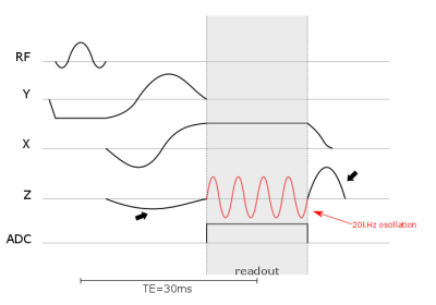 |
42 |
2D k-space waves for silent EPI acquisitions
Jenni Schulz, Riccardo Metere, Dennis Klomp, David Norris
High acoustic noise levels in fMRI-acquisitions are not only problematic in terms of undesired activation patterns in the brain, but also with respect to patient comfort. A 2D-EPI sequence is presented which is capable of acquiring fMRI-data in silent mode by using a head insert z-gradient coil. For silent data acquisition, a wave-like k-space trajectory is then required. The sequence is compared to a standard FLASH and EPI acquisition. The measured acoustic noise of the silent 2D-EPI is in the order of the idle mode of the scanner and arises from the sound of the continuously active helium pump.
|
|
4588.
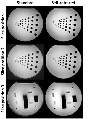 |
43 |
Self-retraced Spiral In-Out 3D Turbo Spin-Echo Imaging
John Mugler III, Craig Meyer, Thomas Benkert, Josef Pfeuffer, Berthold Kiefer
The purpose of this work was to perform a preliminary evaluation of a self-retraced spiral in-out trajectory for 3D turbo/fast spin-echo imaging. By sampling k-space locations twice with a single spiral in-out trajectory, off-resonance effects are robustly attenuated and image quality is improved compared to using a standard spiral in-out trajectory.
|
|
4589.
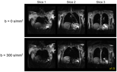 |
44 |
Single Breath-Hold Diffusion MRI utilizing a Spiral TSE with Variable Flip Angle Refocusing
Naoharu Kobayashi, Michael Garwood
A single breath-hold diffusion MRI sequence utilizing turbo spin echo (TSE) with variable flip angle refocusing and spiral readout is introduced. Flip angles of the refocus RF pulses were determined with the prospective extended phase graph method to minimize the impact of fluctuating refocusing echo signals in TSE. Spiral k-space sampling made the sequence tolerant to motion. The feasibility of the proposed sequence was tested in in vivo brain and thoracic imaging. The proposed single breath-hold diffusion sequence achieved diffusion-weighted imaging of the thoracic region without clear cardiac motion artifacts.
|
|
4590.
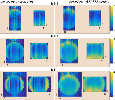 |
45 |
Reduced Noise Enhancement in Whole-Brain Multiband-RASER
Ute Goerke
High acceleration is an effective tool to achieve whole brain coverage with acceptable total acquisition times. A limitation of parallel imaging is high noise amplification at high acceleration factors. In this work, an algorithm for parallel imaging of RASER in two dimensions implemented. The noise enhancement is theoretically derived and experimentally validated. Results show that even at high acceleration noise amplification remains low and high resolution whole brain images can be obtained with RASER.
|
|
4591
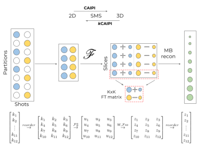 |
46 |
kCAIPI: Reduction of interleaved 3D acquisition into a set of 2D simultaneous multi-slice (SMS) reconstruction problems
Video Permission Withheld
Gilad Liberman, Benedikt Poser
The Fourier Transform (FT) of a vector of N=N1⋅N2 elements is decomposable into N1 FTs of N2-sized vectors followed by N2 FTs of N1-sized vectors, a fact utilized iteratively to produce the Fast FT algorithm. Put in MRI terminology, reconstructing N=k⋅M slices from k-undersampled kz-stacked trajectory can be achieved by FT, followed by solution of the M SMS problems of k slices. This can be used to reduce such 3D reconstruction problems into SMS problems, reducing memory and computational demands. The observation extends to CAIPI patterns. We term this approach kCAIPI.
|
|
4592.
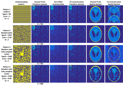 |
47 |
A fast approach for estimation of Spark of the sensing matrix for Compressed Sensing applications.
Bhairav Mehta, Mingrui Yang, Mark Griswold
Compressed sensing (CS) has been extensively used with wide spread application in MRI and other signal processing fields. Spark of the sensing matrix is at the heart of the CS framework for determining the success of the signal recovery for a given designed CS system. However, estimation of Spark of the sensing matrix is a combinatorial process, thus, practically difficult to estimate for realistic sizes of sensing matrices. The purpose of this work is to present a new optimization-problem-based approach for estimation of the Spark of the sensing matrix which will overcome the existing limitations, thereby, a tool to assess and design CS framework based systems.
|
|
4593.
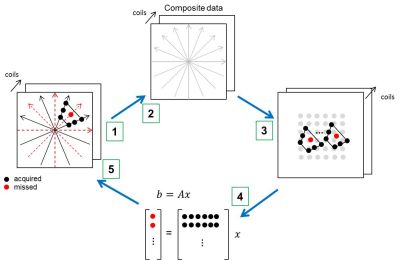 |
48 |
Scalable self-calibrated interpolation of non-Cartesian data with GRAPPA
Seng-Wei Chieh, Mostafa Kaveh, Mehmet Akcakaya, Steen Moeller
Conventional non-Cartesian parallel imaging reconstruction in k-space necessitates large amounts of calibration data for successful estimation of region-specific interpolation kernels. In this work, we propose a self-calibration strategy for obtaining region-specific non-Cartesian interpolation kernels from a single calibration dataset. This enables simple and efficient high-quality reconstruction of non-Cartesian parallel imaging.
|
|
4594.
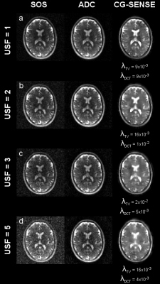 |
49 |
Regularized CG-SENSE for 30-channel 23Na head MRI at 7T
Melanie Schellenberg, Armin Nagel, Peter Bachert, Mark Ladd, Nicolas Behl
23Na MRI provides important information for many pathologies. However, its low SNR entails low spatial resolutions and long acquisition times. The proposed work reconstructs 3D radially undersampled in vivo 30-channel 23Na head data at B0=7T with a sensitivity encoding using a nonlinear conjugate gradient method (CG-SENSE) including a total variation and a discrete cosine transform. With CG-SENSE using iteratively generated Lagrangian coil sensitivities, image quality and contrast within the object are improved compared to sum of squares (SOS) and adaptive combination (ADC) reconstructions.
|
|
| Top |
Artifacts, Implants & Corrections
Digital Poster
Acquisition, Reconstruction & Analysis
Thursday, 16 May 2019
| Exhibition Hall |
09:15 - 10:15 |
| |
|
Computer # |
|
4595.
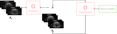 |
51 |
Reconstruction of Undersampled Radial Free-breathing 3D Abdominal MRI using Conditional Generative Adversarial Network
Jun Lv, Jue Zhang
Free-breathing 3D abdominal imaging is challenging since respiratory motion can produce image blurring and ghosting artifact. Our purpose is to employ a novel deep learning method using conditional generative adversarial network (GAN) to reconstruct the undersampled radial 3D abdominal MRI. The whole network combines a generator G consists of 8 convolutional layers and corresponding 8 deconvolutional layers with a discriminator D which is formed using 11 convolutional layers. The GAN-based reconstructed images achieve similar quality to the ground-truth images. Additionally, the average reconstruction time is negligible. Therefore, this method can be adopted for a wide range of clinical applications.
|
|
4596.
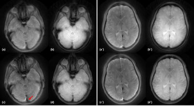 |
52 |
Artifact correction in spiral trajectory with high gradient performance
Daehun Kang, Uten Yarach, Joshua Trzasko, Myung-Ho In, Erin Gray, Ek Tsoon Tan, Nolan Meyer, Thomas Foo, Yunhong Shu, John Huston, Matt Bernstein
A low-cryogen, compact 3T MRI is equipped with high performance gradients, of which increased maximum gradient amplitude and slew rate can improve MR image quality of spiral trajectory to reduce susceptibility and off-resonance effect. However, use of the higher slew rate and gradient strength with an Archimedean spiral trajectory can lead to rotation artifacts and a local blurring. In this work, we corrected those artifacts with using a dynamic field camera and with attention to the azimuthal Nyquist sampling criterion.
|
|
4597.
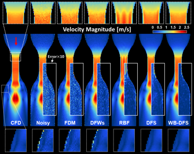 |
53 |
Wall-bounded Divergence-free Smoothing for Denoising of Velocity Data Measured by 4D Flow MRI
Chaehyuk Im, Seungbin Ko, Jeesoo Lee, Jee-Hyun Cho, Doosang Kim, Sang Hyung Lee, Simon Song
Flow data measured by 4D flow MRI often result in inaccurate wall shear stress estimation due to near-wall noise in velocity measurements. We propose wall-bounded divergence-free smoothing (WB-DFS) to denoise the flow data. This method minimizes a residual error under the divergence-free condition for a wall-bounded flow and simultaneously performs data smoothing. The denoising performance of WB-DFS was found to be the best among methods reported in literature. For example, the divergence of the velocity field was reduced by 95.6%. This method is expected to be helpful in accurately estimating wall shear stress in a vascular flow imaging.
|
|
4598.
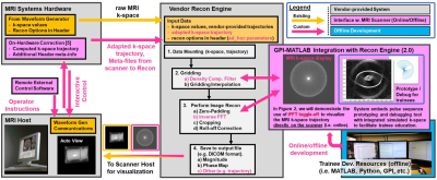 |
54 |
Non-Cartesian MRI Systems Integrated Development using GPI and MATLAB – A New Rosette Pulse Sequence Example
Nanyque Boyd, Alexey Dimov, Amit Patel, Jacob Goes, Hui Wang, Timothy Carroll, Keigo Kawaji
The default MRI scanner systems are not equipped with key technical resources for rapid deployment of novel non-Cartesian pulse sequence approaches. Here, we describe a Graphical Programming Interface-based (GPI) platform that is further embedded into the vendor reconstruction environment. This allows for comprehensive development and validation of new trajectories by integrating on-line MRI systems development with off-line resources such as MATLAB, which also enhances trainee-driven research efforts. This tool offers a set of resources including real-time display of MRI k-space and prototyping/characterization of sampling trajectory corrections that may simplify and streamline these non-Cartesian designs.
|
|
4599.
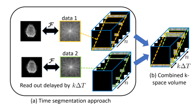 |
55 |
Novel calibration-free correction of field inhomogeneity artifacts in EPI using a structured low rank method
Arvind Balachandrasekaran, Merry Mani, Mathews Jacob
Echo Planar Imaging (EPI) is widely used in many dynamic imaging studies due to its capability to provide very good temporal resolution. However, the off-resonance artifacts due to long read out result in poor correspondence with structural scans and make data interpretation difficult. Here we introduce a novel framework, where the problem of artifact correction is transformed into a recovery of image time series from undersampled measurements. We exploit the exponential structure of the signal at every pixel along with the spatial smoothness of inhomogeneity map to recover the image series. Preliminary results demonstrate the potential of the proposed method.
|
|
4600.
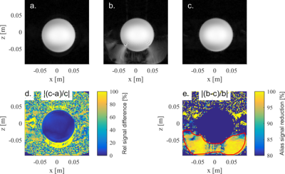 |
56 |
Elimination of fold-in artefacts for gradient inserts by using the existing whole-body gradient in synergy
Edwin Versteeg, Abel Romeijnders, Jeroen Hendrikse, Dennis Klomp, Jeroen Siero
The short encoding field of a gradient insert makes such a coil susceptible to fold-in artefacts, especially when operated along the z-direction. We propose a method that almost completely eliminates this fold-in artefact by using the whole-body z-gradient as pre-winder and gradient insert (also in z) as readout gradient. This causes signal from outside the linear region of the gradient insert to stay dephased, thus suppressing the signal that folds in. The proposed method is validated and quantified in simulation, and in experiments using a lightweight gradient insert that features a short encoding field.
|
|
4601.
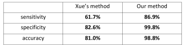 |
57 |
Streak Artifact Suppression in Radial MRI by Automatic Coil Selection
Lexiaozi Fan, Hassan Haji-Valizadeh, Daniel Kim
Streak artifact is very common in radial sampled images. One way we can reduce the artifact is to remove individual streaky coils by visually identification. Although it may not be a hard work, it’s time-consuming, especially when it comes to a large number of images. This abstract aims at developing an algorithm that can automatically detect these streaky coils, and suppress streak artifacts in reconstructed images.
|
|
4602.
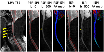 |
58 |
Metal Artifacts Reduction in DWI using Point Spread Function (PSF) Encoding
Sisi Li, Yishi Wang, Zhangxuan Hu, Li Guan, Yong Hai, Xiao Han, Yu Ma, Hanwen Zhang, Xiaolong Chen, Hua Guo
Though metal artifacts have been well-resolved in anatomical imaging by three dimensional multispectral imaging (3D-MSI) methods, diffusion weighted imaging (DWI) near metallic implants still remains a challenge, impeding various clinical applications. Point-Spread-Function encoded EPI (PSF-EPI) combined with Tilted-CAIPI can achieve highly accelerated distortion- and blurring-free high resolution DWI. By using an additional phase encoding, artifacts induced by severe susceptibility inhomogeneity around metal can be reduced even under a high acceleration rate. The reliable performance of PSF-EPI technique in metal artifacts reduction in DWI is demonstrated on phantom, in vitro swine forearm and in vivo patients.
|
|
4603.
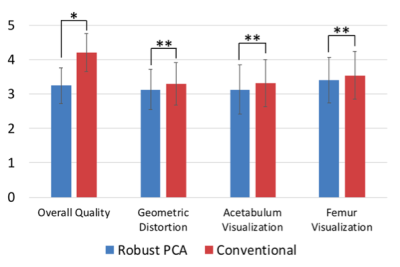 |
59 |
Comparison of Accelerated MAVRIC-SL with Robust-PCA and Conventional MAVRIC-SL in Evaluation of Symptomatic Total Hip Arthroplasties
Zoe Doyle, Daehyun Yoon, Philip Lee, Brian Hargreaves, Christopher Beaulieu, Kathryn Stevens
The substantial reduction of off-resonance artifacts near metal by multi-spectral imaging sequences facilitates the postoperative use of MRI to evaluate total hip arthroplasty patients, but its long scan time can be difficult for patients to tolerate. A novel MAVRIC-SL method using robust principal component analysis (RPCA) recently showed 2.6-fold reduced scan time with comparable artifact suppression. In this study we compare a conventional MAVRIC-SL method with the RPCA-accelerated MAVRIC-SL method in 36 total hip arthroplasty cases. Our data demonstrate nearly equivalent clinical sensitivity of the RPCA MAVRIC-SL method to the conventional method with a mild loss of spatial resolution.
|
|
4604.
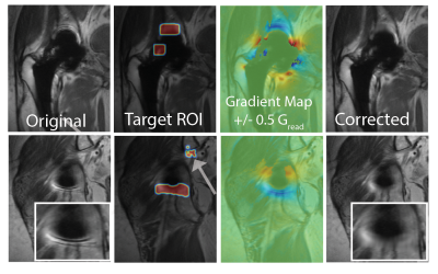 |
60 |
Pileup Artifact Correction Near Metal Implants Using Deep Neural Networks and Spectral K-Space Modulation
Kevin Koch, Robin Karr, Andrew Nencka
Three-dimensional multispectral imaging (3D-MSI) techniques used for metal artifact correction can provide relatively clear images near most metallic implants. However, within localized regions near some implants, 3D-MSI demonstrate residual artifacts that are unlike any other artifact previously seen in MR images. These confounding features in 3D-MSI are known as “pileup” or “ring” artifacts. In this study, we present a novel approach to residual artifact correction in 3D-MSI that relies on 1) deep neural networks, 2) physical modeling of local gradients, and 3) k-space modulation and replacement of spectral data in compromised regions
|
|
4605.
 |
61 |
2D Imaging near Metallic Implants at 0.5T using High Time-Bandwidth Product RF pulses
Chad Harris, Andrew Curtis, Ian Connell, Philip Beatty, Jeff Stainsby, Curtis Wiens
There are several benefits to reducing the main magnetic field strength including a reduction of imaging artefacts near metallic implants and the ability to significantly increase the peak B1+ of the RF pulses due to the reduction in SAR penalty. This enables higher time-bandwidth product (TBP) for a given RF pulse duration. In this work, we utilized high TBP RF pulses on a high-efficiency transmit coil and a 0.5T MR system to reduce through-plane distortions caused by metallic implants. In addition to characterizing through-plane distortions, the impact of these pulses on in-plane distortions and SAR were also characterized.
|
|
4606.
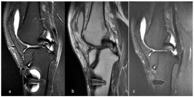 |
62 |
Clinical application of MAVRIC-SL in reducing metal implant artifacts in anterior cruciate ligament reconstruction
Presentation Not Submitted
Jingyi Zhu, Songbai Li, Fei Bie, Lizhi Xie
Some anterior cruciate ligament reconstructions have metal implants, and metal scrap may remain in the surgical procedure. Metals in the conventional magnetic resonance sequence, especially in the fat suppression sequences, produce large artifacts that affect the observation of the surrounding structure. This study performed conventional sequences and MAVRIC-SL sequence scan for patients with metal implants after ACLR and analysis the images. Conclusions that the oblique sagittal MAVRIC-SL PDWI FS sequence can be used to assisting in the diagnosis of traditional oblique sagittal T2WI FS and PDWI sequence.
|
|
4607.
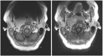 |
63 |
Novel use of the MAVRIC metal artifact reduction technique in MRI of the brain
Nathaniel Swinburne, Regina Loccisano, Duane Nicholson, Norbert Ward, Nayna Patel, Robert Young
While multiacquisition variable-resonance image combination (MAVRIC) is a recognized technique for metal artifact reduction in muskuloskeletal MRI, it is not widely described for MRI of the central nervous system. We investigate the value of this technique for MRI of the brain in patients with MR conditional metal implants and find that MAVRIC-T1 is superior to conventional FSE T1 in both qualitative and quantitative metrics.
|
|
4608.
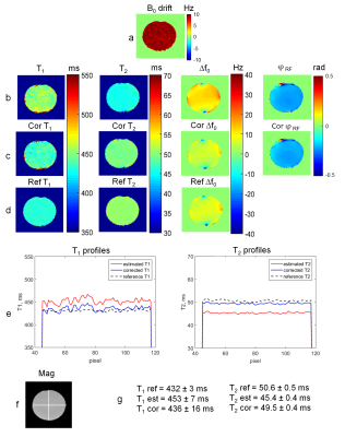 |
64 |
The influence of B0 drift on the performance of the PLANET method and an algorithm for correction
Yulia Shcherbakova, Cornelis van den Berg, Chrit Moonen, Lambertus Bartels
The PLANET method has been recently proposed to quantify the relaxation parameters T1 and T2, the banding free magnitude, the local off-resonance ?f0, and the RF phase from RF phase-cycled balanced steady-state free precession (bSSFP) data. The PLANET model requires a static B0 field over the course of the acquisition. However, due to gradient activity, B0 drift can happen. In this work we present a study of the influence of B0 drift on the performance of the method and we propose a strategy for correction.
|
|
4609.
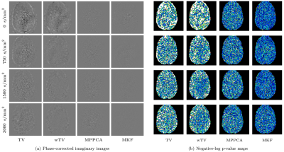 |
65 |
Gaussianization of Diffusion MRI Magnitude Data Using Spatially Adaptive Phase Correction
Feihong Liu, Geng Chen, Jun Feng, Pew-Thian Yap, Dinggang Shen
We present a method of effective phase-correction of diffusion-weighted images with the goal of obtaining real-valued signals with zero-mean Gaussian distributed noise. Our method estimates the noise level locally and is hence well-suited for spatially-varying noise.
|
|
4610.
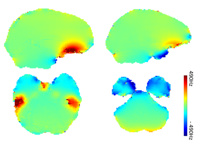 |
66 |
Eliminating susceptibility induced hyperintensities in ultra highd field T1w MPRAGE brain images
Ruoyun Ma, Thomas Henry, Pierre-François Van de Moortele
Ultra high field brain MPRAGE images are commonly affected by local susceptibility induced hyperintensities, which are pronounced in inferior frontal lobe and inferior temporal lobe. In this work, we propose a straightforward approach by applying a frequency offset of 300Hz and widening the bandwidth by 40% to the hyperbolic secant inversion pulse provided by the standard MPRAGE sequence, to eliminate this artefact without introducing additional incomplete inversion through the brain. This approach was tested across different subjects and proven to have robust performance in artefact elimination against variable local frequency offsets.
|
|
4611.
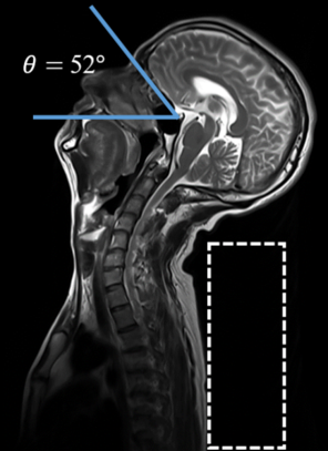 |
67 |
B0 shim improvement in the inferior frontal lobe by head-tilting: feasibility and comparison with 3rd order shimming
Seulki Yoo, Hayoung Song, Won Mok Shim, Seung-Kyun Lee
Susceptibility-induced signal dropout and image quality impairment in the gradient-echo based imaging are well known problems in brain MRI at high fields. Here, we experimentally demonstrate the feasibility and benefit of head-tilted brain scan as a means to reduce B0 inhomogeneity and associated gradient echo signal loss in the inferior frontal lobe (IFL), and compare the shim improvement with simulated 3rd order shimming in the whole brain.
|
|
4612.
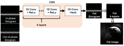 |
68 |
Water-fat separation in spiral readout acquisition for the liver using the convolutional neural network: an approach to reduce blurring artifacts
Daiki Tamada, Ryoichi Kose, Katsumi Kose, Hiroshi Onishi, Utaroh Motosugi
Water/fat separation algorithm for two-echo spiral acquisition in the liver was developed using the convolutional neural network (CNN). The processing in the CNN was performed in the sinogram domain. A Bloch simulator was used to simulate the phase error in the k-space caused by the off-resonance components of background and fat. A volunteer study showed the successful water/fat separation using the proposed method without additional echoes of reference scans.
|
|
4613.
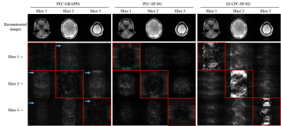 |
69 |
Simultaneous multislice EPI reconstruction by incorporating split slice-GRAPPA with slice-dependent 2D Nyquist ghost correction (PEC-SP-SG)
Yilong Liu, Markus Barth, Mengye Lyu, Ed X. Wu
Simultaneous multislice (SMS) EPI reconstruction is challenging due to slice-dependent 2D phase differences between opposite polarities, which is collapsed across slices. Additionally, slice leakage is one major concern in some applications including diffusion and functional MRI. The proposed SMS EPI reconstruction incorporates phase error correction with split slice-GRAPPA (PEC-SP-SG), and was evaluated using simulation, phantom and in vivo experiments. Results show that the proposed approach can offer a robust SMS EPI reconstruction with slice-dependent 2D Nyquist ghost correction, and provide a balance between slice leakage and in-plane artifacts.
|
|
4614.
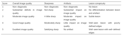 |
70 |
Detection and Correction of MR EPI Data Corrupted by Spike Noise
Ziyi Pan, He An, Elaine Lee, Hua Guo
EPI-based MR images are prone to the spike artifact, which is usually caused by small electrical discharges i.e. sparks that emit radio frequency power within the bandwidth of the scanner receiving system 1 during the MR acquisition. Spike noise causes ripples or stripes covered on the object and can hamper the qualitative or quantitative analysis of the MR images. In this work, we developed a reliable technique that combines Robust Principal Component Analysis 2 (RCPA) with median filtering to robustly detect and correct spike-affected images in human pelvic diffusion weighted imaging (DWI). The overall image quality and the lesion conspicuity were improved after spike removal.
|
|
4615.
|
71 |
Cascaded Deep Learning Networks for Automated Image Quality Evaluation of Structural Brain MRI
SHEEBA SUJIT, REFAAT GABR, IVAN CORONADO, PONNADA NARAYANA
Visual quality assessment of MRI is subjective and impractical for large datasets. In this study, we present a cascaded convolutional neural network (CNN) model for automated image quality evaluation of structural brain MRI. The multisite Autism Brain Imaging Data Exchange dataset of ~1000 subjects was used to train and evaluate the proposed model. The model performance was compared with expert evaluation. The first network rated individual slices, and the second network combined the slice ratings into a final image score. The network achieved 74% accuracy, 69% sensitivity, and 74% specificity, demonstrating that deep learning can provide robust image quality evaluation.
|
|
4616.
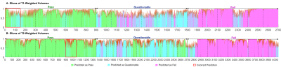 |
72 |
Automatic Quality Assessment of Pediatric MRI via Nonlocal Residual Neural Networks
Siyuan Liu, Kim-Han Thung, Weili Lin, Pew-Thian Yap, Dinggang Shen, UNC/UMN Baby Connectome Project Consortium
Manual MRI quality assessment is time-consuming, subjective, and error-prone. We show that image quality of contrast-varying pediatric MR images can be automatically assessed using deep learning with near-human accuracy.
|
|
4617.
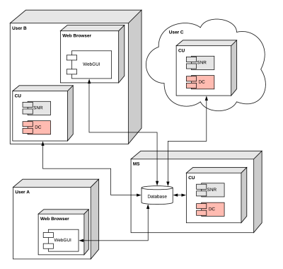 |
73 |
MR Optimum – A web-based application for signal-to-noise ratio evaluation.
Eros Montin, Roy Wiggins, Kai Tobias Block, Riccardo Lattanzi
The signal-to-noise ratio (SNR) is a commonly used metric to evaluate image quality and radiofrequency coil performance in MRI. However, its calculation could be challenging. Here we introduce MR Optimum, a novel web-based application for the evaluation of SNR. By means of a user-friendly web GUI, readily available via any internet browser, it provides access to various methods for SNR calculation. The computing unit can be installed on a local server or distributed over the cloud. Results can be visualized, analyzed and exported in various formats. MR Optimum could help standardizing how SNR is calculated and reported in scientific publications.
|
|
4618.
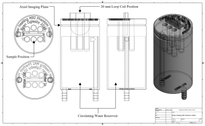 |
74 |
An Investigation into the Origins of an MRI Artifact Induced by Increasing Temperature
Ryan Oglesby, Wilfred Lam, Greg Stanisz
An artifact has been observed in pure water samples after increasing the temperature above 25 °C, regardless of the heating mechanism. This study aims to determine the origins of the temperature-induced artifact by using MR thermometry and T1 relaxation to investigate samples containing increasing concentrations of agar. The addition of a small concentration (0.1%) of agar eliminates the temperature artifact suggesting that the increased viscosity of the samples decreases convection currents. Moreover, this addition of a small concentration of agar study provides a practical means of experimentally scanning samples at physiological temperature.
|
|
4619.
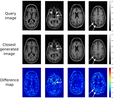 |
75 |
General Abnormality Detection in MR Images using a Generative Adversarial Network
Karsten Sommer, Michael Helle, Axel Saalbach, Marvin Rühe, Heinrich Schulz
In this study, a Generative Adversarial Network is used for detection of abnormalities in MR brain images such as lesions, artifacts etc. Given a query image, a generative model that is trained to create normal appearing brain images is used to find a best match. Since abnormalities cannot be reproduced accurately by the generative model, pathologies and artifacts become apparent.
|
|
| Top |
New RF & Gradient Strategies
Digital Poster
Acquisition, Reconstruction & Analysis
Thursday, 16 May 2019
| Exhibition Hall |
09:15 - 10:15 |
| |
|
Computer # |
|
4620.
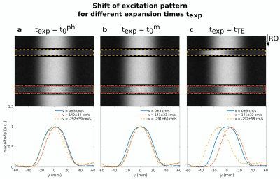 |
76 |
On the Effective Centre of Excitation and the Point of Gradient Moment Expansion for 2D-Selective Excitation in the Presence of Flow
Clarissa Wink, Simon Schmidt, Jean Pierre Bassenge, Sandy Szermer, Giulio Ferrazzi, Bernd Ittermann, Tobias Schaeffter, Sebastian Schmitter
In this work, we demonstrate the distinction and importance of two virtual time points during excitation for correct flow compensation and quantification: the centre of excitation (tm0t0m)
at which spins are excited and thus magnitude is generated, and the isophase time-point (tph0t0ph)
at which all excited spins are in phase. A general method to determine tm0t0m is
presented and tph0t0ph and tm0t0m are
shown to be not necessarily identical. Finally, phantom experiments demonstrate that the knowledge of tm0t0m is
required to remove the displacement artefact in phase-encoding directions to enable correct flow compensation and imaging.
|
|
4621.
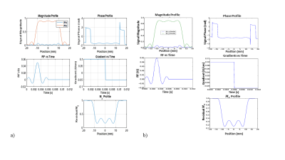 |
77 |
Quantum-Inspired RF Pulse Optimization
Sherry Huang, Darryl Jacob, Michael Beverland, Stephen Jordan, Helmut Katzgraber, Matthias Troyer, Rasim Boyacioglu, Yun Jiang, Dan Ma, Mark Griswold, Julie Love, Debra McGivney
RF pulse design is crucial in creating the desired magnetization profile which is the basis of Magnetic Resonance Imaging. There are various methods to generate the RF pulse and gradient waveforms based on Fourier relationships, filter design, or optimizations. These methods rely on assumptions and approximations due to computational power constraints. Here we present preliminary results of using quantum inspired algorithms for Bloch simulation and RF pulse design optimization.
|
|
4622.
 |
78 |
Improved Accuracy of FLASH-based B1+ Mapping by Optimization of the Fourier Encoding Matrix
Omer Oran, L. Klassen, Ravi Menon
FLASH-B1 mapping with a Fourier-encoding scheme works only for a limited range of flip-angles due to pronounced saturation effects that occur for short TR. The transmit voltage can be adjusted to satisfy this requirement in problematic Fourier combinations that have high dynamic range. However, this results in an unnecessary reduction of the dynamic range in combinations that are already within the accepted range. This study addresses this problem by optimizing the Fourier encoding matrix such that the flip-angle is reduced only in regions of high flip-angle in the problematic combinations.
|
|
4623.
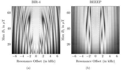 |
79 |
BEEEP: B1-robust Energy Efficient Excitation Pulses
Eric Van Reeth, Hélène Ratiney, Olivier Beuf, Soukaina Kanice, Steffen Glaser, Dominique Sugny
This study introduces a new family of broadband B1-robust excitation (90°) pulses for MRI with large enough bandwidth (+/- 1 kHz) to account for static field inhomogeneities, and minimal energy deposition. RF pulses are designed with a regularized optimal control algorithm, which is able to adapt the pulse B1-robustness range to fit the coil limits in terms of peak amplitude and energy. In vitro acquisitions using an endoluminal-shaped RF transmit coil show comparable excitation profiles than BIR4 pulses, although BEEEP pulses deposit 5.2 times less energy.
|
|
4624.
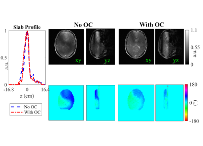 |
80 |
A Simple Method for Constrained Optimal Control RF Pulse Design
Sydney Williams, Jon-Fredrik Nielsen, Jeffrey Fessler, Douglas Noll
Optimal control (OC) methods for RF pulse design are useful in cases where the small-tip angle (STA) approximation is violated. Furthermore, designs with physically meaningful constraints (e.g., RF peak amplitude and integrated power) eliminate the need for parameter tuning to create realizable pulses. In this abstract we introduce a constrained fast OC method that easily generalizes to a variety of RF pulse designs. We demonstrate with examples of SMS and spectral prewinding pulses in simulation and in vivo. The constrained fast OC method guarantees that RF pulses will meet physical constraints while outperforming their non-OC counterparts.
|
|
4625.
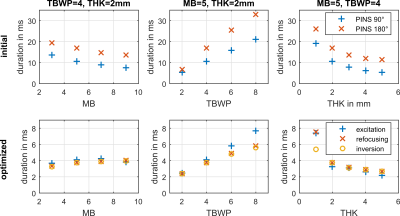 |
81 |
RF pulse design via time optimal control for combined excitation, refocusing and inversion
Christoph Aigner, Armin Rund, Christina Graf, Karl Kunisch, Rudolf Stollberger
This work demonstrates a constrained joint design of minimum duration RF pulse and slice selective gradient waveforms for combined SMS excitation, refocusing and inversion scenarios. A hybrid trust-region semismooth Newton/quasi-Newton method with exact derivatives via adjoint calculus is used to solve the time optimal problem on fine spatial and temporal grids. Specific hardware and safety constraints, including maximal RF, slice selective gradient, slew rate amplitudes as well as global SAR estimates, guarantee practical applicability. High-resolution GRE, crushed SE and inversion recovery GRE slice profile measurements on a 3T MR system validate the numerical results.
|
|
4626.
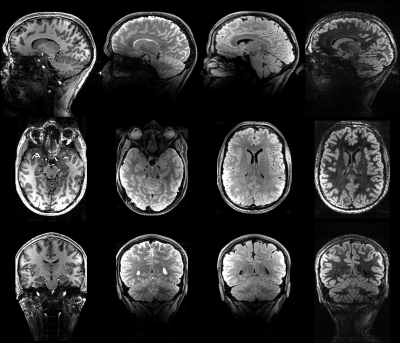 |
82 |
PASTeUR: Package of Anatomical Sequences using parallel Transmission UniveRsal kT-point pulses
Vincent Gras, Franck Mauconduit, Alexandre Vignaud, Caroline Le Ster, Lisa Leroi, Alexis Amadon, Eberhard Pracht, Markus Boland, Rüdiger Stirnberg, Tony Stöcker, Benedikt Poser, Christopher Wiggins, Xiaoping Wu, Kamil Ugurbil, Nicolas Boulant
Despite its power to counteract the inevitable radiofrequency field inhomogeneity problem at ultra-high field, parallel transmission has failed to be embraced by the community in routine due to a cumbersome workflow. Universal pulses have shown great potential to circumvent this problem by providing plug and play solutions. Here we validate a package of 3D anatomical sequences for a given commercial coil covering multiple contrasts for use in clinical routine and including, thanks to their versatility, very few pulse solutions. The utilization of universal kT-points enables direct embedding of these pulses in the sequences and easy handling of the power/SAR limits.
|
|
4627.
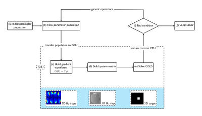 |
83 |
3D Selective RF and Gradient Waveforms designed by using a GPU Accelerated Genetic Algorithm
Christopher Mirfin, Paul Glover, Richard Bowtell
The Genetic Algorithm (GA) is motivated by the process of natural selection, allowing mutliple initialisations. Due to the stochastic nature of genetic algorithms they are beneficial in avoiding local minima, although they can require significantly more function evaluations to run than a traditional solver. In this work, motivated by the field of shape optimisation, an approach is taken to perform the joint design of RF and gradient waveforms using a GA with a GPU-accelerated iterative solver.
|
|
4628.
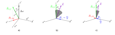 |
84 |
Spin Lock Adiabatic Correction (SLAC) of BIR4 pulses for increased B1-insensitivity at 7T
Edward Green, Yasmin Blunck, James Korte, Bahman Tahayori, Peter Farrell, Leigh Johnston
Inhomogeneous B1 excitation impedes image quality, particularly at high field. Adiabatic pulse modulation ameliorates this effect, however super-adiabatic properties can be exploited to further improve performance. Spin Lock Adiabatic Correction (SLAC) pulses can be applied to any adiabatic pulse shape, through reduction of flip angle inaccuracies induced by B1 variability. In this work, SLAC is derived for BIR4 pulse shapes, and the superior performance of SLAC-BIR4 is demonstrated in both simulation and phantom experiments at 7T. The SLAC procedure is an attractive analytical alternative to numerical optimisation of adiabatic pulses.
|
|
4629.
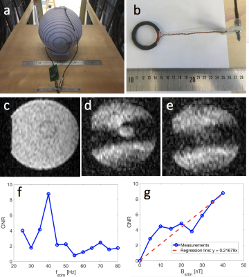 |
85 |
Sensing low-frequency, low-amplitude AC magnetic fields at ultra-low field with steady-state SIRS
Bragi Sveinsson, Neha Koonjoo, Thomas Witzel, Matthew Rosen
In this work, we demonstrate a method to detect low-frequency, low-amplitude AC magnetic fields in an ultra-low-field (ULF) MRI system using a steady-state implementation of the Stimulus-Induced Rotary Saturation (SIRS) method. The method optimizes SNR efficiency by applying the SIRS mechanism in a bSSFP scan. This approach takes advantage of the low SAR and small absolute B0 deviations of the ULF system. We describe simulation results, show a clear signal response in phantoms, and describe an in vivo protocol for using the method to detect response from an auditory stimulus.
|
|
4630.
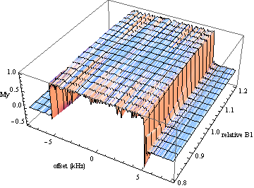 |
86 |
PAREWISE: High-bandwidth, reduced-rephase, slice-selective excitation pulses for high-field spectroscopy
James Murdoch
To meet the localization needs of high-field spectroscopy, new high-bandwidth computer-optimized RF excitation pulses have been generated, featuring phase modulation, t=0 points near the end of the pulse duration, B1 insensitivity over a ±15% range, and sharp excitation profiles. In the design process, a maximum amplitude of 1 kHz was imposed, and bandwidth was increased by stepping up the pulse length from 5 to 11 ms. Both 90° and 60° flip angles have been investigated thus far.
|
|
4631.
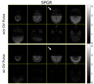 |
87 |
3D Inner Volume Imaging with 3D Tailored Outer Volume Suppression RF pulses
Tianrui Luo, Douglas Noll, Jon-Fredrik Nielsen
3D inner-volume (IV) steady-state imaging is a candidate for, e.g., high-resolution BOLD fMRI, but it can be challenging to achieve sufficient outer-volume (OV) signal suppression. This is particularly true for 3D tailored RF pulses that excite an arbitrary 3D IV (e.g., a cylinder of finite height) thus enabling fast non-Cartesian readouts, as 3D IV pulses are more difficult to design than 2D tailored pulses. We propose to insert a 3D tailored OV suppression pulse to help suppress OV steady-state signal in 3D IV imaging sequences that use 3D tailored IV excitation pulses. We show that this strategy can substantially improve the IV signal profile for commonly used and emerging steady-state sequences such as spoiled gradient-echo (SPGR), balanced SSFP, and small-tip fast-recovery (STFR).
|
|
4632.
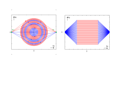 |
88 |
Spiral RARE with annular segmentation
Juergen Hennig, Marius Menza, Antonia Barghoorn, Bruno Riemenschneider, Stefan Kroboth, Maxim Zaitsev
We present a new approach to spiral RARE with annular segmentation. Annular segmentation leads to monotonous T2-dependent weighting of signal amplitudes across k-space and thus to very benign artifact behavior. Preliminary results show that single shot images (128x128) of decent quality can be acquired without fat suppression and without field inhomogeneity correction. By cyclic shifting of the spiral segments quantitative T1- and/or T2- images can be acquired in a few seconds. Sequence implementation was performed swiftly and efficiently in MatLab with the vendor independent Pulseq sequence development environment.
|
|
4633.
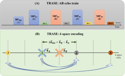 |
89 |
A Novel Approach to Investigate 1D TRASE MRI Pulse Sequence Performance in Imperfect B1 Fields
Pallavi Bohidar, Hongwei Sun, Gordon Sarty, Jonathan Sharp
The Transmit Array Spatial Encoding (TRASE) MRI technique uses transmit radio-frequency (RF) magnetic field (B1) phase gradients for spatial encoding. Imaging performance is reliant on |B1| homogeneity. This study investigates the performance of a set of variants of 1-dimensional TRASE under conditions of |B1| inhomogeneity. Both Bloch equation simulations and experimental confirmations are presented. Results show that one variant of the sequence allows imaging over ~90% of the FOV for Δ|B1| = ± 15% allowing the use of practical B1 transmit coil designs. This improves upon the previous sequence which generated ~75% usable field-of-view (FOV).
|
|
4634.
 |
90 |
AZTEK: Adaptive Zero TE K-space trajectories
Tanguy Boucneau, Brice Fernandez, Anne Menini, Florian Wiesinger, Luc Darrasse, Xavier Maître
Because of short signal lifetimes and respiratory motion, 3D MR lung imaging is still challenging today. Zero TE (ZTE) pulse sequences are promising as they overcome the problem of short T2*. Nevertheless, because of the continuous readout gradients they require, their k-space trajectories are non-optimal for retrospective gating. We propose AZTEK, a 3D radial trajectory featuring several tuning parameters to adapt the acquisition to any moving organ while keeping a smooth transition between consecutive spokes. The increase in image quality was validated with static and moving phantom experiments, and demonstrated with dynamic thoracic imaging performed on a human volunteer.
|
|
4635.
 |
91 |
Redesigned Variable-Density Cones Trajectory for High Resolution MR Imaging
Kwang Eun Jang, Srivathsan Koundinyan, Dwight Nishimura, Shreyas Vasanawala
The 3D cones trajectory has been employed for various applications. In this work we transform the task of designing the cones trajectory into a generic procedure of discretizing an analytic coordinate. We present a new discretization scheme with a spiral path on a unit sphere, which enables allocation of readout interleaves on distinct conic surfaces for any given number of readouts. Subsequently, we derive the relationship between the sampling density of each interleaf and that of overall interleaves, which allows us matching the sampling density of the cones trajectory to the 1/f-model of 3D images.
|
|
4636.
 |
92 |
Silent Structural Imaging and T1-mapping with a Rapid-Radial Twice-Prepared (R2P2) Sequence
Emil Ljungberg, Florian Wiesinger, Tobias Wood
We combined the MP2RAGE sequence with a silent radial ZTE readout and acquired a high-contrast, high-SNR T1-weighted image and quantitative T1 map at 0.9mm isotropic resolution at 3T free from B1-inhomogeneity. This has potential for high resolution structural imaging of populations that would not otherwise tolerate MRI due to the acoustic noise of standard sequences.
|
|
4637.
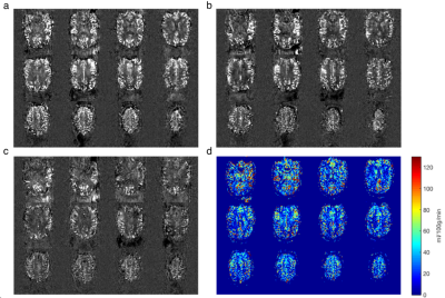 |
93 |
Improving pseudo-continuous arterial spin labelling at ultra-high field using a VERSE-guided parallel transmission strategy
Yan Tong, Peter Jezzard, Thomas Okell, William Clarke
Implementing ASL at ultra-high field is challenging due to increased specific absorption rate (SAR), along with B1+ and B0 inhomogeneity. Parallel transmission (pTx) provides additional degrees of freedom to mitigate B1+ inhomogeneity. Among various pTx strategies, RF shimming is a simple formulation that modulates the complex weights of each RF channel. In this study, we explored the possibility of using VERSE to further improve PCASL at 7T, and VERSE-guided RF shimming was shown to achieve improved SNR in perfusion-weighted images over power-matched Circularly-Polarised (CP) mode and RF-shimmed Gaussian labelling schemes.
|
|
4638.
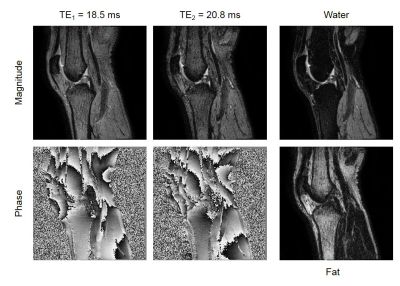 |
94 |
Quiet Dixon Imaging with Looping Star Sequence
Holger Eggers, Kay Nehrke, Peter Börnert, Johan van den Brink
Dixon imaging with a conventional bipolar multi-gradient-echo sequence is often loud, mainly because of rapid switching of the strong readout gradient. In this work, the feasibility of using the Looping Star sequence instead is explored, which was recently introduced for quiet radial multi-gradient-echo imaging. Different variants of a dual-acquisition Looping Star sequence are proposed and demonstrated to allow a robust water-fat separation in phantom and volunteer experiments.
|
|
4639.
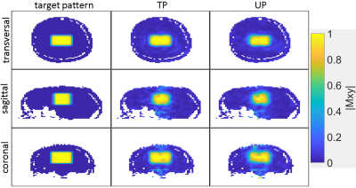 |
95 |
Universal Parallel Transmit Pulse Design for 3-Dimensional Local-Excitation – A 9.4T Simulation Study
Ole Geldschläger, Tingting Shao, Anke Henning
This study introduces a parallel-transmission (pTx) radio-frequency (RF) pulse-design-method to create an universal pTx RF-pulse that excites the same 3-dimensional local excitation pattern with a desired flip-angle in different human heads at 9.4T. Thus, it prospectively abandons the need for time-consuming subject specific B1+ mapping and pTx-pulse calculation during the scan session. The resulting universal pulses created magnetization profiles with an only marginal worse Normalized-Root-Mean-Square-Error (NRMSE) compared to the magnetization profiles produced by the pulses tailored to individual heads.
|
|
4640.
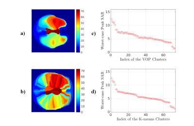 |
96 |
A K-means Clustering Algorithm for MRI Virtual Observation Points Compression in Local SAR Supervision
Xianglun Mao, Joseph Rispoli, David Love
Virtual observation point (VOP) compression has become a standard technique to address SAR and temperature constraints in MRI parallel transmission (pTx) design. SAR matrices need to be pre-averaged for the regions of interest, and finally be conservatively compressed to a much smaller set of SAR matrices (i.e. VOPs) that is still capable of reliably calculating a peak spatial SAR. We demonstrated a new approach that used a k-means algorithm for VOP compression. The new VOP compression method does not yield any under-estimation but allows for a lower over-estimation in the peak local SAR prediction.
|
|
4641
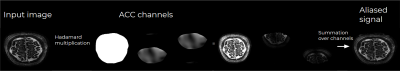 |
97 |
Aliased Coil Compression
Video Permission Withheld
Gilad Liberman, Benedikt Poser
Cartesian sub-sampling patterns play a major role in routine MRI, usually reconstructed using GRAPPA or SENSE and image based regularization. Coil compression is commonly applied to reduce computational load and noise. Software coil compression achieves only mediocre compression factors without compromising signal. Geometrical/ESPIRiT coil-compression use fully-sampled axes, when availables, to improve compression factors without reducing signal or reconstruction level. In this work we present Aliased Coil Compression for Cartesian subsampling patterns, achieving optimal compression without any signal loss. The method is especially useful for alleviating fast image-domain regularization (compressed sensing or deep learning) for available sequences.
|
|
4642.
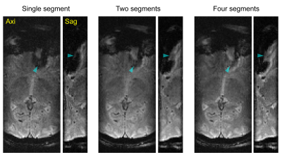 |
98 |
Zoomed 3D GRE EPI utilizing a Segmented 2D Selective RF Pulse Excitation
Naoharu Kobayashi, Michael Mullen, Kamil Ugurbil, Michael Garwood
Segmented 2D selective RF pulse excitation is introduced in 3D-EPI for zoomed imaging. The feasibility of the 2D selective pulse segmentation was tested in phantom and in vivo brain measurements. The segmented pulse excitation provided nearly identical excitation profile to a non-segmented pulse excitation. In zoomed in vivo brain imaging, the segmented pulse excitation showed conspicuous improvement of susceptibility artifacts around the frontal sinus.
|
|
4643.
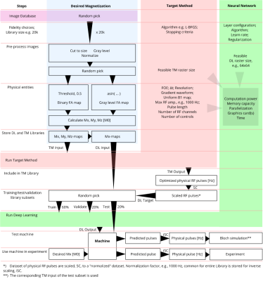 |
99 |
Rapid multi-dimensional RF pulse design with deep learning
Mads Vinding, Birk Skyum, Ryan Sangill, Torben Lund
For multi-dimensional RF pulses, neural networks and deep learning may boost the clinical applicability by allowing very rapid pulse predictions, based on offline training and offline generated training libraries. This can potentially offer opportunities, for example, to revive slow, abandoned pulse design techniques, or to include many more constraints or complexities into the pulse designs that until now were infeasible to bring into a clinical setting, since the neural network will simply learn the features of the training library. We are demonstrating the principle with numerical simulations, and phantom and in vivo experiments.
|
|
| Top |
Machine Learning for Image Reconstruction: A New Frontier
Digital Poster
Acquisition, Reconstruction & Analysis
Thursday, 16 May 2019
| Exhibition Hall |
09:15 - 10:15 |
| |
|
Computer # |
|
4644.
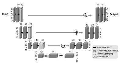 |
101 |
An Unsupervised Deep Learning Approach for Reconstructing Arterial Spin Labeling Images from Noisy Data
Kuang Gong, Paul Han, Debra Horng, Georges El Fakhri, Chao Ma, Quanzheng Li
Recently convolutional neural networks (CNNs) have been successfully applied to computer vision tasks and attracted growing interests in medical imaging. One barrier for the application of deep neural networks is the need of large amounts of training pairs, which are not always available in clinical practice. Inspired by the deep image prior method, this work presents a new image reconstruction framework based on CNN representation where no training pairs and pre-training are needed. We demonstrate the effectiveness of the proposed method by performing denoising and image reconstruction using noisy arterial spin labeling (ASL) data with and without undersampling.
|
|
4645.
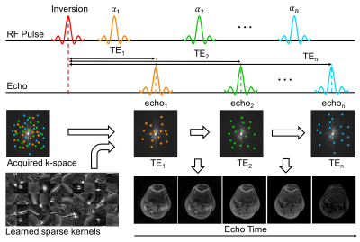 |
102 |
Unsupervised Learning for Improved Fidelity Multi-contrast MRI
Ke Wang, Frank Ong, Jonathan Tamir, Michael Lustig
Multi-contrast MRI acquisitions from a single scan have tremendous potential to streamline exams and reduce imaging time. However, maintaining clinically feasible scan times necessitates significant undersampling, pushing the limits on compressed sensing and other low-dimensional techniques. While learning methods have been proposed to overcome this limitation, they rely on fully sampled data for training, which are difficult to obtain for multi-dimensional imaging. Here, we present an unsupervised learning approach based on convolutional sparse coding, which learns a structured convolutional dictionary directly from undersampled k-space datasets. We apply the proposed method to T2 Shuffling knee datasets and demonstrate improvements to image sharpness and relaxation dynamics compared to the locally low-rank reconstruction.
|
|
4646.
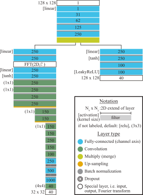 |
103 |
Signal Stability and Sensitivity of Referenceless Reconstructions by a Neural Network in Simultaneous Multi-Slice Imaging
Klaus Eickel, Martin Blaimer, Matthias Günther
A deep neural network for reconstruction of SMS data without the need of additional reference data to calibrate for the spatial encoding information of the multi-coil receiver is presented. Noise-propagation through the reconstruction process is investigated in form of g-factors. Simulations with pseudo-multiple replicas showed robustness and stability of this new method. In addition, the sensitivity for physiological signal variations of this approach is considered in BOLD-signal dynamics. Results are compared to conventional methods like split slice-GRAPPA.
|
|
4647.
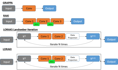 |
104 |
LORAKI: Reconstruction of Undersampled k-space Data using Scan-Specific Autocalibrated Recurrent Neural Networks
Tae Hyung Kim, Pratyush Garg, Justin Haldar
We introduce LORAKI, a novel MRI reconstruction method that bridges two powerful existing approaches (LORAKS and RAKI). Like RAKI (a deep learning extension of GRAPPA), LORAKI trains a scan-specific autocalibrated convolutional neural network (which only relies on autocalibration data, and does not require external training data) to interpolate missing k-space samples. However, unlike RAKI, LORAKI is based on a recurrent convolutional neural network architecture that is motivated by the iterated convolutional structure of a certain LORAKS algorithm. LORAKI is very flexible and can accommodate arbitrary k-space sampling patterns. Experimental results suggest LORAKI can have better reconstruction performance than state-of-the-art methods.
|
|
4648.
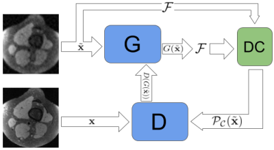 |
105 |
Unpaired Super-Resolution GANs for MR Image Reconstruction
Ke Lei, Morteza Mardani, Shreyas Vasawanala, John Pauly
While undersampled MRI data is easy to obtain, lack of high-quality labels for dynamic organs impedes the common supervised training of deep neural nets for MRI reconstruction. We propose an unpaired training super-resolution model with pure GAN loss to use a minimal amount of labels but all available low-quality data for training. Leveraging Wasserstein-GANs with gradient penalty followed by a data-consistency refinement high-quality Knee MR images are recovered from 3-fold undersampled single coil measurements using 20% of the labels compared with a paired training model.
|
|
4649.
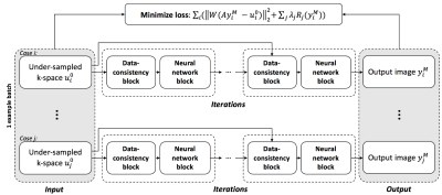 |
106 |
Semi-Supervised Learning for Reconstructing Under-Sampled MR Scans
Feiyu Chen, Joseph Cheng, John Pauly, Shreyas Vasanawala
Supervised deep-learning approaches have been applied to MRI reconstruction, and these approaches were demonstrated to significantly improve the speed of reconstruction by parallelizing the computation and using a pre-trained neural network model. However, for many applications, ground-truth images are difficult or impossible to acquire. In this study, we propose a semi-supervised deep-learning method, which enables us to train a deep neural network for MR reconstruction without using fully-sampled images.
|
|
4650.
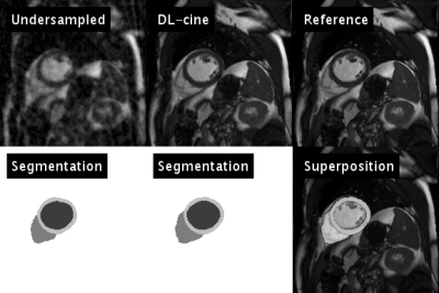 |
107 |
Deep learning aided compressed sensing for accelerated cardiac cine MRI
Tobias Wech, Thorsten Bley, Herbert Köstler
A reconstruction technique for accelerated functional cardiac MRI is presented that exploits a convolutional neural network trained for semantic segmentation of undersampled data. The idea is inspired by the experience that the human eye is capable of distinguishing between typical undersampling artifacts and cardiac shape and/or motion, even for high acceleration factors. The temporal courses of the segmentations determined by the network are used for an efficient sparsification within a compressed sensing algorithm.
|
|
4651.
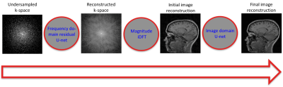 |
108 |
W-net: A Hybrid Compressed Sensing MR Reconstruction Model
Roberto Souza, Richard Frayne
Compressed sensing (CS) magnetic resonance (MR) imaging acquisitions reduce MR exam times by decreasing the amount of data acquired during acquisition, while still reconstructing high quality images. Deep learning methods have the advantage of reconstructing images in a single step as opposed to iterative (and slower) CS methods. Our proposal aims to leverage information from both k-space and image domains, in contrast to most other deep-learning methods that only use image domain information. We compare our W-net model against four recently published deep-learning-based methods. We achieved second best results in the quantitative analysis, but with more visually pleasing reconstructions.
|
|
4652.
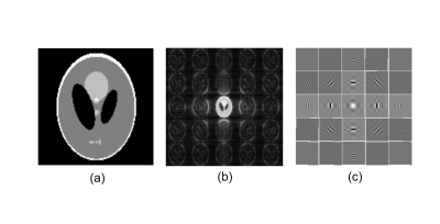 |
109 |
Deep Scaled Domain Learning for Compressed MRI using Optional Scaling Transform
Satoshi ITO, Kohei SATO
Image domain learning designed for image denoiser has superior performance when aliasing artifacts are incoherent; however, its performances will be degraded if the artifacts show small incoherency. In this work, a novel image domain learning CNN is proposed in which images are transformed to scaled space to improve the incoherency of artifacts. Simulation and experiments showed that the quality of obtained image was fairly improved especially for lower sampling rate and the quality was further improved by cascaded network. It was also shown that the resultant PSNR exceeded one of the transform learning method.
|
|
4653.
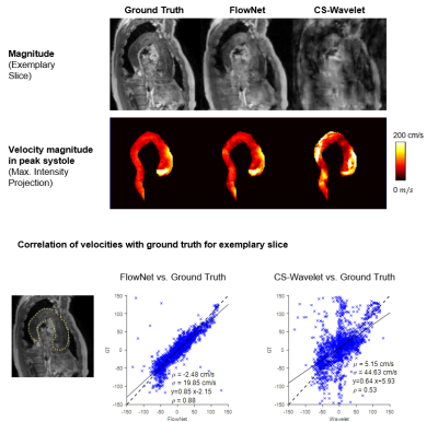 |
110 |
FlowNet: High-Speed Compressed Sensing 4D Flow MRI Image Reconstruction using Loop Unrolling
Jonas Walheim, Valery Vishnevskiy , Sebastian Kozerke
A variational neural network for the reconstruction of compressed sensing 4D flow MRI is presented. Nine iterations of an iterative reconstruction are unfolded in a neural network which was trained using eight retrospectively undersampled datasets. A phase-invariant network architecture was designed with two types of filter operations, one with equal real and imaginary component and the other operating on image magnitude only. The method is shown to outperform spatial regularization in the Wavelet domain. A retrospectively undersampled patient scan demonstrates that the network can reconstruct pathologies based on healthy training samples. Reconstruction of prospectively undersampled 4D flow MRI shows good agreement of peak velocities and peak flow.
|
|
4654.
 |
111 |
Multi-scale Unrolled Deep Learning Network for Accelerated MRI
Ukash Nakarmi, Joseph Cheng, Edgar Anselmo Piedra, Morteza Mardani, John Pauly, Leslie Ying, Shreyas Vasanawala
Model prior based reconstruction and data-centric prior reconstruction are two strong paradigms in image reconstruction inverse problems. In this abstract, we propose a framework that integrates the model prior and data-centric multi-scale deep learning priors for reconstructing magnetic resonance images (MRI) from undersampled k-space data. The proposed framework brings together the best of both paradigms and has proven superior to conventional accelerated MRI reconstruction techniques.
|
|
4655.
 |
112 |
Dual-domain Generative Adversarial Model for Accelerated MRI Reconstruction
Guanhua Wang, Enhao Gong, Suchandrima Banerjee, Karen Ying, Greg Zaharchuk, John Pauly
Previous CS frameworks based on Deep Learning like GANCS have demonstrated improved quality and efficiency. To further improve the restoration of the high-frequency details and the suppression of aliasing artifacts, a data-driven regularization is explicitly added on the k-space, in the form of an adversarial loss (GAN). In this work, the cross-domain generative adversarial model is trained and evaluated on diverse datasets and show decent generalization ability. For both quantitative comparison and visual inspection, the proposed method achieves better reconstruction than previous networks.
|
|
4656.
 |
113 |
Dynamic Multi-Coil Reconstruction using Variational Networks
Kerstin Hammernik, Matthias Schloegl, Erich Kobler, Rudolf Stollberger, Thomas Pock
In this work, we present a variational network for reconstructing dynamic multi-coil data. Incorporation of parallel imaging increases the acceleration potential due to additional spatial information, but was not considered so far in other learning-based reconstruction approaches for dynamic MRI. We show that variational network reconstructions with learned spatio-temporal regularization lead to further improvements in image quality compared to state-of-the-art Compressed Sensing approaches for different CINE cardiac datasets and acceleration factors with 10-times faster reconstruction time.
|
|
4657.
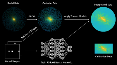 |
114 |
Pseudo-Cartesian k-Space Interpolation Using Artificial Neural Networks
Nikolai Mickevicius, Eric Paulson, Andrew Nencka
This work aims to extend the RAKI method for artificial intelligence-based k-space interpolation to non-Cartesian acquisitions. It was tested in radial acquisitions up to acceleration factors of 7. This method performs similarly well, or better than total-variation regularized sensitivity encoding.
|
|
4658.
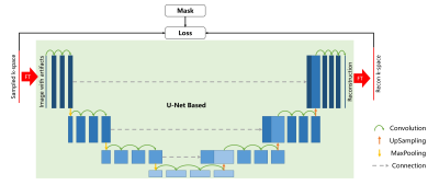 |
115 |
A Reference-Free Convolutional Neural Network Model for Magnetic Resonance Imaging Reconstruction from Under-Sampled k-Space
Yang Song, Yida Wang, Xu Yan, Minxiong Zhou, Bingwen Hu, Guang Yang
We used a reference-free model based on convolutional neural network (RF-CNN) to reconstruct the under-sampled magnetic resonance images. The model was trained without fully sampled image (FS) as the reference. We compared our model with the traditional compressed sensing reconstruction (CS) and the CNN model trained by FS. Mean square error and structure similarity were used to evaluate the model. Our RF-CNN model performed better than CS, but did not perform as good as usual CNN model.
|
|
4659.
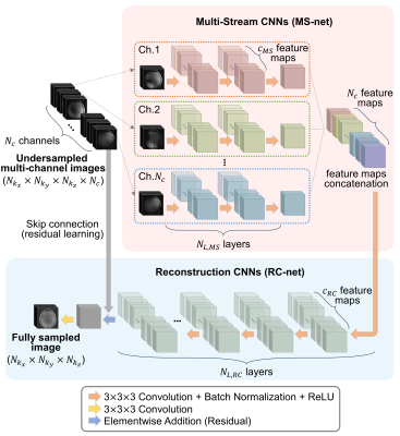 |
116 |
Parallel Imaging in Time-of-Flight Magnetic Resonance Angiography Using Deep Multi-Stream Convolutional Neural Networks
Yohan Jun, Taejoon Eo, Hyungseob Shin, Taeseong Kim, Hojoon Lee, Dosik Hwang
A deep parallel imaging network (“DPI-net”) was developed to reconstruct 3D multi-channel MRA from undersampled data. It comprises two deep-learning networks: a network of multi-stream CNNs for extracting feature maps of multi-channel images and a network of reconstruction CNNs for reconstructing images from the multi-stream network output feature maps. DPI-net was effective in reconstructing 3D time-of-flight MRA from highly undersampled multi-channel MR data, achieving superior performance, both quantitatively and qualitatively, over conventional parallel imaging and other deep-learning methods.
|
|
4660.
 |
117 |
Parallel Imaging based on k-x Domain Interpolation using Deep Neural Networks
Hyungseob Shin, Taejoon Eo, Yohan Jun, Taeseong Kim, Dosik Hwang
In this study, we compare two deep learning approaches to reconstruct multi-channel magnetic resonance (MR) images subsampled along phase-encoding (PE) direction. They are both based on the Fully-Connected (FC) layers but are performed in two different domains : Image domain, and k-x domain which is 1D inverse Fourier transformed (IFT) k-space. We demonstrate that the latter method shows superior performance to the former one in terms of removing the aliasing artifacts and recovering the details of MR images. The performance of the proposed method to the conventional MR reconstruction on image domain was qualitatively and quantitatively evaluated.
|
|
4661.
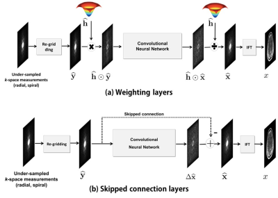 |
118 |
Non-Cartesian k-space Deep Learning for Accelerated MRI
Yoseob Han, Jong Chul Ye
The annihilating filter-based low-rank Hankel matrix approach (ALOHA) [1] is one of the most recent compressed sensing (CS) approaches that directly interpolates the missing k-space data using low-rank Hankel matrix completion. Inspired by the recent low-rank Hankel matrix decomposition using data-driven framelet basis [2], we propose a completely data-driven deep learning algorithm for k-space interpolation. In particular, our method can be applied directly by simply adding an additional re-gridding layer to non-Cartesian k-space trajectories such as radial trajectories.
|
|
4662.
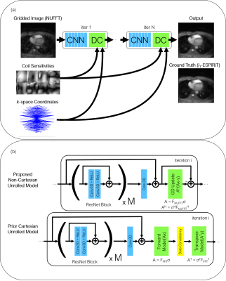 |
119 |
Accelerated 3D Non-Cartesian Reconstruction with Deep Learning
Mario Malavé, Srivathsan Koundinyan, Christopher Sandino, Frank Ong, Joseph Cheng, Dwight Nishimura
In this work, we demonstrate the application of a non-Cartesian unrolled architecture in reconstructing images from undersampled 3D cones datasets. One shown application of this method is for reconstructing undersampled 3D image-based navigators (iNAVs), which enable monitoring of beat-to-beat nonrigid heart motion during a cardiac scan. The proposed non-Cartesian unrolled network architecture provides similar outcomes as l1-ESPiRIT in one-twentieth of the time, and the reconstructions exhibit robustness when using an undersampled 3D cones trajectory.
|
|
4663.
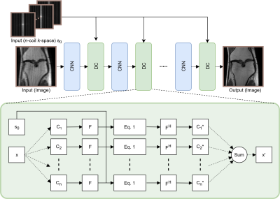 |
120 |
Data Consistency Networks for (Calibration-less) Accelerated Parallel MR Image Reconstruction
Jo Schlemper, Jinming Duan, Cheng Ouyang, Chen Qin, Jose Caballero, Joseph Hajnal, Daniel Rueckert
We present simple reconstruction networks for multi-coil data by extending deep cascade of CNN’s and exploiting the data consistency layer. In particular, we propose two variants, where one is inspired by POCSENSE and the other is calibration-less. We show that the proposed approaches are competitive relative to the state of the art both quantitatively and qualitatively.
|
|
4664
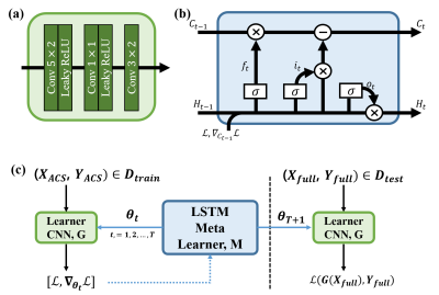 |
121 |
Fast estimation of GRAPPA kernel using Meta-learning
Video Permission Withheld
Dongwook Lee, Jong Chul Ye
This paper proposes an accelerated MR reconstruction method for parallel imaging from uniformly undersampled k-space data by learning scan-specific GRAPPA kernel using the long short-term memory network (LSTM). In particular, the meta-leaner LSTM is redesigned to quickly estimate the GRAPPA kernel for each k-space from its auto-calibration signals (ACS). The proposed method shows improved reconstruction performance with minimum error.
|
|
4665.
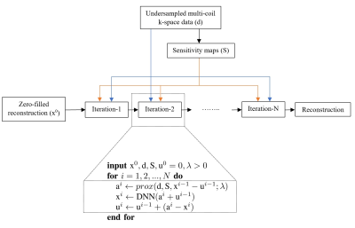 |
122 |
Deep Plug-and-Play Prior for Parallel MRI Reconstruction
Ali Pour Yazdanpanah, Onur Afacan, Simon Warfield
Fast data acquisition in Magnetic Resonance Imaging (MRI) is vastly in demand and scan time directly depends on the number of acquired k-space samples. Conventional MRI reconstruction methods for fast MRI acquisition mostly relied on different regularizers which represent analytical models of sparsity. However, recent data-driven methods based on deep learning has resulted in promising improvements in image reconstruction algorithms. In this paper, we propose a deep plug-and-play prior framework for parallel MRI reconstruction problems which utilize a deep neural network (DNN) as an advanced denoiser within an iterative method. We demonstrate that a deep plug-and-play prior framework for parallel MRI reconstruction with a regularization that adapts to the data itself results in excellent reconstruction accuracy and outperforms the clinical gold standard GRAPPA method.
|
|
4666.
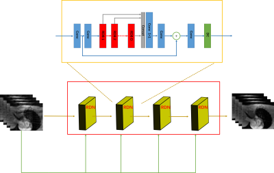 |
123 |
A Cascaded Residual Dense Network for Cardiac MR Imaging
Ziwen Ke, Shanshan Wang, Cheng Li, Huitao Cheng, Leslie Ying, Xin Liu, Hairong Zheng, Dong Liang
Cardiac MR imaging plays an important role in clinical diagnosis. But the long scan time limits its wide applications. To accelerate data acquisition, deep learning based methods have been applied to effectively reconstruct the undersampled images. However, current deep convolutional neural network (CNN) based methods do not make full use of the hierarchical features from different convolutional layers, which impedes their performances. In this work, we propose a cascaded residual dense network (C-RDN) for dynamic MR image reconstruction with both local features and global features being fully explored. Our proposed C-RDN achieves the best performance on in vivo datasets compared to the iterative optimization methods and the state-of-the-art CNN method.
|
|
4667.
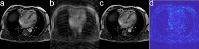 |
124 |
Reconstruction of high undersampling rate images using a cascade of convolutional neural networks
Presentation Not Submitted
Xi Chen, Shuo Chen, Rui Li
Imaging speed is important in many magnetic resonance imaging (MRI) applications because long scan time increases the risk of artifacts. At present, reconstruction method based on compressed sensing and deep learning significantly increases the speed of MRI scan. However, the performance of current models is not good at high undersampling rate. Here we used a large dataset to improve the undersampling rate of a CNN based MR reconstruction while maintaining high image quality. Our results showed an average 2.6% root-mean-square error in reconstructing from 16-fold undersampling k-space, which outperforms traditional method.
|
|
4668.
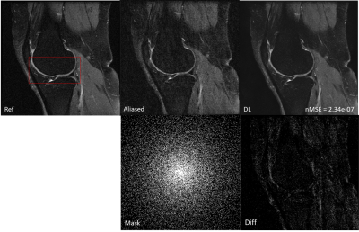 |
125 |
Deep MRI Reconstruction without Ground Truth for Training
Peizhou Huang, Chaoyi Zhang, Hongyu Li, Sunil Kumar Gaire, Ruiying Liu, Xiaoliang Zhang, Xiaojuan Li, Leslie Ying
Deep learning has recently been applied to image reconstruction from undersampled k-space data with success. Most existing works require both undersampled data and ground truth image as the training pair. It is not practical to obtain a large number of ground truth images for training in some MR applications. Here a novel deep learning network is studied for image reconstruction using only undersampled data for training. Experiment results demonstrate the feasibility of training without the ground truth images for image reconstruction.
|
|
| Top |
Improving Definition & Reducing Artifacts
Digital Poster
Acquisition, Reconstruction & Analysis
Thursday, 16 May 2019
| Exhibition Hall |
09:15 - 10:15 |
| |
|
Computer # |
|
4669.
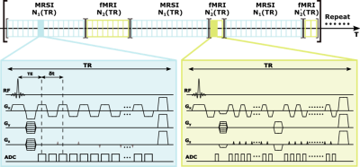 |
126 |
High-Resolution Simultaneous Mapping of Brain Function and Metabolism
Rong Guo, Yibo Zhao, Yudu Li, Yao Li, Zhi-Pei Liang
We present a new method for simultaneous mapping of brain function and metabolism. This method provides an unprecedented capability to simultaneously obtain high-resolution metabolic maps (2.4×2.4×3.0 mm3) and brain functional maps (3.0×3.0×2.6 mm3) of the whole brain coverage (230×230×120 mm3) in 8 minutes. The proposed method extends the subspace-based imaging framework of the SPICE technique with a new data acquisition scheme and exploits the complementary information between MRSI and fMRI signals for high-quality image reconstruction. Brain imaging experiments have been carried out, demonstrating the impressive capability of our method. With further improvement, the method can provide an unprecedented tool for mapping brain function and metabolism simultaneously.
|
|
4670.
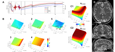 |
127 |
CSF-suppressed T2 weighted imaging at 7T
Jullie Pan, Chan Moon, Arun Antony, Frank Lieberman, Vikas Agarwal, Hoby Hetherington
T2-weighted lesional imaging is most commonly performed using inversion recovery turbo spin echoes. At 7T, however, this acquisition is limited for specific absorption rate and resolution. We implement a strategy that uses a driven equilibrium spin-echo preparation within an inversion recovery with multiple 3D gradient-echo imaging blocks to generate CSF-suppressed T2 weighted sensitivity. Images are combined using the self-normalization approach. Data acquired with an 8x2 transceiver array are shown to demonstrate sensitivity in brain tumors and epilepsy.
|
|
4671.
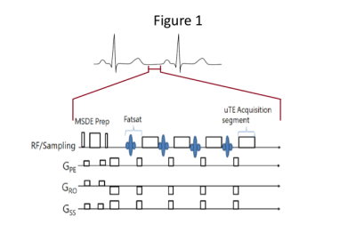 |
128 |
Visualizing the Myocardium in-vivo with a 3D uTE Acquisition
Larry Kasuboski, Jason Ortman, Sho Tanaka, Bharath Ambale Venkatesh, Joao Lima
A dark blood 3D uTE acquisition scheme with an MSDE pre-pulse is shown to provide suppression of flowing blood, while maintaining good definition of the myocardium.
|
|
4672.
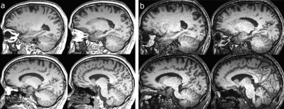 |
129 |
Optimize Wave-CAIPI MPRAGE protocol for the study of Short-term Apparent Change (SAC) of Grey Matter in Motor Training
Presentation Not Submitted
Tie-Qiang Li, Tobias Granberg, Sven Petersson
To assess the extent and dynamics of short-term apparent change (SAC) of GM in motor training we investigated how the VBM results are affected by the different levels of acceleration of the MP-RAGE sequence using the wave-CAIPI technique which provides highly accelerated MPRAGE imaging and retain high image quality. The optimized wave-CAIPI MPRAGE imaging protocol overcomes the g-factor noise amplification penalty and allows for over an order of magnitude acceleration of MPRAGE imaging in VBM studies. The standard and wave-CAIPI MPRAGE sequences have different sensitivity in detecting SAC of GM likely due to their differences in noise and contrast characteristics.
|
|
4673.
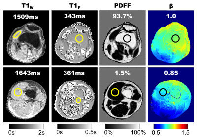 |
130 |
Simultaneous B1- and Fat-Corrected T1 Mapping Using Chemical-Shift Encoded MRI
Nathan Roberts, Diego Hernando, Timothy Colgan, Xiaoke Wang, Scott Reeder
Spatially varying B1 inhomogeneities and tissue fat are known to be confounders of quantitative T1 mapping methods that use multiple flip-angle techniques. Separately acquired B1 calibration maps can be used to correct flip angle errors caused by B1 inhomogeneities, but this requires an additional acquisition. In this work we propose a comprehensive approach that combines concepts from actual flip-angle imaging with variable flip-angle imaging to simultaneously estimate B1 inhomogeneity, T1, proton density fat-fraction and R2*. The feasibility and noise performance of this joint acquisition and fitting approach are evaluated using Cramer-Rao Lower Bound analysis, simulations, phantom experiments, and preliminary in vivo examples.
|
|
4674.
 |
131 |
Robust R1rho asymmetry performed with optimal B1 selection
Baiyan Jiang, Jian Hou, Weitian Chen
The measurement of R1rho (1/T1rho) spectrum and its asymmetry have several advantages over Chemical Exchange Saturation Transfer (CEST) and Chemical Exchange Spin-lock (CESL). It is recently becoming one of the important approaches for probing the chemical exchange process. In this work, we demonstrated the advantage of R1rho asymmetry over CESL, and proposed a way to predict the optimal B1 for R1rho asymmetry.
|
|
4675.
 |
132 |
Super-resolution MRI with 2D Phaseless Encoding
Rui Tian, Franciszek Hennel, Klaas Pruessmann
Super-resolution MRI with 1D phaseless encoding achieves high-resolution with immunity to shot-dependent phase fluctuation by simultaneously acquiring multiple k-space bands. We now explore a 2D extension of this technique to facilitate more k-space sampling strategies. Two distinct encoding schemes were analyzed and tested with EPI acquisition. By properly adjusting the overlapping of the mixed k-space bands, the 2D phaseless encoding could also be combined with the spiral acquisition. The amplitude modulation caused by band overlapping was eliminated by an inverse filter during reconstruction. The overlapped bands regions were also exploited to provide information about unexpected bands errors for post-processing corrections.
|
|
4676
 |
133 |
Super-resolution based on the Signal Extrapolation in Phase scrambling Fourier Transform Imaging using Deep Convolutional Neural Network
Video Permission Withheld
Satoshi ITO
The signal obtained in phase scrambling Fourier transform imaging can be extrapolated beyond sampling length after data acquisition like Half-phase encoding method. To realize the method for phase varied images, precise phase distribution map is required. In this paper, a new post-processing super resolution in PSFT imaging is proposed in which deep convolution neural network (CNN) is used and phase map is not required. Simulation and experimental results showed that spatial resolution was fairly improved with signal extrapolation and the improvement of spatial resolution is proportional to the strength of phase scrambling coefficient.
|
|
4677
 |
134 |
Semi-Classical Signal Analysis Method with Soft-Thresholding for MRS denoising
Video Permission Withheld
Abderrazak Chahid, Sourav Bhaduri, Malik Wali, Eric Achten, Hacene Serrai , Taous-Meriem Laleg-Kirati
A Semi-Classical Signal Analysis (SCSA) method with soft thresholding is proposed for MRSI denoising. The SCSA takes advantage of the pulse-shaped MRS spectrum to decompose both real and imaginary parts, into localized basis given by squared eigenfunctions of the Schrödinger operator. An optimization-based soft-threshold is provided to find optimal semi-classical parameters, for both the real and imaginary parts of the MRS signal. The optimal SCSA parameters discard the eigenfunctions representing noise from the noisy spectrum, and conserve the eigenfunctions representing the useful information. The obtained in-vivo results show the efficiency of the SCSA with soft thresholding in removing noise and conserving metabolite signals.
|
|
4678.
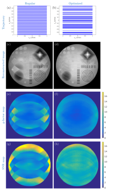 |
135 |
Encoding trajectory optimization based on the pixel variance using graphics processing units
Ali Sadr, Stefan Kroboth, Elmar Fischer, Feng Jia, Sebastian Littin, Huijun Yu, Jürgen Hennig, Maxim Zaitsev
Undersampled trajectories were optimized for parallel imaging with explicit consideration of the RF coil sensitivities in order to complement the RF coil elements. A second-order approximation of pixel variance was used as a metric to evaluate encoding trajectories and also serves as the cost function in the optimization problem, solved using Simulated Annealing. The metric was implemented on a Graphical Processing Unit (GPU) to accelerate computations. The developed method was evaluated on two test cases with Cartesian and radial sampling for isotropic and anisotropic fields-of-view. Resulting optimized trajectories led to improved image quality, more uniform SNR and reduced g-factors.
|
|
4679.
 |
136 |
Online compressed sensing MR image reconstruction for high resolution T∗2T2∗ imaging
Loubna El Gueddari, Emilie Chouzenoux, Jean-Christophe Pesquet, Alexandre Vignaud, Philippe Ciuciu
Compressed sensing theory reduces lengthy acquisition time in MRI at the expense of computationally demanding iterative reconstruction. Usually, reconstruction is performed offline once all the data have been collected. Here, we introduce an online CS reconstruction framework that interleaves acquisition and reconstruction steps in a convex setting and permits the delivery of intermediate images on the scanner console during acquisition. In particular, the sum of acquisition and reconstruction times is reduced without compromising image quality. The gain of this strategy is shown both on retrospective Cartesian and prospective non-Cartesian under-sampled ex-vivo baboon brain data at 7T with an in-plane resolution of 400μμm.
|
|
4680.
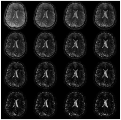 |
137 |
Radial Single-Shot Fast Spin Echo: Toward Fast, Motion-Robust, Multi-Contrast Imaging
Daniel Litwiller, Kang Wang, Lloyd Estkowski, Ali Ersoz, Ty Cashen, Ersin Bayram
We present a radial Single-Shot Fast Spin Echo pulse sequence that is capable of generating multiple contrasts from a single spin echo train via temporal filtering. Undersampling artifacts are minimized by using a long echo train and an aggressive variable refocusing flip angle scheme. In vivo feasibility is demonstrated in the brain.
|
|
4681.
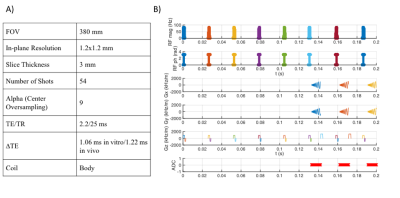 |
138 |
Rapid Prototyping of Spiral Based Three-Points Dixon Acquisition and Reconstruction Using Pulseq
Marina Manso Jimeno, Sairam Geethanath, John Vaughan
Spiral imaging possesses special characteristics that would benefit both research and clinical MR fields. However, its use is often limited due to the difficulties its implementation on a scanner entails. This problem frequently limits the spread and evolution of spirals and consequently their potential and multiple applications. The current study proposes a solution to this problem by demonstrating a rapid prototyping of a three-points Dixon spiral sequence. Results agree with the standard sequence used by the system manufacturer for the same purpose. The software used is open-source and makes possible the sequence implementation in multiple vendor scanners.
|
|
4682.
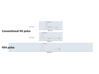 |
139 |
Improvement of 3D-STIR for TRANCE non-contrast MR angiography at 3T using stretched adiabatic inversion pulse
Kosuke Morita, Masami Yoneyama, Masanobu Nakamura, Takeshi Nakaura, Seitaro Oda, Masahiro Hatemura, Yasuyuki Yamashita
The purpose of our study was to improve 3D-STIR for TRANCE non-contrast MR angiography in clinical 3.0T MR system using modified hyperbolic secant (HS4) pulses. the higher field strength poses additional challenges to 3D STIR, including wider offset frequency between water and fat combined with larger B0 and B1 inhomogeneities, which reduce the reliability of fat suppression. 3D STIR (TRANCE) with HS4 pulse has clearly improved fat suppression due to B0/B1 inhomogeneous compared with conventional HS pulse in clinical.
|
|
4683.
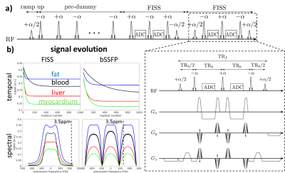 |
140 |
3D Cartesian Fast-interrupted Steady-state Sequence (FISS) with Intrinsic Fat Suppression
Thomas Kuestner, Aurelien Bustin, Olivier Jaubert, Radhouene Neji, Claudia Prieto, René Botnar
Fat-suppressed balanced steady-state free precession (bSSFP) is a rapid imaging sequence often used in cardiovascular MRI. Recently a fast interrupted steady-state (FISS) sequence was proposed which periodically interrupts the steady-state of the bSSFP. The resulting frequency response modulation can be leveraged for suppression of the off-resonant fat signal without the need of fat preparation pulses. FISS was demonstrated for 2D radial acquisitions, however challenges to apply this approach to 3D Cartesian have been reported. Here we propose to extend FISS to 3D Cartesian imaging and investigate its behavior by extended phase graph simulations and in-vivo leg, abdominal measurements.
|
|
4684.
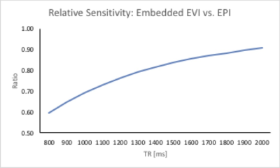 |
141 |
Simultaneous Functional MRI and Proton Echo-Planar Spectroscopic Imaging in Human Brain (fPEPSI)
Stefan Posse, Bruno Sa De La Rocque Guimaraes , Kishore Vakamudi, Steen Moeller
This study introduces simultaneous fMRI and MRSI integrates multi-slab echo-volumar-imaging (MEVI) into the water suppression module of Proton-Echo-Planar-Spectroscopic-Imaging to simultaneously acquire fMRI, water suppressed and water reference data in a single scan (fPEPSI). FMRI image quality and BOLD sensitivity acquired with 4x4x6 mm3 resolution was comparable to our recently developed MEVI method. Spectral quality and sensitivity of 3D metabolite maps acquired with 4x4x7 mm3 resolution were comparable to conventional PEPSI. This hybrid fMRI/MRSI approach considerably reduces scan times in multi-modal clinical research studies and it is applicable to characterizing neurotransmitter and lactate concentration changes in relation to BOLD signal changes.
|
|
4685.
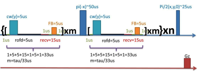 |
142 |
Fixed-Point MR Imaging for Early Gliobrastoma Detection
Presentation Not Submitted
Zhao Li, Chao-Hsiung Hsu, Huiyuan Zheng, Yung-Ya Lin
Problem Early detection of high-grade malignancy, such as GBM, remains challenging using MRI. Methods A new approach using continuous-wave and feedback field to reach “fixed-point spin dynamics” was developed to enhances the local magnetic-field gradient variations due to irregular water contents and deoxyhemoglobin concentration in early GBM.
Results In vivo MR images and mappings acquired on orthotopic GBM mice using “fixed-point pulse sequence” shows 3-4 times of enhancement in GBM contrast than the best conventional images acquired.
Conclusion Simulations and in vivo GBM mouse models validated the superior contrast/sensitivity and robustness of fixed-point spin dynamics method towards early GBM detection.
|
|
4686.
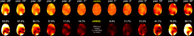 |
143 |
Implications of within-scan patient head motion on B1+ homogeneity and Specific Absorption Rate at 7T
Emre Kopanoglu, Alix Plumley, M. Arcan Erturk, Cem Deniz, Richard Wise
Parallel-transmit pulses are commonly used to improve B1+-homogeneity at higher field strengths, while local-SAR constraints are applied to ensure safety. However, patient motion may become unavoidable with longer scans or less cooperative patients, and motion may affect B1+-homogeneity and local-SAR. We investigated the effect of all 6 degrees-of-freedom of head motion on B1+-homogeneity and local-SAR for parallel-transmit multi-spoke pulses using simulations. We observed more than a 2-fold increase in local-SAR due to motion for some pulses. We also investigated the changes in B1+-homogeneity of spokes pulses using in-vivo B1+-maps and showed regional variations between 12%-22% in the excitation profile.
|
|
4687.
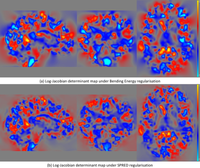 |
144 |
Symmetric Priors for Regularisation of Elastic Deformations (SPRED) - efficient GPU-accelerated enforcement of diffeomorphism in B-spline parametrised 3D nonlinear registration
Frederik Lange, Stephen Smith, Jesper Andersson
We present a method for regularising nonlinear registration which produces deformations which are biologically more plausible than conventional techniques. Our method, the Symmetric Prior for Regularisation of Elastic Deformations (SPRED), not only enforces diffeomorphism, but additionally penalises linear, planar and volumetric changes. Application of SPRED to the high quality NIREP dataset produced results whose quality matches that of established registration methods. The resulting deformations show significantly more plausible Jacobian distributions, both in terms of spatial locality and intensity. Future work will look to extend SPRED to include variable spatial priors, allowing different brain regions freedom to deform by varying amounts.
|
|
4688.
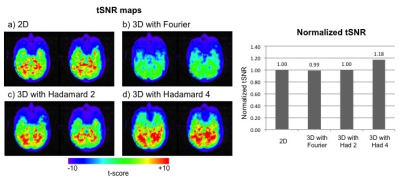 |
145 |
Hadamard Encoding Compared with Fourier Encoding in Three-dimensional (3D) Functional MRI
Seul Lee, Gary Glover
Three-dimensional (3D) acquisition is beneficial for functional MRI (fMRI) compared to two-dimensional (2D) acquisition since it can provide higher spatial resolution, resulting from potentially higher temporal signal-to-noise ratio (tSNR) and thinner slices. However, 3D has higher physiological noise due to higher signal at the center of k-space, which results in lower tSNR. The number of slices can be decreased to reduce physiological noise. However, a small number of slices in Fourier encoding results in Gibbs ringing. In this study, we show that 3D Hadamard acquisition avoids Gibbs artifacts while increasing SNR compared with conventional 2D and 3D methods.
|
|
4689.
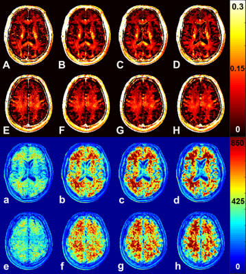 |
146 |
Myelin Water Fraction Maps with improved Fit to Noise using TGV and conventional filters
René Schranzer, Günther Grabner, Alexander Weber, Kristian Bredies, Gernot Reishofer, Alexander Rauscher
Myelin Water Imaging is the technique of choice to measure myelination changes in healthy and abnormal situations in the brain. However, calculation of myelin water fraction (MWF) maps is challenging due to the low signal-to-noise ratio in the acquired data. Here, we demonstrate different filter methods, such as TGV, Gaussian and Wiener to overcome this problem. 3D GRASE images filtered with all three methods show significant enhanced fit-to-noise (FNR) values compared to unfiltered, while TGV preserves sharper edges and detailed structures. Finally, noise reduction and thus more reliable MWF maps can lead to certain advantages in the field of MS.
|
|
4690.
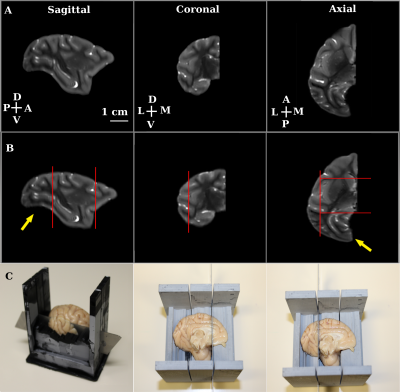 |
147 |
MRI guided hierarchical sectioning and stitching of brain blocks for alignment of digitized histology to corresponding MR images
Sethu Boopathy Jegathambal, Kelvin Mok, David Rudko, Amir Shmuel
Ex-vivo MRI of brain tissue can provide important morphological and microstructural information. MR images can also serve as undistorted references for reconstruction of digitized histology, immunohistochemistry or clearing techniques. However, due to space constraints imposed by small animal scanners, imaging a whole human brain generally requires a large bore scanner with limited gradient system performance and constraints on achievable spatial resolution. Importantly, large histology sections cut from large brains are prone to distortions, tears and folding. Here we present a method for MRI guided, hierarchical sectioning and stitching of brain blocks for alignment of digitized processed tissue to corresponding MRI volumes.
|
|
4691.
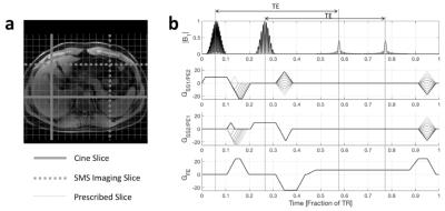 |
148 |
Simultaneous Acquisition of Orthogonal Plane Cine Imaging and Isotropic 4D-MRI Using Super-Resolution
Nikolai Mickevicius, Eric Paulson
The super-resolution-based, isotropic 4D-MRI, and orthogonal cine imaging pulse sequence (SR-4D-SOPI) was developed and evaluated in this work. Cine imaging was acquired at more than 3 frames per second in sagittal and coronal planes simultaneously while also acquiring two orthogonal 4D-MRI volumes. The volumes were combined using super-resolution methods to create an isotropic 4D-MRI to be used for dose reconstruction following each fraction of abdominal or thoracic MR-guided radiation therapy.
|
|
4692.
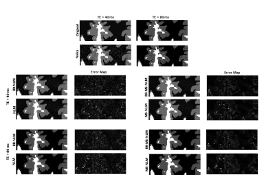 |
149 |
Multispectral nonlocal means filters incorporating rotations and reflections for improved noise reduction with edge preservation in magnetic resonance imaging
Nikkita Khattar, Mustapha Bouhrara, Richard Spencer
Image denoising is used extensively for MR image post-processing. The nonlocal means (NLM) filter shows excellent noise reduction while preserving detail. NLM takes advantage of the structural redundancy in MR images by comparing local neighborhoods of voxels throughout the image, and estimating the intensity of an index voxel to be denoised through a weighted average of voxel intensities. However, this excludes patches that may be similar except for rotation or reflection, and therefore does not make full use of image redundancy. We introduce a multispectral implementation of NLM incorporating rotations and reflections, finding improved performance compared to conventional non-multispectral filtering.
|
|
4693.
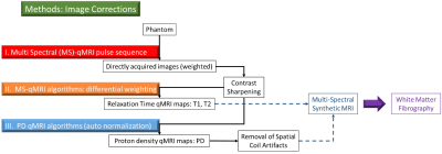 |
150 |
Effects of Image Sharpening on the Accuracy of quantitative-MRI (qMRI) Maps.
Ryan McNaughton, Mina Botros, Xin Zhang, Hernan Jara
Purpose: To study the effects of image sharpening and low spatial frequency removal on the quality of qMRI maps of T1, T2, and proton density (PD). Methods: Previously developed qMRI algorithms, augmented with specialized image filters, were tested with a gel based phantom containing three distinct solutions of variable gadolinium, sucrose, and agarose concentrations. Results: Images were successfully sharpened without significantly effecting pixel values of T1 and T2 weighted maps, while removing PD map spatial artifacts in the gadolinium vials. Conclusion: Unsharp masking and spatial flattening algorithms are effective methods for enhancing qMRI quality toward generating more accurate Synthetic-MRI maps.
|
|
| Top |
Machine Learning for Image Reconstruction: Breakthroughs
Digital Poster
Acquisition, Reconstruction & Analysis
Thursday, 16 May 2019
| Exhibition Hall |
09:15 - 10:15 |
| |
|
Computer # |
|
4694.
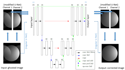 |
151 |
EPI artifacts reduction using deep learning
Christophe Schülke, Karsten Sommer, Peter Börnert
The inherent speed of EPI is penalized by the calibration prescans necessary to suppress N/2 ghosts. Here, we propose a deep neural network with a novel architecture that suppresses N/2 ghosts in a post-processing step starting from magnitude images, thereby eliminating the necessity of a prescan. The proposed network achieves better results than more classical networks of the same size by taking into account the N/2 structure of ghosts. The network architecture could easily be adapted to also correct for ghosts of higher order in multishot EPI.
|
|
4695.
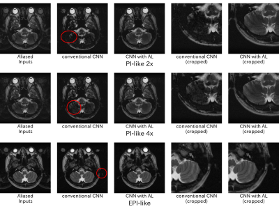 |
152 |
Convolutional Neural Networks with Aliasing Layers for Correcting Parallel Imaging and EPI Ghost Artifacts
Hidenori Takeshima
The author proposes a new layer named aliasing layer (AL) for effectively correcting MR-specific aliasing artifacts using convolutional neural networks. In MR images acquired using parallel imaging (PI) and/or echo-planar imaging (EPI), the locations of aliasing artifacts and/or N/2 ghost artifacts can be analytically calculated. The AL preprocesses MR images by moving the calculated locations to the locations accessible through summations over all channels in a convolution layer. The experimental results demonstrate that the correction method using the proposed AL could effectively remove PI aliasing and EPI ghosting artifacts.
|
|
4696.
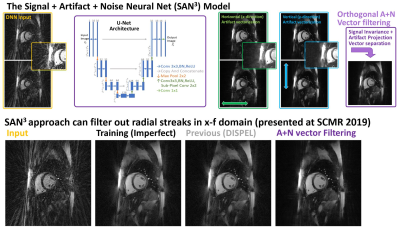 |
153 |
A Validation Approach for Imperfect Training Data Fidelity using Signal + Artifact + Noise-based Neural Net (SAN3)-derived Directionalized Streaking Removal
Nanyque Boyd, Yudai Suzuki, Amit Patel, Jacob Goes, Marcella Vaicik, Satoru Hayamizu, Satoshi Tamura, Keigo Kawaji
While Deep Neural Network (DNN)-based sub-Nyquist reconstruction approaches are well-suited for high-fidelity static imaging targets such as the brain, temporally constrained (i.e. dynamic) sequences may potentially be ill-suited for DNN as these would often embed unresolved MR artifacts into the Training Data. Here, we describe an assessment approach for a generalizable DNN-based dynamic MRI reconstruction method that outputs such artifacts as characterizable and filterable streaks. This work further validates the DNN-model coding process to ensure the desired artifact/noise properties into the DNN output. Using Fourier properties, we demonstrate such validation of streaking directionalization using DNN.
|
|
4697.
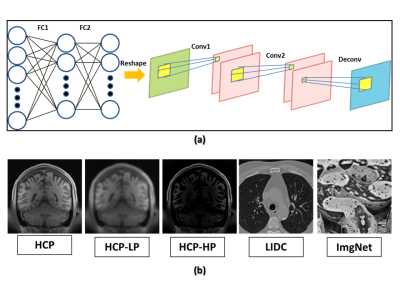 |
154 |
Comparison of Quality Assessment Methods for Deep-Learning-Based MR Image Reconstruction
Mohammadhassan Izady Yazdanabady , Kyoko Fujimoto, Benjamin Berman, Matthew Rosen, Neha Koonjoo, Bo Zhu, Christian Graff, Aria Pezeshk
The proper methodology to perform rigorous quantitative task-based assessment of image quality for deep learning based MR reconstruction methods has not been devised yet. In this study we reconstructed T1-weighted brain images using neural networks trained with five different datasets, and explored the consistency and relationship between rankings of image quality using three different assessment metrics and FreeSurfer-based quantitative analysis. Our study indicates that assessment of image quality for a data-driven reconstruction algorithm may require several types of analysis including using different image quality assessment metrics and their agreement with clinically relevant tasks.
|
|
4698.
 |
155 |
Conditional generative adversarial network for three-dimensional rigid-body motion correction in MRI
Patricia Johnson, Maria Drangova
In this work we present a deep learning solution for motion correction in brain MRI; specifically we approach motion correction as an image synthesis problem. Motion is simulated in previously acquired brain images; the image pairs (corrupted + original) are used to train a conditional generative adversarial network (cGAN), referred to as MoCo-cGAN, to predict artefact-free images from motion-corrupted data. We also demonstrate transfer learning, where the network is fine-tuned to apply motion correction to images with a different contrast. The trained MoCo-cGAN successfully performed motion correction on brain images with simulated motion. All predicted images were quantitatively improved, and significant artefact suppression was observed.
|
|
4699.
 |
156 |
Volumetric real-time imaging with deep-learning reconstruction
Jiahao Lin, Fadil Ali, Kyunghyun Sung
We propose a deep-learning reconstruction pipeline for 3D real-time imaging. We use a 3D golden-angle GRE sequence, and a deep-learning network based reconstruction. Gadgetron framework is used for real-time pipelining. Using 320 images in total, our network is trained with decaying data fidelity update, and deployed without it. Dilated convolution and skip concatenation improve the image quality. We achieved a Matrix size of 192x192x8 pixels, a temporal resolution of 889ms, a reconstruction time of 300-350ms, and our image quality is comparable to iGRASP.
|
|
4700.
 |
157 |
Diffusion-weighted MR Image Reconstruction using Automated Transform by Manifold Approximation (AUTOMAP) on Human Brains
Koonjoo Neha, Bo Zhu, Matthew Christensen, John Kirsch, Bragi Sveinsson, Matthew Rosen
Low intrinsic Signal-to-Noise Ratio (SNR) in diffusion-weighted (DW) images are recurrent issues especially at high b-values. Here, we apply the deep neural network image reconstruction technique, AUTOMAP (Automated Transform by Manifold Approximation) to in-vivo diffusion-weighted MR data acquired at 1.5 T with varying b-values. In addition, apparent diffusion coefficient (ADC) maps were assessed. We also compared the reconstruction of the images using two different training corpura. The results for AUTOMAP reconstruction showed a significant increase in SNR.
|
|
4701
 |
158 |
Deep Learning Based Adaptive Noise Reduction in Multi-Contrast MR Images
Video Permission Withheld
Kensuke Shinoda, Kenzo Isogawa, Masahito Nambu, Yuichi Yamashita, Mika Kitajima, Hiroyuki Uetani, Yasuyuki Yamashita
We have proposed a deep learning-based approach for MR image denoising that can adapt to the input noise power. We compare the performance of the proposed denoise approach with Deep Learning based Reconstruction (dDLR) method with state-of-the art image denoising method called Block-matching and 3D filtering method (BM3D) on multiple contrast MR images. Our experiments demonstrate that the proposed method outperforms the state-of-the art BM3D image denoising method.
|
|
4702.
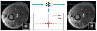 |
159 |
Deep Partial Fourier Reconstruction
Alexander Toews, Marcus Alley, Shreyas Vasanawala, Brian Hargreaves, Joseph Cheng
Standard methods for partial Fourier (PF) reconstruction do not perform well in the presence of significant phase variations. In this study, we propose a deep-learning-based approach for PF reconstruction (DPFR) to mitigate this issue. We compare DPFR results against standard methods (Homodyne, POCS) for in vivo images of the foot, leg, and abdomen. We demonstrate that DPFR achieves superior reconstruction quality, especially near phase boundaries, across a range of partial sampling parameters. Ultimately this may extend the applicability of partial Fourier reconstruction to instances where it is not commonly used.
|
|
4703.
 |
160 |
Multi-supervised Learning in Cross-domain Networks for Cardiac Imaging
Ziwen Ke, Shanshan Wang, Huitao Cheng, Leslie Ying, Xin Liu, Hairong Zheng, Dong Liang
Dynamic MR image reconstruction from incomplete k-space data is an important technique for reducing its scan time. Deep learning has shown great potential in assisting this task. Nevertheless, most frameworks only adopt a final loss for network training and the intermediate results generated during the network forward pass haven't been considered for the network training. This work proposes a multi-supervised learning strategy, which constrains the frequency domain information and reconstruction results at different levels. Improved reconstruction results have been achieved with the proposed strategy.
|
|
4704.
 |
161 |
Ultra-low-dose Amyloid PET/MRI Reconstruction by Generative Adversarial Network
Jiahong Ouyang, Kevin Chen, Enhao Gong, John Pauly, Greg Zaharchuk
Amyloid PET is widely used in the early diagnosis of dementia. However, the injection of the radiotracer will lead to radiation exposure to the subject. We proposed a novel method based on Generative Adversarial Network (GAN) with perceptual loss to achieve diagnostic image quality PET images using ultra-low-dose PET images with or without additional MR contrasts as inputs.
|
|
4705.
 |
162 |
Improved TWIST Imaging using k-Space Deep Learning
Eunju Cha, Eung Yeop Kim, Jong Chul Ye
Time-resolved angiography with interleaved stochastic trajectories(TWIST) has been widely used for dynamic contrast enhanced (DCE) MRI. To achieve highly accelerated acquisitions for improved temporal and spatial resolution, the high frequency region is randomly sub-sampled at each time frame. Therefore, the periphery of the k-space data from multiple time frames are combined to obtain the uniformly sub-sampled k-space data so that the temporal resolution of TWIST is limited. The purpose of this research is to improve the temporal resolution of TWIST by reducing the view-sharing. Furthermore, we proposed the algorithm that can reconstruct the imagesat various number of view sharing using k-space deep learning.
|
|
4706.
 |
163 |
Contrast Transfer Learning for Reconstruction of Undersampled Dynamic Contrast-Enhanced MRI
Li Feng, Fang Liu, Lihua Chen, Ricardo Otazo
The application of deep learning for reconstruction of dynamic contrast-enhanced MRI presents significant challenges caused by the rapid passage of the contrast agent, which makes it difficult to acquire fully-sampled images to train a neural network. This work proposes to use images from a delayed contrast phase, where contrast changes are in a relatively steady state, for training, and to apply the trained neural network for reconstruction of undersampled data acquired in other contrast phases. The proposed contrast transfer learning reconstruction was trained on 55 post-contrast liver cases and tested on a first-pass liver DCE-MR acquisition.
|
|
4707.
 |
164 |
Development of a deep learning method for phase unwrapping MR images
Kanghyun Ryu, Sung-Min Gho, Yoonho Nam, Kevin Koch, Dong-Hyun Kim
MRI phase images are increasingly used for susceptibility mapping and distortion correction in function and diffusion MRI. However, acquired values of phase maps are wrapped between [-π π ] and require an additional phase unwrapping process. Here we developed a novel deep learning method that can learn the transformation between the wrapped phase images and the corresponding unwrapped phase images. The method was tested for numerical simulations and on actual MR images.
|
|
4708.
 |
165 |
Ultrafast 3D Partial Fourier Reconstruction with Well-Preserved Phase using DNN
Guangliang Ju, Aiqi Sun, Mingliang Chen, Yu Wang, Dong Han, Feng Huang
Partial Fourier (PF) is a widely used fast imaging scheme. Since phase information is crucial in many applications, such as SWI, it is necessary that PF can preserve phase well. Many PF methods cannot preserve phase well especially at locations with rapid phase change. DPA is a method can recover both magnitude and phase well, but suffers from low speed for two-directional PF acquisition. Considering recent advances in deep learning, we proposed a DNN-based framework for two-directional PF reconstruction. Preliminary experiments demonstrate that the proposed method is almost 50 times faster while restores magnitude and SWI even better than DPA.
|
|
4709.
 |
166 |
Accelerated MR image reconstruction using iterative feasible set projection
Doohee Lee, Jaeyeon Yoon, Jingyu Ko, Jingu Lee, Yoonho Nam, Jongho Lee
We proposed a new deep learning architecture for the reconstruction of highly undersampled data. The new architecture combines an iterative generative adversarial network (GAN) with a shared discriminator and interacts with data consistency blocks. The algorithm was applied to accelerate the data acquisition of the routine clinical protocols, particularly 2D Cartesian sampling sequences. The new method was tested to explore generalizability of the algorithm in in-vivo data under various conditions (difference pulse sequences, organs, coil types, sites, and health condition).
|
|
4710.
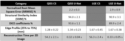 |
167 |
Rapid Image Reconstruction of Single-Shot Coronary Quiescent-Interval Slice-Selective (QISS) MRA and Late Gadolinium-Enhanced MRI using Deep Learning
Daming Shen, Hassan Haji-Valizadeh, Daniel Kim
While compressed sensing (CS) enables highly-accelerated cardiac MRI acquisitions, its lengthy image reconstruction may limit clinical translation. Deep learning (DL) is capable reconstructing undersampled images with clinically acceptable reconstruction times. The purpose of this study was to build, train, and validate a deep learning framework for rapidly reconstructing highly-accelerated cardiac MR images, where CS reconstructed images are used as reference.
|
|
4711.
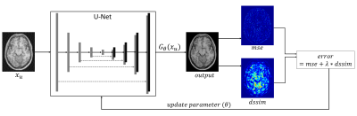 |
168 |
Enforcing Structural Similarity in Deep Learning MR Image Reconstruction
Kamlesh Pawar, Zhaolin Chen, N Shah, Gary Egan
Deep Learning (DL) MR image reconstruction from undersampled data involves minimization of a loss function. The loss function to be minimized drives the DL training process and thus determine the features learned. Usually, a loss function such as mean squared error or mean absolute error is used as the similarity metric. Minimizing such loss function may not always predict visually pleasing images required by the radiologist. In order to predict visually appealing MR images in this work, we propose to use the difference of structural similarity as a regularizer along with the mean squared loss.
|
|
4712.
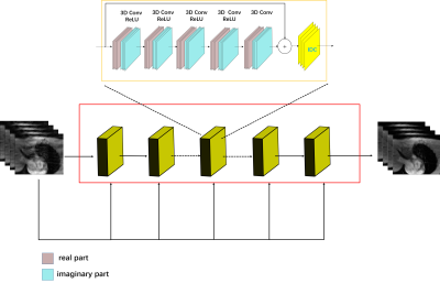 |
169 |
Edge-enhanced Loss Constraint for Deep Learning Based Dynamic MR Imaging
Shanshan Wang, Ziwen Ke, Huitao Cheng, Leslie Ying, Xin Liu, Hairong Zheng, Dong Liang
Cardiac magnetic resonance (MR) imaging provides a powerful imaging tool for clinical diagnosis. However, due to the constraints of magnetic resonance (MR) physics and reconstruction algorithms, dynamic MR imaging takes a long time to scan. Recently, deep learning has achieved preliminary success in MR reconstruction. Compared with the classical iterative optimization algorithms, the deep learning based methods can get improved reconstruction results in shorter time. However, most current deep convolutional neural network (CNN) based methods use mean square error (MSE) as the loss function, which might be a reason for image smooth in the reconstruction. In this work, we propose to employ edge-enhanced constraint for loss function and explore different types of total variation on network training. Encouraging performances have been achieved.
|
|
4713.
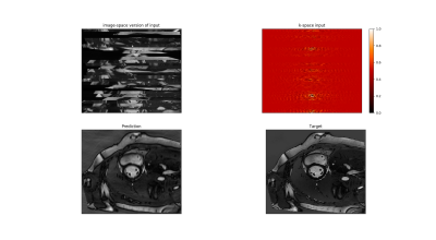 |
170 |
Exploration on deep-learning based sorting of k-space data for ECG-free cardiac cine-MRI
Seb Harrevelt, Tim Leiner, J.P.W. Pluim, A.J.E. Raaijmakers
Cine-cardiac MRI reconstruction relies on the ECG signal to sort k-space data. However, ECG triggering comes with disadvantages among which increased setup time. Here we suggest an alternative method of sorting cine MRI k-space data using deep-learning. An explorative study has been performed using an encoder-decoder network with Sinkhorn layer to sort k-space data that was randomly disordered in one spatial dimension. Good reconstructions were obtained using a group size of 8 or more k-space lines during randomization. These results hold promise for subsequent application in the time dimension.
|
|
4714.
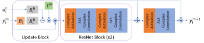 |
171 |
Complex-Valued Convolutional Neural Networks for MRI Reconstruction
Elizabeth Cole, John Pauly, Shreyas Vasanawala, Joseph Cheng
To improve MRI reconstruction accuracy, we propose various complex-valued frameworks for reconstructions using convolutional neural networks. By introducing complex-valued convolution and activation functions, we improve reconstruction of our subsampled images and achieve competitive results compared to the real-valued counterpart of our model.
|
|
4715.
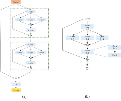 |
172 |
Deep Inception Residual Network (DIRN) for Reconstruction of Undersampled Brain MR Image
Presentation Not Submitted
Sekeun Kim, Yeonggul Jang, Hackjoon Shim, Hyukjae Chang
Acquiring the full-sampling k-space magnetic resonance imaging (MRI) data for detailed anatomical information is ideal. We propose the Deep Inception Residual Network (DIRN) based on a deep convolutional neural network (DCNN) consisting of inception blocks and residual blocks for the reconstruction of the MR image from undersampled k-space data. The experimental results on an ADNI dataset demonstrate that DIRN is appropriate for a reconstruction of brain MR images.
|
|
4716.
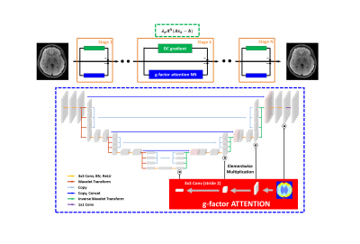 |
173 |
g-factor attention model for deep neural network powered parallel imaging: gANN
Jaeyeon Yoon, Doohee Lee, Jingyu Ko, Jingu Lee, Yoonho Nam, Jongho Lee
In this study, we proposed a new concept of an attention model for deep neural network based parallel imaging. We utilized g-factor maps to inform the neural network about the location containing high possibility of aliasing artifact. Also the proposed network used sensitivity maps and acquired k-space data to ensure the data consistency. Since the g-factor attention deep neural network considered both multi-channel information and spatially variant aliasing condition, our proposed network successfully removed aliasing artifacts up to factor 6 in uniform under-sampling and showed high performance when compared to conventional parallel imaging methods.
|
|
4717.
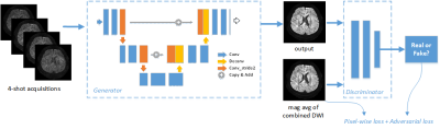 |
174 |
Accelerating high resolution DWI via deep learning
Da Zou, Ruibo Song, Dong Han, Feng Huang
The conventional multi-shot diffusion weighted imaging (DWI) techniques, such as MUSE, have not been widely adopted clinically due to long scan time. In this study, an accelerated multi-shot DWI method based on deep learning is proposed. By learning a fully convolutional neural network to enhance DWI images, more structural details and less noise can be achieved, especially when with fewer shots or NSA (Number of Signal Average), in the meantime the reconstruction time can be reduced by over 200 times. It means the proposed approach reduces the scan and reconstruction time dramatically while keeping high quality of the images, which makes it a potential technique for high resolution multi-shot DWI in routine clinical study.
|
|
4718.
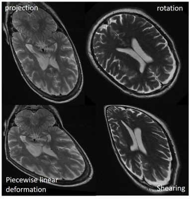 |
175 |
A Generative Adversarial Network with a Progressively Growing Training Strategy for MRI Dataset Augmentation
Vahid Ghodrati, Haotian An, Zihao Xiong, Jiaxin Shao, Mark Bydder, Peng Hu
For medical imaging applications, it is not straightforward to create a large database due to high costs associated with acquiring the data, patent privacy issues, and challenges in pooling data from multiple medical institutions. Generating high-resolution medical images from the latent noise vector could potentially mitigate training data size issues in applying DNN to medical imaging. This could facilitate objective comparisons between the different machine learning algorithms in medical imaging. In this study, progressive growing strategy is considered to train the GAN stably and generate super resolution brain datasets from noise vector.
|
|
Back to Program-at-a-Glance |  Back to Top Back to Top |
| The International Society for Magnetic Resonance in Medicine is accredited by the Accreditation Council for Continuing Medical Education to provide continuing medical education for physicians. |
