Digital Poster Session
Contrast Mechanisms Back to Program-at-a-Glance Back to Program-at-a-Glance
|
Thursday, 16 May 2019
Digital PosterContrast Mechanisms
4894 -4918 Imaging Myelin+
4919 -4943 QSM
4944 -4968 Pseudo-Continuous & Velocity-Selective ASL
4969 -4993 Relaxometry
4994 -5018 Novel Developments in CEST
5019 -5043 Perfusion & Permeability
5044 -5068 Electromagnetic Tissue Mapping |
| |
Imaging Myelin+
Digital Poster
Contrast Mechanisms
Thursday, 16 May 2019
| Exhibition Hall |
14:45 - 15:45 |
| |
|
Computer # |
|
4894.
 |
1 |
Inversion Recovery Pointwise Encoding Time Reduction with Radial Acquisition (IR-PETRA) for Direct Myelin Imaging in Human Brain

Hyungseok Jang, Michael Carl, Yajun Ma, Yanjun Chen, Saeed Jerban, Eric Chang, Jiang Du
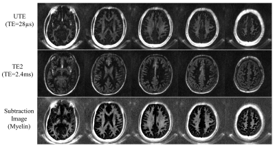
Due to very low proton density and rapid signal decay (T2*<300µs at 3T), it is challenging to directly image myelin in the white matter of the brain using MRI. The literature demonstrates that direct myelin imaging is feasible using inversion recovery (IR) preparation followed by dual echo ultrashort echo time (UTE) MRI, allowing direct capture of the rapidly-decaying myelin signal with greatly improved dynamic range. In this study, we show the efficacy of IR prepared Pointwise Encoding Time Reduction with Radial Acquisition (IR-PETRA) for direct myelin imaging in the human brain.
|
|
4895.
 |
2 |
Myelin UTE imaging, to be or not to be?
Kevin Harkins, Mark Does
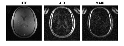
This work attempts to directly image ultrashort T2 myelin signals using ultra short echo time (UTE) MRI. Long T2 water signals were suppressed using either adiabatic inversion recovery (AIR) to null signal of a single T1, or multiple adiabatic inversion recovery (MAIR) to null signal over a range of T1s. AIR-UTE showed contrast in white matter, but no such signal was observed in MAIR-UTE. These findings indicate that the AIR-UTE white matter signals are unsuppressed water signals and that the solid proton signals of myelin decay too quickly to be observed by UTE MRI.
|
|
4896.
 |
3 |
Silent Myelin Imaging with a dipolar-coupled/inhomogeneous MT-Prepared ZTE Radial Sequence
Tobias Wood, Emil Ljungberg, Ana-Beatriz Solana Sanchez, Florian Wiesinger
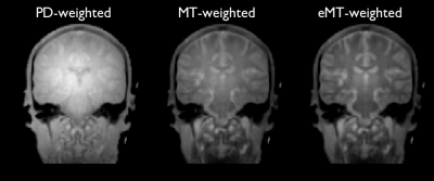
We generated myelin-specific contrast in a silent radial ZTE sequence using a dipolar-coupled MT-prep module. This sequence has great potential for visualising myelin in patient cohorts that do not tolerate the noise from standard MRI, such as infants.
|
|
4897.
 |
4 |
Improved estimates of the g-ratio by modelling its contribution to complex signal evolution in GRE data
Mark Drakesmith, Elena Kleban, Fabrizio Fabrizio, Derek Jones
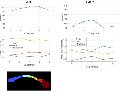
g-ratio is an important parameter of axon physiology and there is great interest in estimating it non-invasively in MRI. Existing approaches rely on fitting to a multi-compartment model and calculating g-ratio from the estimated volume fractions (Stikov et al, 2015). Here, we show that we can get improved estimates of the g-ratio by modelling its contribution to frequency offsets in GRE data using a hollow cylinder fibre model. Through simulations and model fitting to in vivo human GRE data we show g-ratio estimates are improved and closer to values obtained from histology compared with the existing approach.
|
|
4898.
 |
5 |
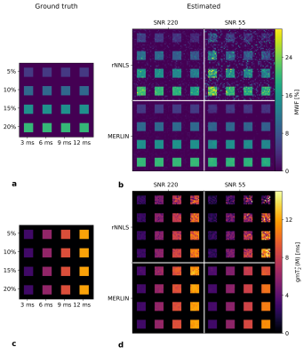
Multi-exponential Relaxometry using ℓ1ℓ1-regularized
Iterative NNLS (MERLIN) for Accurate and Robust Myelin Water Fraction Imaging
Markus Zimmermann, Ana-Maria Oros-Peusquens, Zaheer Abbas, Elene Iordanishvili, Seonyeong Shin, Seong Dae Yun, N. Jon Shah
A new parameter estimation algorithm, MERLIN, is presented for accurate and robust multi-exponential relaxometry using MRI. Multi-exponential relaxometry is fundamentally ill-conditioned, and as such, is extremely sensitive to noise. MERLIN is a fully automated, multi-voxel approach that incorporates ℓ1ℓ1-regularization
to enforce sparsity and spatial consistency of the estimated distributions. The proposed method is compared to the conventional ℓ2ℓ2-regularized
NNLS (rNNLS) in simulations and in vivo experiments, using a multi-echo gradient-echo (MEGE) sequence at 3T. The estimated water fraction maps from MERLIN are spatially more consistent, more precise, and more accurate, reducing the root-mean-squared-error by up to 90 percent in simulations.
|
|
4899.
 |
6 |
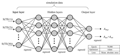
Feasibility study on artificial neural network based myelin water fraction mapping
Soozy Jung, Hongpyo Lee, Kanghyun Ryu, Jaeeun Song, Yoonho Nam, Hojoon Lee, Donghyun Kim
We developed an artificial neural network (ANN) using magnitude 3-pool signal model based training sets. Simulations were performed for evaluation with various SNR and slice inhomogeneity (GZ) levels. Two in-vivo data sets were tested. The results show decreased mean error and standard deviation when using the ANN model. The ANN model was more stable than the fitting method for different GZ values. Moreover, the processing time of the ANN model took 140 times less than the fitting method.
|
|
4900.
 |
7 |
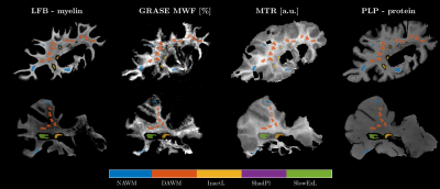
Sensitivity of in vivo myelin imaging techniques to detect subtle changes in myelin lipid and protein content in post-mortem multiple sclerosis brain tissues
Vanessa Wiggermann, Verena Endmayr, Enedino Hernandez-Torres, Romana Höftberger, Gregor Kasprian, Alexander Rauscher, Simon Hametner
Previous post-mortem single-slice myelin water fraction (MWF) measurements have shown good correlations with the myelin lipid fraction across tissue types. However, the role of protein content was not assessed nor have validations been performed for the whole brain 3D-Gradient and Spin Echo (GraSE) technique that has been employed in recent studies. We showed that 3D-GraSE based MWF measurements reliably distinguished regions of different myelin integrity reflective of difference in myelin lipid and protein content. In contrast, subtle variations in MWF within tissue classes or between persons may relate to differences in protein content.
|
|
4901.
 |
8 |
Robust myelin water imaging from multi-echo T2 data using second-order Tikhonov regularization with control points
Erick Canales-Rodriguez, Marco Pizzolato, Gian Piredda, Tom Hilbert, Kunz Nicolas, Tobias Kober, Jean-Philippe Thiran, Caroline Pot, Alessandro Daducci
Myelin water imaging is an MRI technique used to quantify myelination in the brain. The state-of-the-art reconstruction method is based on non-negative least squares optimization with zero-order Tikhonov regularization. In this study, a second-order Tikhonov regularization approach with control points was examined. This penalty term is more efficient for promoting smooth solutions while minimizing the contamination between myelin and non-myelin components. The performance of the proposed algorithm was investigated on in-vivo and ex-vivo multi-echo T2 data. It exhibited a higher correlation with histology than the state-of-the-art method. Its stability was studied using scan-rescan data.
|
|
4902.
|
9 |
A Model-Based Method for Estimation of Myelin Water Fractions
Yudu Li, Rong Guo, Yibo Zhao, Yang Chen, Bryan Clifford, Tianyao Wang, Chenyan Wang, Yiping Du, Yao Li, Zhi-Pei Liang
Quantitative mapping of myelin water fractions (MWF) can substantially improve our understanding of the progression of several demyelination white matter diseases such as multiple sclerosis. While MWF can be determined from both T2-weighted and T2*-weighted imaging data, it is much faster to collect T2*-weighted imaging data. However, estimation of MWF from T2*-weighted imaging data using a multi-exponential component model is an ill-conditioned problem whose solutions are often very sensitive to noise and modeling errors. In this work, we address this problem using a new model-based method. This method is characterized by: a) absorbing the spectral priors using the Bayesian-based statistical framework, and b) absorbing the spatial priors using a reproducing kernel based model. Both simulation and experimental results show the proposed method significantly outperforms the conventional parameter estimation methods currently used for MWF estimation.
|
|
4903.
 |
10 |
Analysis of Gradient Echo Myelin Water Imaging (GRE-MWI) for water exchange and scan parameters
Hyeong-Geol Shin, Se-Hong Oh, Joon Yul Choi, Kyeongseon Min, Hyunsung Eun, Jongho Lee
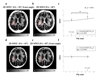
In this study, we investigate the effects of the compartmental water exchange on gradient echo myelin water imaging (GRE-MWI). We simulate MWF variation from different scan parameters (flip angle and TR) using a four pool white matter model and compare the simulation results with the in-vivo measurements. The results demonstrate that 1) the simulation with the water exchange better explains the in-vivo results and 2) GRE-MWI with a long TR can provide robust myelin water quantification regardless of changes in flip angle. Therefore, our results suggest GRE-MWI with a long TR as a robust option for myelin water imaging.
|
|
4904.
 |
11 |
Influence of model settings on myelin water fraction and frequency distribution for gradient-echo MRI at 7 Telsa
Kiran Thapaliya, Viktor Vegh, Steffen Bollmann, Markus Barth
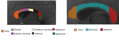
Quantitative assessment of model parameters (water fraction and frequency shift) estimated using a multi-compartment model can be useful to study tissue properties in white matter. In this work, we utilise multi-compartment models for multi-echo gradient echo data acquired at 7T. We investigate the variation of model parameters that could potentially be affected by differences in tissue microstructure in the corpus callosum. We further study the effect of different models (number of compartments and parameters) on the estimation of tissue parameters. We show that the tissue parameters vary across the sub-regions of the corpus callosum and are effected by different modelling choices.
|
|
4905.
 |
12 |
Gradient echo modelling with macroscopic field variations and large flip angles
Martin Soellradl, Stefan Ropele, Christian Langkammer
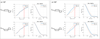
The signal decay of a 2D gradient echo sequence is substantially influenced by macroscopic field variations along the slice profile. Here we propose a numerical model describing the signal decay due to a macroscopic field gradient for arbitrary excitation pulses with large flip angles. Phantom and in-vivo experiments show that accurate modelling requires inclusion of the phase along the slice profile and the polarity of the slice selection gradient. Additionally, we show that applying the model yields better results for R2*-mapping and myelin water fraction estimation.
|
|
4906.
 |
13 |
In vivo assessment of the anisotropy of R2* maps in white matter
Renat Sibgatulin, Andreas Deistung, Daniel Güllmar, Christoph Birkl, Stefan Ropele, Jürgen Reichenbach
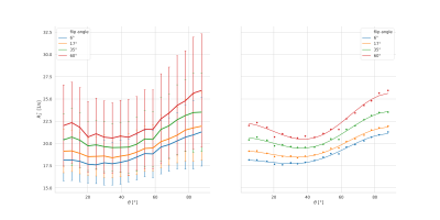
The effective transverse relaxation rate (R2*) is increasingly used in quantitative MRI, and its dependence on the orientation of white matter fibers in the brain has received significant attention. In this contribution, we assess the effect of the flip angle of a multi-echo gradient-echo sequence on the orientation dependence of the derived R2*map and suggest a simplified explanation to the observed R2*(θ; FA) behavior.
|
|
4907.
 |
14 |
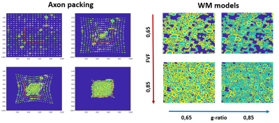
Estimation of microstructural properties of white matter from multiple orientation GRE signal simulations of realistic models
Renaud Hedouin, Kwok-Shing Chan, Riccardo Metere, Jose Marques
This study presents the creation of 2D white matter models, based on real histologically derived axon shapes, with large range of microstructure parameters (FVF, g-ratio). These models are used to simulate the complex gradient echo signal evolution under different main magnetic field orientations for (amongst other parameters) varying magnetic susceptibility and water density in the myelin compartment. A deep learning network, trained from those data, shows its capacity to recover parameter microstructure properties as g-factor and susceptibility on test data.
|
|
4908.
 |
15 |
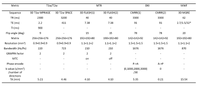
Evaluating the sensitivity of T1w/T2w, MTR, MWF and DKI to variation of myelin content
Run Pu, Hongjian He, Zhe Wu, Yi Sun, Jianhui Zhong
MRI metrics such as T1w/T2w ratio, magnetization transfer ratio (MTR), myelin water faction (MWF) and diffusion kurtosis imaging (DKI) indices have been used to detect myelin content. To assess the sensitivity of above metrics to variation of myelin content, in vivo human corpus callosum is used as a test case in the study. The results suggest that MTR varies least but MWF varies the most as myelin content changes.
|
|
4909.
 |
16 |
Stain-free histology to validate quantitative MRI
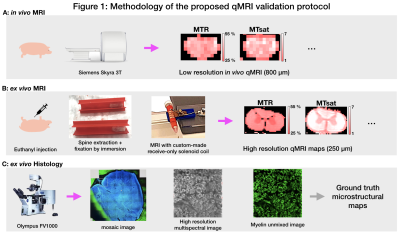
Gabriel Mangeat, Harris Nami, Nicolas Pinon, Alexandru Foias, Nikola Stikov, Tobias Granberg, Julien Cohen-Adad
Quantitative MRI (qMRI) is reproducible but often lacks calibration and/or specificity to the underlying microstructure. Light transmission optical histology of stained tissue is a popular method for validation, however, it is hampered by calibration issues and inhomogeneous penetration of staining agents. We propose a method to validate quantitative MRI metrics using stainless histology by utilizing the innate autofluorescence spectra of tissues when excited with ultraviolet laser. We demonstrate a proof-of-concept application of a qMRI validation pipeline on a pig spinal cord section with in vivo and ex vivo qMRI followed by histological autofluorescence microscopy to quantify myelin content.
|
|
4910.
 |
17 |
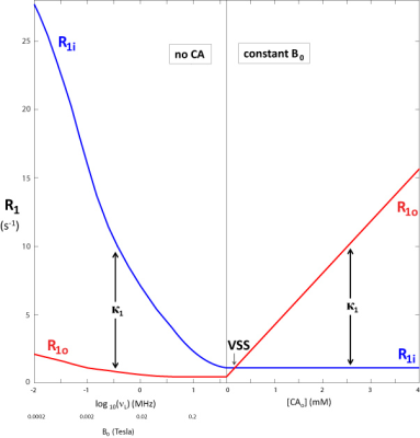
Apparent Population Inversion Due to Steady-State Transcytolemmal Water Exchange
Xin Li, Silvia Mangia, Jing-Huei Lee, Ruiliang Bai, Charles Springer
The homeostatic cellular water efflux rate constant, kio, has a significant contribution from cell membrane sodium pump activity previously unmeasurable. With high extracellular contrast agent concentration or ultra-low magnetic field, kio can be precisely determined by two-site-exchange analysis of in vivo 1H2O longitudinal relaxation data. With the low field case, there is an inversion of the apparent tissue compartmental contributions from the true values. The NMR shutter-speed organizing principle informs an analysis spanning the entire range of conditions.
|
|
4911.
 |
18 |
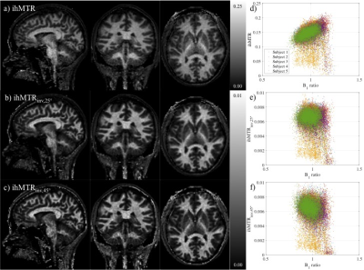
An inhomogeneous magnetization transfer (ihMT) quantification method robust to B1 and T1 variations in magnetization prepared acquisitions
Gopal Varma, Fanny Munsch, Olivier Girard, Guillaume Duhamel, David Alsop
Standard MT and ihMT ratio (ihMTR) measures can be sensitive to B1 and T1, making them less specific to tissue microstructure. Using the inverse signal, i.e. one divided by the signal, and a high flip-angle reference image in calculation of an ihMTR metric has been proposed as a metric with improved insensitivity to T1 and B1 in steady-state gradient-echo sequences. We present a modified method for use in prepared sequences such as magnetization prepared rapid gradient echo (MPRAGE). The sensitivity of ihMT MPRAGE metrics to T1 and B1 was tested using simulations and acquisitions in brains of healthy volunteers.
|
|
4912.
 |
19 |
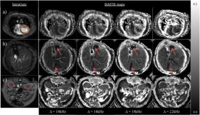
In vivo inhomogeneous magnetization transfer (ihMT) outside the brain using radial ultra-short echo-time acquisitions
Gopal Varma, Cody Callahan, Olivier Girard, Guillaume Duhamel, Aaron Grant, David Alsop
Inhomogeneous magnetization transfer (ihMT) effects have been readily observed in myelinated structures. The advent of low duty-cycle ihMT to increase the signal allows application of ihMT in other tissues. In this work, we explore the feasibility of applying ihMT in non-myelinated tissues such as the heart, liver, and kidneys of mice. This is achieved using a radial, ultra-short echo-time acquisition for greater motion robustness. The results demonstrate a measurable ihMT signal outside the central nervous system. Thus the microstructure of such tissues might be assessed based on the dipolar order contribution to ihMT.
|
|
4913.
 |
20 |
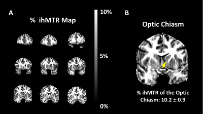
Myelin-sensitive imaging of the optic chiasm and optic nerve at 3T using inhomogeneous magnetization transfer (ihMT) with high B1 pulses
Ece Ercan, Fang Yu, Ivan Dimitrov, Gopal Varma, David Alsop, Robert Lenkinski, Elena Vinogradov
Inhomogeneous magnetization transfer (ihMT) imaging is a novel enhanced magnetization transfer contrast, which has been shown to originate from long-lived dipolar couplings in the tissue (e.g. dipolar couplings between the methylene molecules of the myelin phospholipid bilayer). In this study, we optimized an ihMT scan protocol for imaging the optic nerve and chiasm for the first time. This method may potentially be used for quantitative evaluation of patients with multiple sclerosis (MS), as well as other diseases affecting the visual pathway.
|
|
4914.
 |
21 |
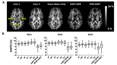
Investigating the Influence of Adipose Fat on the Inhomogeneous Magnetization Transfer (ihMT) Images
Ece Ercan, Gopal Varma, Ivan Dimitrov, Marco Pinho, Shu Zhang, Xinzeng Wang, Ananth Madhuranthakam, David Alsop, Robert Lenkinski, Elena Vinogradov
Inhomogeneous Magnetization Transfer (ihMT) imaging is a novel enhanced magnetization transfer technique. In this study, we investigated the influence of fat (i.e. adipose tissue) and echo time on the ihMT ratio through simulation, phantom, and in vivo studies. A substantial variation on the ihMTR values in the presence of fat is illustrated, depending on the echo times used.
|
|
4915.
 |
22 |
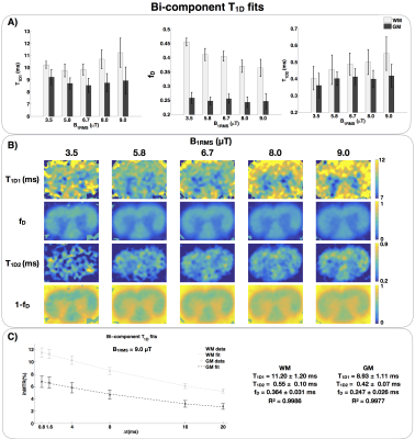
Assessment of two T1D components within myelinated tissue with ihMT MRI
Victor Carvalho, Olivier Girard, Andreea Hertanu, Samira Mchinda, Lucas Soustelle, Axelle Grélard, Antoine Loquet, Erick Dufourc, Gopal Varma, David Alsop, Pierre Thureau, Guillaume Duhamel
T1D, the relaxation time of the dipolar order, is a probe for membrane dynamics and organization that could be used to assess myelin integrity. A single-component T1Dmodel associated with a modified ihMT sequence had been proposed for in vivo evaluation of T1D with MRI. However, experiments and simulations revealed that myelinated tissues exhibit multiple T1D components. A bi-component T1D model is proposed and validated. Fits in a rat spinal cord yield two T1Ds of about 10 ms and 400 μs. The results suggest that myelin has a dynamically heterogeneous organization.
|
|
4916.
 |
23 |
Reproducibility of inhomogeneous magnetization transfer (ihMT): a test-retest, multi-site study
Lei Zhang, Huipeng Ren, Qing Fan, Xiaocheng Wei, Zhuanqin Ren
Derived from conventional magnetization transfer, inhomogeneous magnetization transfer (ihMT) has been shown to be a promising method for myelin imaging in recent studies. In the present study, the test-retest reproducibility and multi-site variability of ihMT in measuring major white matter fibers were evaluated. Good test-retest reproducibility and multi-site agreements were obtained. These findings support the use of ihMT measurements as biomarkers in multicenter and/or longitudinal studies.
|
|
4917.
 |
24 |
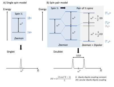
On the dipolar order underlying broad macromolecular lines
Olivier Girard, Victor Carvalho, Ludovic de Rochefort, Andreea Hertanu, Pierre Thureau, Gopal Varma, David Alsop, Guillaume Duhamel
Dipolar order has recently regained attention in MRI to analyze dipolar broadened lines in CEST and inhomogeneous Magnetization Transfer (ihMT), leading to new frequency irradiation patterns for enhanced saturation and access to an unexplored degree of freedom. A better understanding of dipolar order is of great interest to guide intuition and may lead to fundamental optimization of the ihMT technique, which is a promising tool providing new tissue contrasts. In this contribution we propose to review this concept, considering a simplified model of isolated proton pairs and the general Provotorov theory of RF saturation which applies to an ensemble of coupled spin.
|
|
4918.
 |
25 |
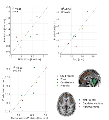
Non-invasive detection of molecular profiles in the human brain.
Shir Filo, Oshrat Shtangel, Aviv Mezer
Lipids makes more than 40% of the human brain in dry weight, and have broad information carrying roles in the CNS. In-vivo quantitative MRI (qMRI) aims at characterizing the biological properties of brain tissue. However, it lacks specificity to the molecular environment. Here, we present a novel biophysical framework that allows to decode the lipid composition of brain tissue from the MRI signal. First, we tested our approach on lipid samples of known composition. Next, by comparing the our molecular-specific measures to postmortem histological data, we were able to predict in-vivo lipidomic profiles in the human brain.
|
|
| Top |
QSM
Digital Poster
Contrast Mechanisms
Thursday, 16 May 2019
| Exhibition Hall |
14:45 - 15:45 |
| |
|
Computer # |
|
4919.
 |
26 |
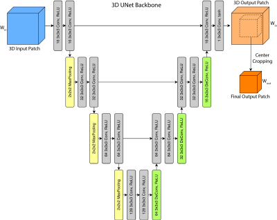
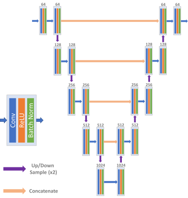
Global Information Matters in Quantitative Susceptibility Mapping Using 3D Fully Convolutional Neural Networks
Yicheng Chen, Angela Jakary, Christopher Hess, Janine Lupo
Recent research has shown that deep convolutional neural networks (DCNNs) have the potential to solve the ill-posed dipole inversion problem in quantitative susceptibility mapping (QSM). This study investigates the effects of patch-based QSM reconstruction by modifying a DCNN to take global susceptibility-phase relation into consideration.
|
|
4920.
 |
27 |
Magnitude and phase based mapping of particle concentrations - effects of diffusion
Lukas Buschle, Christian Ziener, Sabine Heiland, Martin Bendszus, Heinz-Peter Schlemmer, Felix Kurz
The magnitude and phase of the gradient echo signal in biological tissue highly depend on its iron concentration. A quantitative evaluation of the iron concentration, however, is complicated due to the complex interplay between susceptibility and diffusion effects. In this work, we analyze the gradient echo signal as well as the spin echo signal of uniformly distributed particles, with inclusion of diffusion and susceptibility effects, and provide analytical relations that connect magnitude, phase and iron concentration. This allows a quantitative description of the iron concentration based on magnitude or phase images.
|
|
4921.
 |
28 |
Quantitative Susceptibility Mapping of the Brain – A Comparative In vivo Study of Humans and Nonhuman Primates
Rakshit Dadarwal, Luzia Hintz, Amir Moussavi, Susann Boretius
Quantitative susceptibility mapping of the brain was performed in healthy humans and cynomolgus monkeys at comparable age using almost identical MR parameters, including the magnetic field strength. This comparative study revealed very similar values of magnetic susceptibility in gray matter structures between the two species, but a significantly lower magnetic susceptibility in parts of the corpus callosum of monkeys compared to the humans. This difference may be related to differences in the position of fiber tracts relative to the magnetic field lines, but it may also reflect differences in iron content, fiber density, and myelination.
|
|
4922.
 |
29 |
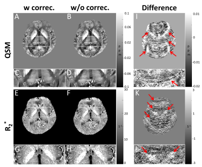
Improvement of Reproducibility in Quantitative Susceptibility Mapping (QSM) and Transverse Relaxation Rates (R2*) after Physiological Noise Correction
Joon Yul Choi, Jingu Lee, Yoonho Nam, Woojin Jung, Jongho Lee, Se-Hong Oh
Respiration-induced local magnetic field variation makes artifacts in gradient echo based images and reduces reproducibility of QSM and R2*. This study investigated reproducibility after respiration-induced error correction. The results showed a significantly improved reproducibility in QSM and R2* mapping.
|
|
4923.
 |
30 |
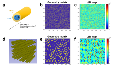
R2, R2* and quantitative susceptibility mapping (QSM) changes of corpus callosums in aging rats: Possible contributions from myelin thickness
HwaPyeong Cho, Seokha Jin, HyungJoon Cho
Myelin is essential component for complex motor, sensory and cognition function.1 Among many quantitative magnetic resonance imaging (MRI) techniques investigating myelin structure, direct MR parameter influenced by the myelin thickness is rarely investigated.1 Here, we study the effect of myelin thickness on R2, R2* and susceptibility using the finite perturber method (FPM)-based simulation and post-mortem aging rat brains. It is observed from simulations that both myelin thickness and myelin volume fraction (MVF) affects R2 and R2* values, whereas the phase change (QSM) showed a significant change only by the change of MVF. In preliminar experiments, consistent results were observed.
|
|
4924.
 |
31 |
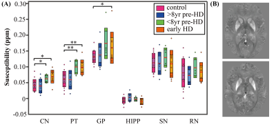
Altered Brain Iron Content and Deposition Rate in Huntington Disease Indicated by Quantitative Susceptibility MRI
Lin Chen, Jun Hua, Christopher Ross, Shuhui Cai, Peter van Zijl, Xu Li
We investigated the natural history of brain iron content at different stages of premanifest and manifest Huntington disease (HD) as indicated by changes of magnetic susceptibility values measured by quantitative susceptibility mapping (QSM). Higher susceptibilities were observed in striatum and globus pallidus of closer-to-onset premanifest HD and early HD patients, but not in the further-from-onset premanifest HD group as compared to controls using 1-way MANCOVA. Analysis using a general linear model showed significantly higher iron deposition rates (11.9%/yr in caudate and 6.1%/yr in globus pallidus) in closer-to-onset premanifest HD and early HD as compared to controls over a one-year follow-up.
|
|
4925.
 |
32 |
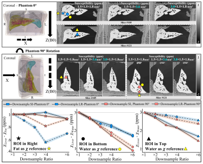
Characterization of Bias in Quantitative Susceptibility Mapping with Anisotropic Imaging Resolution: Studies in a Numerical Phantom, 3D Printed Liver Phantom, and In Vivo Patient Scans
Ante Zhu, Timothy Colgan, Scott Reeder, Diego Hernando
Quantitative susceptibility mapping (QSM) is a promising technique for measuring iron concentration in patients with liver iron overload. In liver QSM, the constraints of scan time in a breadth-hold and the requirement of a short first echo time lead to limited imaging resolution, with anisotropic voxels. In this work, we characterized bias in liver QSM with anisotropic imaging resolution in simulation, a 3D printed liver phantom and patients. Our study shows that resolution-induced bias is related to the downsampling direction and is spatially-varying. In vivo results suggest the liver imaging resolution along the left-right dimension may affect liver susceptibility measurements.
|
|
4926.
 |
33 |
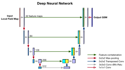
QSM Inversion Through Parcellated Deep Neural Networks
Juan Liu, Robin Karr, Brad Swearingen, Andrew Nencka, Kevin Koch
Quantitative Susceptibility Mapping (QSM) can estimate tissue susceptibility distributions and reveal pathology in conditions such as Parkinson's disease and multiple sclerosis. QSM reconstruction is an ill-posed inverse problem due to a mathematical singularity of the requisite dipole convolution kernel. State-of-art QSM reconstruction methods either suffer from image artifacts or long computation times. To overcome the limitations of these existing methods, a deep-learning-based approach is proposed and demonstrated in this work. 200 QSM datasets were utilized to compare current QSM reconstruction methods (TKD, closed-form L2, and MEDI) with the proposed deep-learning approach using visual scoring assessment of streaking artifacts and image sharpness. These multi-reader study results showed that the deep learning solution can produce QSM images with improved scores in both streaking artifacts and image sharpness evaluation while providing an almost instantaneous inversion computation through neural network inferencing.
|
|
4927.
 |
34 |
Quantitative Susceptibility Mapping (QSM) MRI in a Collagenase Rat Model of Intracerebral Hemorrhage (ICH)
*Kimmo Lehtimäki, Artem Shatillo, Elina Latonummi, Antti Nurmi
Management of ICH is critical for the recovery and appropriate imaging methods to follow the process are needed. ICH was induced by intra-striatal infusion of collagenase IV. Study consisted T2/diffusion-maps at 6 hours, 1, 3 and 14 days and ex vivo QSM at D1 and D3. QSM revealed large ICH lesions with low susceptibility core and high susceptibility outer rim. Histogram comparison showed modulation in susceptibilities; D1 with higher proportion edema processes and iron than D3. QSM method seems particularly suitable for in vivo applications to study ICH in rats due to proper lesion size and clear presence of iron.
|
|
4928.
 |
35 |
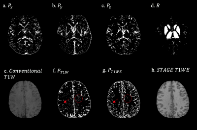
Structurally Constrained Quantitative Susceptibility Mapping
Sara Gharabaghi, Saifeng Liu, Ying Wang, Yongsheng Chen, Thomas Wischgoll, Nasser Kashou, E. Haacke
In this study, a structurally constrained susceptibility reconstruction method, SCSWIM, is proposed. This method employs the unique contrast of STAGE imaging and segmented basal ganglia and vessels. It is tested on both simulated and in vivo human brain data. Evaluations show the improved reliability of the geometry information, reduced streaking artifacts, and increased accuracy of the susceptibilities of both basal ganglia and veins in the SCSWIM compared to other methods.
|
|
4929.
 |
36 |
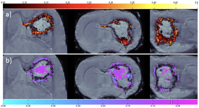
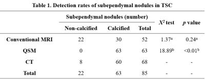
Investigation of quantitative susceptibility mapping (QSM) in diagnosis of tuberous sclerosis complex (TSC) and assessment of associated brain injuries at 1.5 Tesla
Lei Zhang, Zhuanqin Ren, Xiaocheng Wei
Tuberous sclerosis complex (TSC) often progress to serious clinical consequences which had close relationship with cortical/subcortical tubers and white matter lesions. Quantitative susceptibility mapping (QSM) is capable of quantitatively measure the susceptibility. However, little is known about the susceptibility changes of brain damage caused by TSC. This study aims to investigated the diagnostic value of QSM in TSC. Our results suggest that QSM can shown subependymal calcified nodules and provided the quantitative information of white matter damage. So, QSM sequence may have a complementary role in the conventional MRI evaluation of tuberous sclerosis patients.
|
|
4930.
 |
37 |
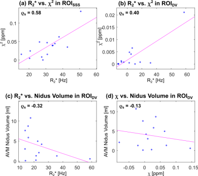
Evaluating the Relationship Between the Venous Magnetic Susceptibility (χχ)
and R∗2R2∗ of
Brain Arteriovenous Malformations (AVMs)
Emma Biondetti, Alvaro Rojas Villabona, Hans Jäger, David Thomas, Karin Shmueli
Arteriovenous malformations (AVMs) are characterised by arteriovenous shunting, which increases oxygenation in the veins draining the AVM compared to healthy veins. In healthy veins, a quadratic relationship is expected between the transverse relaxation rate (R∗2R2∗)
and the magnetic susceptibility (χχ).
By calculating χχ and R∗2R2∗ we
investigated whether this relationship holds in the AVM draining veins and superior sagittal sinuses of fourteen patients. We found a significant positive correlation between R∗2R2∗ and χ2χ2in
the healthy veins, but not in the AVM draining veins where the quadratic relationship is disrupted and χχ values
can be used to measure oxygenation.
|
|
4931.
 |
38 |
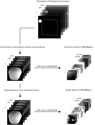
SHARQnet - Sophisticated Harmonic Artifact Reduction in Quantitative Susceptibility Mapping using a Deep Convolutional Neural Network
Mathias Olsen, Morten Larsen, Matilde Kristensen, Mads Pedersen, Lasse Østergaard, Kieran O'Brien, Christian Langkammer, Markus Barth, Steffen Bollmann
We propose a fully convolutional neural network for background field removal in MR phase images for Quantitative Susceptibility Mapping. Our proposed method, SHARQnet, learns to solve the background field problem from theoretical simulations of background field distributions, and the results are compared to current state-of-the-art methods like SHARP, V-SHARP, and RESHARP.
|
|
4932.
 |
39 |
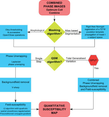
Quantitative susceptibility mapping for routine clinical use – An inline automated QSM reconstruction pipeline
Ashley Stewart, Kieran O'Brien, Jinsuh Kim, Bénédicte Maréchal, Fatimah Nasrallah, Michael Kean, Markus Barth, Steffen Bollmann
Quantitative susceptibility mapping (QSM) is a post-processing technique for gradient-recalled-echo (GRE) phase data, which provides information about tissue composition complementary to common Susceptibility Weighted Imaging (SWI). To date, QSM’s multiple complex processing steps has limited its clinical application. In this work, we present an automated and robust inline QSM post-processing pipeline compatible with flow-compensated GRE and VIBE sequences. The QSM pipeline includes morphological and atlas-based segmentation, two different QSM algorithms and is compatible with SWI processing. Two clinical cases of QSM in Traumatic Brain Injury and Multiple Sclerosis are presented.
|
|
4933.
 |
40 |
Quantitative Susceptibility Mapping using a Deep Learning prior
Zhe Liu, Jinwei Zhang, Shun Zhang, Pascal Spincemaille, Thanh Nguyen, Yi Wang
A Bayesian method is proposed by formulating deep learning outcome as a regularization in QSM reconstruction. It enforces the fidelity between the network generated QSM and the measured inhomogeneity field. Preliminary results indicate both quantitative and qualitative improvement over QSM by deep learning alone.
|
|
4934.
 |
41 |
A Comparsion Study of Ultrashort Echo Time Quantitative Susceptibility Mapping (UTE-QSM) with Different Sampling Trajectories
Xing Lu, Hyungseok Jang, Yajun Ma, Wenhui Yang, Eric Chang, Jiang Du
The ability to accurately and non-invasively quantify IONPs is desirable for many emerging applications, including for the evaluation of iron overload in the human body. 3D UTE Cones has demonstrated ability to detect high iron concentration with shorter echo times. In this study, we aimed to make clear whether the non-Cartesian sampling of Cones trajectory affects the accuracy of QSM. By comparing three different kinds of UTE sampling trajectory, as well as different stretch factors of Cones, the results show that no significant differences between these UTE QSM results were found.
|
|
4935.
 |
42 |
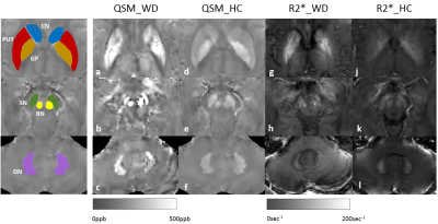
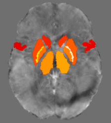
Measurement of Copper and Iron Accumulation in the Deep Gray Matter Nuclei of patients with Wilson Disease Using Quantitative Susceptibility Mapping and R2* Mapping
Presentation Not Submitted
Gaiying Li, Rong Wu, Rui Tong, Zhe Liu, Pascal Spincemaille, Kelly Gillen, Yi Wang, Xiaoping Wang, Jianqi Li
The objective of this study was to evaluate magnetic susceptibility and R2* values from QSM and R2* for differentiating Wilson Disease (WD) from healthy controls (HC). 14 WD and 14 HC subjects were scanned using a 3D multi-echo GRE sequence. The results indicated that susceptibility values in the caudate nucleus (CN), putamen (PUT), globus pallidus (GP), substantia nigra (SN), red nucleus (RN) were significantly higher in patients with WD as compared to those of HC. R2* values were significantly higher in WD patients in all ROIs. Receiver operating characteristic analysis showed that QSM provided the highest AUC=0.888 at SN.
|
|
4936.
 |
43 |
Quantitative Analysis of QSM Image for PD Basal-Cortico Circuit
Zhaoyu Lai, Chenfei Ye, Junyan Sun, Tao Wu, Junjie Liu, Pengzheng Zhou, Chushu Yang, Yuan Fang, Ting Ma
Parkinson’s disease is associated with iron accumulation, while quantitative susceptibility mapping can provide quantitative measures of magnetic susceptibility. To investigate the connection about iron deposition and PD etiology or progression, we focused on 16 regions in Basal-Cortico motional circuit by using quantitative susceptibility mapping. Combined with a series of Parkinson’s disease scale score, we derived the relationship between iron content and the scale scores.
|
|
4937.
 |
44 |
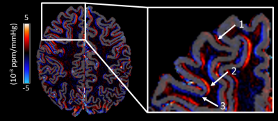
Functional quantitative susceptibility mapping (fQSM) during auditory stimulation
Mauro Costagli, Marta Lancione, Luca Cecchetti, Pietro Pietrini, Mirco Cosottini, Emiliano Ricciardi, Michela Tosetti
Functional Quantitative Susceptibility Mapping (fQSM) has two very appealing and promising features: it is a quantitative way of mapping brain function and it is considerably less affected by the non-local effects typical of the Blood Oxygenation Level-Dependent (BOLD) signal. Here, the response of the auditory cortex to the presentation of relatively short acoustic stimuli has been studied. The majority of voxels with positive BOLD responses exhibited negative fQSM responses, while some other voxels exhibited positive fQSM repsonses, which might reflect different interplays among changes in fractional oxygen saturation, cerebral blood flow and volume.
|
|
4938.
 |
45 |
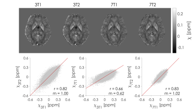
Echo time-dependent reproducibility of Quantitative Susceptibility Mapping at different field strength
Marta Lancione, Graziella Donatelli, Paolo Cecchi, Mirco Cosottini, Michela Tosetti, Mauro Costagli
The assessment of reproducibility of Quantitative Susceptibility Mapping (QSM) is critical in multi-center studies and clinical follow-ups. However, many experimental factors and acquisition parameters may compromise quantification accuracy. In this work, we analyze the impact of echo time on intra-scanner repeatability and inter-scanner reproducibility of QSM using a 3D multi-echo GRE sequence on MRI scanners of different field strength (3T and 7T) from the same vendor.
|
|
4939.
 |
46 |
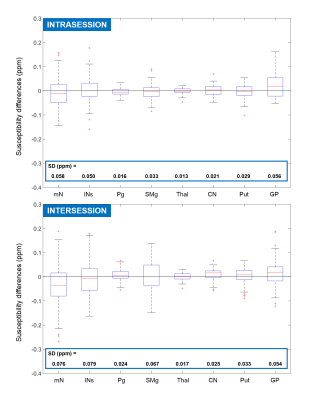
High Repeatability of Quantitative Susceptibility Mapping (QSM) in the Head and Neck With a View to Detecting Hypoxic Cancer Sites
Anita Karsa, Shonit Punwani, Karin Shmueli
As hypoxic tumours in the head-and-neck are more resistant to radiation therapy, there is a pressing clinical need to measure tumour oxygenation non-invasively. Since deoxyhemoglobin in the blood, which indicates hypoxia, is paramagnetic, QSM is a candidate technique. Here, we tested QSM’s repeatability in various head-and-neck regions in ten healthy volunteers to investigate the feasibility of detecting the susceptibility difference expected to result from hypoxia. We found low minimum detectable effect sizes in the lymph nodes (0.12 ppm), submandibular glands (0.08 ppm), and parotid glands (0.04 ppm). This high QSM repeatability paves the way for clinical studies.
|
|
4940.
 |
47 |
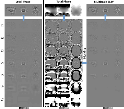
Multiscale Spherical Mean Value based background field removal method for Quantitative Susceptibility Mapping
Carlos Milovic, Christian Langkammer, Sergio Uribe, Pablo Irarrazabal, Julio Acosta-Cabronero, Cristian Tejos
We present a multiscale SMV implementation (MSMV) for background field removal in QSM. We use a combined redundant Laplacian decomposition and Laplacian pyramid approach with fuzzy masks to remove background fields and reconstruct the local field. We tested this algorithm against PDF, LBV, ESHARP and VSHARP in analytic phantom and in vivo experiments. Experiments show MSMV’s accuracy to be in par with VSHARP, with an order of magnitude speed gain. MSMV achieved results with less harmonic remnants in our in vivo test. This multiscale approach may also be extended to the susceptibility inversion problem, with adaptive preconditioners and weighting.
|
|
4941.
 |
48 |
Dynamic quantitative susceptibility mapping to assess vascular compliance in the brain
Christoph Birkl, Christian Langkammer, Pascal Sati, Christian Enzinger, Franz Fazekas, Stefan Ropele
In this study we explored if quantitative susceptibility mapping (QSM) allows assessing blood pressure induced changes of the magnetic susceptibility in the brain as consequence of cerebral autoregulation. Eight healthy subjects underwent fast QSM at 3.0-T and simultaneous measurement of the mean arterial pressure (MAP) following a small drop in MAP caused by a change in posture. A linear relationship between MAP and susceptibility was observed, where the slope represents a measure of the cerebral vascular compliance with different signs for arterial and venous blood vessels.
|
|
4942.
 |
49 |
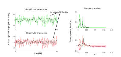
Correlation and frequency based analyses between functional QSM and fMRI
Pinar Ozbay, Lars Kasper, Burak Akin, Klaas Pruessmann, Daniel Nanz
Earlier works demonstrated applications of Quantitative Susceptibility Mapping (QSM) in functional MRI, including both task- and resting-state experiments. The focus had been mostly on the bi-directional activations consistently observed in fQSM. In this work, our aim was to compare the temporal and frequency characteristics of susceptibility and magnitude time-course signals. Importantly, we also included cardiac and respiration signals, and showed that the global susceptibility signal might inherently include more physiological information than the magnitude.
|
|
4943.
 |
50 |
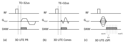
Readout Duration-dependent Bias on R2* Mapping and Quantitative Susceptibility Mapping Using 3D Radial and Cones Acquisitions at 3.0T
Shihan Qiu, Timothy Colgan, Ante Zhu, Kevin Johnson, Scott Reeder, Diego Hernando
Ultra-short TE (UTE) R2* mapping and Quantitative Susceptibility Mapping (QSM) are emerging techniques for quantifying iron deposition in various organs, including the brain and liver. In tissues with short T2* values (high R2*), the fast signal decay-induced errors during the relatively long readout in typical UTE acquisitions, i.e., 3D radial and cones UTE, may confound R2* and susceptibility measurements. In this study, we characterized the readout duration effects on R2* and susceptibility estimation in 3D radial and cones UTE-acquisitions at 3.0T. Simulation and phantom studies showed bias in the estimated R2* and susceptibility when long readout durations were used.
|
|
| Top |
Pseudo-Continuous & Velocity-Selective ASL
Digital Poster
Contrast Mechanisms
Thursday, 16 May 2019
| Exhibition Hall |
14:45 - 15:45 |
| |
|
Computer # |
|
4944.
 |
51 |
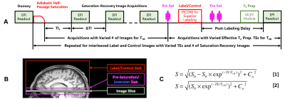
Cerebral spin compartmentalization based on biexponential modeling of T2-prepared pCASL 3D GRASE data
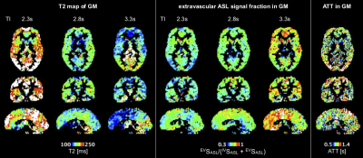
Martin Schidlowski, Markus Boland, Theodor Rüber, Tony Stöcker
In this work, a pCASL sequence with T2 preparation module and 3D GRASE readout was developed. We present a novel approach to estimate the spin compartment of labeled protons by a voxel-wise and biexponential fit to whole-brain ASL data. This method allows for the spatial quantification of intra- and extravascular spin fractions of the ASL signal as well as their temporal evolution.
|
|
4945.
 |
52 |
Combined estimation of dispersion and macrovascular signal in multi-PLD pCASL data using a two-component model
Merlijn van der Plas, Michael Chappell, Matthias van Osch
In pCASL a well-defined, box-shaped bolus is created at the labeling plane and for quantification this shape is assumed to be preserved, however, in reality this shape will be dispersed. With multi-timepoint data, the effects of dispersion can be observed in the macrovascular component, which can be separated from the tissue component using a two-component model. In this study the combined estimation of dispersion and macrovascular signal was investigated. When a gamma distribution dispersion kernel was incorporated into the two-component model, a significant decrease in CBF values was found, while a significant increase in macrovascular signal was observed.
|
|
4946.
 |
53 |
Robust arterial transit time estimation using combined acquisition of Hadamard-encoded pCASL and long-labeled long-delay pCASL: a simulation and in vivo study
Shota Ishida, Hirohiko Kimura, Naoyuki Takei, Yasuhiro Fujiwara, Tsuyoshi Matsuda, Masayuki Kanamoto, Nobuyuki Kosaka, R Lebel, Toshiki Adachi
A combination scan of 3-delay Hadamard-encoded pseudo-continuous ASL (H-pCASL) and single-delay pCASL with long labeling duration and long post-labeling delay was proposed as the robust arterial transit time (ATT) estimation for prolonged ATTs. Simulation showed that the mean normalized error of the proposed method was small for a wide range of ATTs compared to that of H-pCASL alone. In in vivo experiments, ATTs were not significantly different among the methods. However, 7-delay H-pCASL presented a lower ATT and larger variance. The proposed method improves the robustness of ATT estimation for prolonged ATTs with practical acquisition times in the clinical framework.
|
|
4947.
 |
54 |
Assessment of Hepatic Perfusion Before and After a Meal Challenge Using Pseudo-Continuous Arterial Spin Labeling in MRI: Comparison with Intravoxel Incoherent Motion and Phase Contrast
Saori Watanabe, Takashi Hamaguchi, Naoki Ohno, Yudai Shogan, Yu Ueda, Tadanori Takata, Satoshi Kobayashi, Tosiaki Miyati, Toshifumi Gabata
To assess hepatic blood flow (HBF) with a noninvasive method, we acquired HBF flow before and after meal ingestion using the pCASL method. In addition, we investigated the relationship of HBF, perfusion-related diffusion coefficient (D*) with intravoxel incoherent motion and portal vein blood flow (PVBF) with phase contrast. For each value of HBF, D*, and PVBF following meal ingestion increased significantly compared with the values prior to ingestion. However, there were no correlations between hepatic blood flow, perfusion-related diffusion coefficient, or portal flow with either pre- or post-ingestion.
|
|
4948.
 |
55 |
Influence of labeling parameters of velocity selective arterial spin labeling for renal perfusion imaging
Isabell Bones, Suzanne Franklin, Anita Harteveld, Matthias van Osch, Jeroen Hendrikse, Chrit Moonen, Marijn van Stralen, Clemens Bos
Velocity selective arterial spin labeling (VSASL) is a spatially non-selective method that labels spins based on their flow velocity, thereby labeling closer to the target tissue, reducing the influence of arterial transit time (ATT) and requiring no planning. In the abdomen, motion and complex vascular anatomy might, however, require dedicated VS-labeling parameters. We assessed the feasibility of VSASL for renal perfusion measurement by investigating its dependency on essential labeling parameters, and by comparing it with pseudo-continuous ASL (pCASL) as a spatially-selective reference ASL-technique. Our results show, that with carefully chosen sequence parameters, VSASL is feasible for renal perfusion measurement.
|
|
4949.
 |
56 |
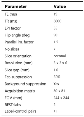
Enabling free-breathing renal pCASL with background suppression and motion correction: a comparison with paced-breathing
Isabell Bones, Anita Harteveld, Suzanne Franklin, Matthias van Osch, Jeroen Hendrikse, Chrit Moonen, Clemens Bos, Marijn van Stralen
Renal perfusion imaging using arterial spin labeling (ASL) is challenged by respiratory motion and physiologic noise, often dealt with by breathing instructions requiring patient cooperation. We investigated if background suppression (BGS) combined with image registration, guided by the ASL-images themselves or additionally acquired fat-images, would enable free-breathing renal ASL. To this end, free-breathing ASL was compared with paced-breathing ASL, both including BGS and image registration. BGS and registration improved the quality of free-breathing renal pCASL, showing increased temporal SNR similar to paced-breathing ASL, without reducing perfusion-weighted signal. In conclusion, free-breathing renal pCASL is possible when employing BGS and image registration.
|
|
4950.
 |
57 |
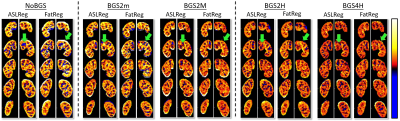
Quantification of intracranial vascular compliance using multi-PLD pseudo-continuous arterial spin labeling with retrospective cardiac gating
Soroush Heidari Pahlavian, Mayank Jog, Samantha Ma, Danny Wang, Lirong Yan
Intracranial vascular compliance (IVC) is an important factor in regulating the cerebral perfusion pressure and is believed to be linked to multiple neurological disorders. In this study, a retrospectively-gated multi-PLD pCASL technique was used to estimate arterial cerebral blood volume (aCBV) and compliance. Our results showed that this technique can quantify cardiac-induced variations of aCBV as well as IVC distribution maps.
|
|
4951.
 |
58 |
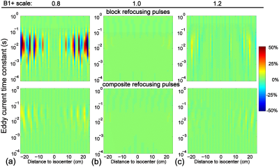
Exploring dynamic RF shimming for labelling in PCASL at 7T
Christopher Mirfin, Paul Glover, Richard Bowtell
Despite the intrinsic SNR gains at 7T, pseudo-continuous arterial spin labeling (PCASL) is limited by poor |B+1| coverage
in the labelling plane and the associated high local SAR of the sequence. In this work we perform simulations to consider the usefulness of dynamic RF shimming using a commercially available head-only RF coil equipped with 8-transmit channels, for labelling in PCASL.
|
|
4952.
 |
59 |
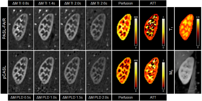
Comparison of multi-delay renal PASL-FAIR and pCASL perfusion quantification at 3T
Anita Harteveld, Anneloes de Boer, Suzanne Franklin, Tim Leiner, Marijn van Stralen, Clemens Bos
ASL has emerged as a non-invasive tool for measuring renal perfusion. Whereas in the brain consensus leans towards pCASL as the preferred labeling strategy, in the kidney PASL-FAIR has been reported on most. A systematic comparison of renal PASL-FAIR and pCASL perfusion measurement was performed at 3T in 16 volunteers, with separate visits to assess repeatability. PASL-FAIR perfusion values were significantly higher than those obtained with pCASL. Moreover, at 3T PASL-FAIR had approximately 2-3 times better repeatability compared to pCASL.
|
|
4953.
 |
60 |
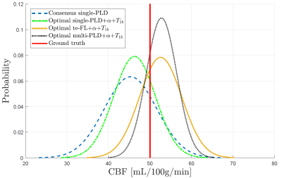
Beyond the consensus: what to include when 5 minutes are available for perfusion imaging by PCASL?
Piet Bladt, Matthias van Osch, Eric Achten, Arnold den Dekker, Jan Sijbers
While the consensus statement on the recommended implementation of arterial spin labeling (ASL) has advanced ASL to clinical application, variations in labeling efficiency, longitudinal relaxation time of blood and arterial transit times can cause significant quantification errors. With simulation experiments, it is shown that sacrificing ASL scan time for measurements of these parameters improves the estimation reproducibility of the cerebral blood flow on a population level. Furthermore, multi-delay ASL modalities in combination with these extra measurements can compete with or outperform the single-delay consensus implementation in terms of estimation accuracy and precision, depending on the underlying distribution of transit times.
|
|
4954.
 |
61 |
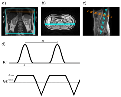
Optimization of Pseudo Continuous Arterial Spin Labeling for renal ASL
Rebeca Echeverria-Chasco, Marta Vidorreta, Verónica Aramendía-Vidaurreta, Gorka Bastarrika, María A. Fernández-Seara
Arterial Spin Labeling (ASL) is a non-contrast MR perfusion imaging technique. Pseudo continuous ASL (pCASL) is one of its recomended implementations. The efficiency of pCASL has been shown to be dependent on velocity and magnetic field variations. pCASL was assessed through simulations for the measured velocities in the aorta and including off-resonance effects. Five volunteers were imaged with different average gradient to ratio combinations. The results showed that aorta velocities and off-resonance effects shifted the efficiency towards lower ratios and to a constrained smaller range of gradients. A p-value of 0.04 demonstrated that differences in efficiency were significant across Gave values.
|
|
4955.
 |
62 |
Alternative Slice Acquisition Orders for High-Resolution MB-EPI PCASL Imaging with Background Suppression
Xiufeng Li, Dingxin Wang, Gregory Metzger, Essa Yacoub, Kamil Ugurbil
Relative static tissue signal differences between neighboring slices across slice bands in MB-EPI PCASL imaging with background suppression (BS) are dramatically larger than those in MB-EPI PCASL imaging without BS, and can result in severe subtraction errors/artifacts for imaging data with large subject motion that sometimes cannot be corrected or removed by motion correction. To resolve this issue, alternative slice acquisition orders are proposed and evaluated. Our results suggest that the proposed alternative slice acquisition orders can improve the robustness of MB-EPI PCASL imaging with BS, providing comparable CBF estimates with minimized subtraction errors.
|
|
4956.
 |
63 |
Implementation and validation of ASL perfusion measurements for population-based imaging
Esther Warnert, Rebecca Steketee, Meike Vernooij, Mika Vogel, Juan-Antonio Hernandez-Tamames, Gyula Kotek
Pseudocontinuous ASL (pCASL) is an ideal tool for non-invasive perfusion measurements in population-based imaging studies, which require longitudinal scanning with an unchanging MRI hardware and software set-up. Herein we present the results of the implementation and validation of a 3D pCASL sequence for use in the Rotterdam Study, running since 2005.
|
|
4957.
 |
64 |
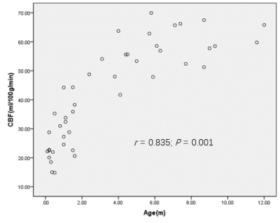
Investigation of the effects of age and gender on normal cerebral blood flow in infants using arterial spin labeling MRI
KEKE ZHAO, ZHUANQIN REN, Xiaocheng Wei
This study systematically revealed normal values of cerebral blood flow (CBF) in different age groups of infants using three-dimensional pseudocontinuous arterial spin labeling (3D PCASL) technique. Our results demonstrated a significantly lower CBF value in neonates than in other age groups. We also found a significant positive correlation between age and various regional mean gray matter (GM) and white matter (WM) CBF values in infants. Taken together, our findings demonstrated benefits of the application of the infants perfusion imaging technology to the clinical field by using arterial spin labeling (ASL) to provide information of metabolic status and neurodevelopmental outcomes.
|
|
4958.
 |
65 |
Arterial spin labeling reveals altered cerebral vascular reactivity to carbon dioxide challenge in Q175 mouse model of Huntington's disease
Somaie Salajeghe, Johan Van Audekerke, Verdi Vanreusel, Dorian Pustina, Longbin Liu, Mette Skinbjerg, Celia Dominguez, Ignacio Munoz-Sanjuan, Annemie Van der Linden, Marleen Verhoye
CVR deficits can cause a negative effect on neurovascular coupling leading to blood delivery impairment in activated brain regions. As such, impaired CVR may lead to neural degeneration over a period of time. We measured CBF and CVR using pCASL in Q175 mouse model of Huntington’s disease (11 transgenic and 10 wild-type at 15 month). In order to measure CVR, we measured changes in CBF during a 10% CO2 vascular challenge. The results indicated an overall decreased CVR in transgenic compared to wild-type mice.
|
|
4959.
 |
66 |
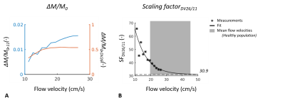
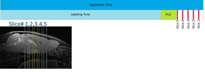
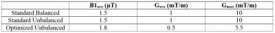
Pseudo-continuous arterial spin labeled renal perfusion imaging at 3T with improved robustness to off-resonance
Joshua Greer, Yiming Wang, Ivan Pedrosa, Ananth Madhuranthakam
Pseudo-continuous arterial spin labeling (pCASL) has been applied for renal perfusion imaging, where inflowing blood is labeled in the descending aorta, just above the kidneys. However, in some cases when the labeling plane is positioned close to the lungs, significant decreases in SNR have been observed. We hypothesized that this was due to decreased labeling efficiency caused by the off-resonance effects near the lungs. In this study, an unbalanced pCASL gradient scheme that was optimized to be more robust to B0 inhomogeneities was compared with default implementations of pCASL at different labeling locations along the descending aorta.
|
|
4960.
 |
67 |
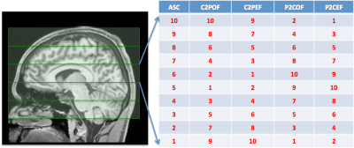
Convolutional Neural Network based Automatic Planning for Pseudo-Continuous Arterial Spin Labeling
Michael Helle, Thomas Lindner, Karsten Sommer
Pseudo-continuous arterial spin labeling (pCASL) requires careful planning of the labeling plane to achieve high labeling efficiency, which makes the quality of the imaging results dependent on the experience of the operator. Here we demonstrate the feasibility of using a convolutional neural network to automatically predict an appropriate labeling position based on angiography images, thereby allowing for fully automatic pCASL perfusion scans.
|
|
4961.
 |
68 |
Assessing Morphology of Cerebral Macro- and Microvasculature Using Dynamic Perfusion Tensor Imaging ASL
Leonie Petitclerc, Suzanne Franklin, Lydiane Hirschler, Matthias van Osch
Time-encoded pseudo-continuous ASL was combined with bipolar crusher gradients to measure a time-resolved perfusion tensor of the brain vasculature. Gradients provided a high degree of attenuation of the intravascular signal which increased with greater gradient strength and decreased (down to 25%) at long post-labeling delays (PLDs). Perfusion tensor images showed correspondence with known structures such as the anterior cerebral artery and the circle of Willis. Fractional anisotropy of perfusion remained elevated and increased with longer PLDs. Adjustments in gradient strength and time-encoding scheme may permit the imaging of microvascular structure.
|
|
4962.
 |
69 |
Compensating T2 blurring in 3D TSE with Cartesian acquisition based arterial spin labeled MRI
Yiming Wang, Joshua Greer, Trevor Wigal, Marco Pinho, Joseph Maldjian, Ananth Madhuranthakam
3D fast/turbo spin echo (FSE/TSE) acquisitions are preferred for arterial spin labeled (ASL) MRI due to their higher SNR and compatibility with background suppression. However, 3D TSE suffers from T2 blurring caused by the T2 decay of the ASL signal along the prolonged echo train lengths, which may degrade image quality. This is often more noticeable in 3D TSE with Cartesian acquisitions. In this study, a truncated k-space filter is designed to compensate the T2 blurring of 3D TSE with Cartesian acquisitions and improve sharpness of ASL brain perfusion images.
|
|
4963.
 |
70 |
Robust and SAR-efficient whole-brain pseudo-continuous ASL at 7T
Markus Boland, Rüdiger Stirnberg, Eberhard Pracht, Tony Stöcker
In this work, a modified pseudo-continuous ASL sequence is presented, which reduces the SAR deposition by ~50% and provides robust labeling efficiency in the presence of off-resonances between -300Hz and 300Hz. The sequence was successfully tested on two coils with different coverage of the neck region at two labeling positions. The method allows PCASL experiments at UHF without a pre-scan in significantly reduced scan time and, therefore, exploits the advantage of UHF for perfusion imaging.
|
|
4964.
 |
71 |
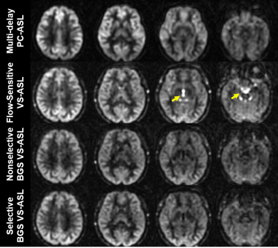
Optimization of Velocity-Selective-Inversion Arterial Spin Labeling (VSI-ASL) with 3D Whole-Brain Coverage
Dapeng Liu, Wenbo Li, Doris Lin, Peter Zijl, Qin Qin
Velocity-selective arterial spin labeling (VSASL) has the advantage of insensitivity to transit time delay compared to the spatially selective method, thus potentially providing more accurate and robust blood flow measurements in cerebrovascular diseases. Fourier-transform based velocity-selective inversion (FT-VSI) prepared ASL has higher sensitivity to perfusion signal than conventional velocity-selective saturation (VSS) prepared methods. To date, VSASL has largely been implemented with 2D EPI acquisitions. However, a 3D readout is preferred for ASL techniques. This study demonstrated the feasibility of FT-VSI prepared VSASL with 3D whole-brain coverage and compared it with conventional VSS ASL and pseudo-continuous ASL (PCASL) at 3T.
|
|
4965.
 |
72 |
Optimal Strategies for CSF and Tissue Suppression in Velocity-Selective Arterial Spin Labeling
Mu-Lan Jen, James Holmes, Kevin Johnson
Velocity-selective arterial spin labeling (VS-ASL) inherently suffers from low signal-to-noise ratio (SNR) and contamination from cerebrospinal fluid (CSF) motion. This study aims to develop and evaluate optimal strategies for inversion based background suppression (BGS). Specifically, we investigate the influence of the timing of signal nulling and inflow from outside the region of interest. Our results suggest an optimized BGS which allows VS-ASL based measurement of cerebral blood flow maps with reduced CSF contamination while preserving sufficient perfusion signal.
|
|
4966
 |
73 |
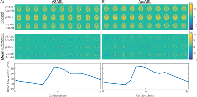
The influence of the cardiac cycle on Velocity Selective and Acceleration Selective Arterial Spin Labeling, using retrospective triggering.
Video Permission Withheld
Suzanne Franklin, Sophie Schmid, Clemens Bos, Matthias Van Osch
In this study, the influence of the cardiac cycle on the amount of label produced by velocity-selective (VSASL) and acceleration-selective arterial spin labeling (AccASL) was investigated. A sequence combining pCASL and VSASL(AccASL) was developed to isolate the arterial blood pool. Results showed significant arterial signal fluctuations in the amount of label produced by VSASL, AccASL and pCASL over the cardiac cycle. Hence, in order to become independent of the cardiac cycle, sufficient averages need to be taken when applying these techniques. Alternatively, these findings could be highly interesting for the purpose of quantifying pulsatility higher up in the vascular tree.
|
|
4967.
 |
74 |
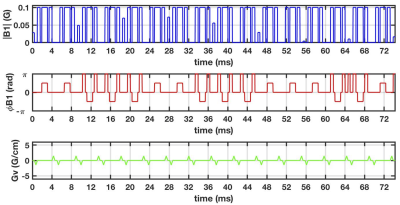
Improved Velocity-Selective Labeling Pulses for Myocardial ASL
Vanessa Landes, Terrence Jao, Ahsan Javed, Krishna Nayak
Velocity selective ASL is an exciting option for myocardial perfusion imaging as it does not require any contrast agents and is insensitive to coronary arterial transit times. Feasibility in humans was recently demonstrated with performance primarily limited by 1) spurious labeling of moving myocardium, and 2) low labeling efficiency. We present improvements to the velocity selective labeling pulse that overcome these limitations, leveraging recent developments in velocity-selective MRA. Specifically, we use Fourier Velocity Encoding to reduce spurious labeling of moving myocardium and use inversion to increase labeling efficiency.
|
|
4968.
 |
75 |
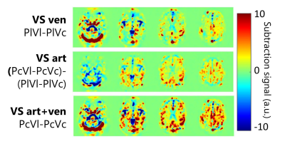
Venous Velocity Selective Inversion for improved selection of the venous blood pool for oxygen extraction fraction determination
Sophie Schmid, Suzanne Franklin, Merlijn van der Plas, Matthias van Osch
By combining pulsed ASL and Velocity Selective Inversion it is possible to selectively label the venous blood pool. This new method, dubbed venous velocity selective inversion (vVSI) could be used to measure the oxygen extraction fraction in the venous and arterial blood with a single scan.
|
|
| Top |
Relaxometry
Digital Poster
Contrast Mechanisms
Thursday, 16 May 2019
| Exhibition Hall |
14:45 - 15:45 |
| |
|
Computer # |
|
4969.
 |
76 |
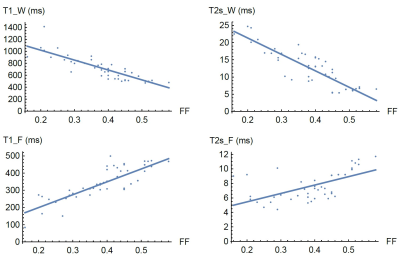
Correlation between Fat Fraction and MR relaxation times in the vertebral bone marrow at 1.5 T.
Louis Marage, Jérémy Lasbleiz, Mathieu Lederlin, Giulio Gambarota, Hervé Saint-Jalmes
The aim of this study was to investigate the in-vivo correlation between Fat Fraction and T1, T2* for both water and fat compartments, in vertebral bone marrow at 1.5T. A fast chemical-shift-encoded 3D multi-gradient-echo sequence and a B1-mapping sequence were acquired at two different flip angles. Fat Fraction, T1 of water, T2* of water, T1 of fat and T2* of fat were obtained using a previously published method. The results of the current study show strong correlation between Fat Fraction and the relaxation times.
|
|
4970.
 |
77 |
Rapid, whole-brain T1 mapping using inversion recovery EPIK (ir-EPIK): a quantitative assessment with a group of subjects
N. Jon Shah, Seong Dae Yun
Due to their relative insensitivity to B1 inhomogeneities, Look-Locker methods are widely used for the quantification of T1 relaxation time. One such Look-Locker method, TAPIR, has been demonstrated with several clinical applications and has been shown to be faster than conventional gradient-echo sequences. However, it still requires a considerable acquisition time for whole-brain imaging. To overcome this limitation, a much faster method, ir-EPIK, has been presented in our earlier work. This work aims to perform a quantitative assessment of ir-EPIK in comparison to TAPIR using phantom data and twenty data sets from subjects. All data were acquired at 3T.
|
|
4971.
 |
78 |
Observation and Mitigation of Magnetization Transfer Effects in the two-point 3D Variable Flip Angle T1 Mapping Technique at 3T
Jean-David Jutras, Atefeh Kordzadeh, Nicola De Zanche
The Variable Flip Angle (VFA) T1 mapping technique has been employed extensively in the past given its high contrast-to-noise ratio per unit scan time. However, its sensitivity to B1 field inhomogeneity and imperfect spoiling hinders its reproducibility among different scanners and imaging centers. In this study, we investigate the impact of magnetization transfer (MT) effects in a 3D VFA technique using fixed flip angles, while varying B1 amplitudes and durations, following corrections for non-ideal RF spoiling and RF inhomogeneity. We show that via careful tuning of the RF pulse amplitudes and durations, MT effects can be mitigated, yielding T1 measurements that match closely with a gold-standard IR-EPI technique.
|
|
4972.
 |
79 |
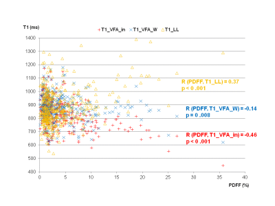
The influence of fat on T1 mapping of the liver: a comparison of Look-Locker and variable-flip-angle techniques
Claudia Fellner, Niklas Verloh, Dominik Nickel, Stephan Kannengießer, Christian Stroszczynski, Michael Haimerl
In 383 patients two methods for T1 mapping – 2D Look-Locker (LL), and 3D variable-flip-angle (VFA) combined with a 2-point-Dixon technique – were compared and their correlation with the intrahepatic proton density fat fraction (PDFF) was evaluated. T1_LL showed a moderate positive correlation with PDFF, while there was an intermediate negative correlation between T1_VFA_in (T1 calculated from water and fat in-phase signal) and PDFF; T1_VFA_W (T1 calculated from water only signal) was nearly independent of PDFF. In patients with PDFF above 5%, LL, VFA_in, and VFA_W yielded significantly different results for T1.
|
|
4973.
 |
80 |
Time efficient T1 measurement of Cortical Bone using a Three-Dimensional Ultrashort Echo Time Cones Variable Flip Angle-Actual Flip Angle Imaging (3D UTE-Cones VFA-AFI) method
Zepeng Wang, Yajun Ma, Lidi Wan, Saeed Jerban, Yanjun Chen, Eric Chang, Jiang Du
To reduce scan time and maintain the accuracy of T1 measurement for cortical bone, we propose a novel T1 measurement approach using three-dimensional ultrashort echo time cones variable flip angle (3D UTE-Cones VFA) with actual flip angle imaging (AFI) technique for the correction of B1 inhomogeneity. The results show the similar bone T1 values were obtained by the proposed fast 3D UTE-Cones AFI-VFA method compared with the previous UTE-Cones AFI and variable TR method.
|
|
4974.
 |
81 |
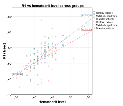
Investigating the T1 of human venous blood at 7T in patients with diabetes, metabolic syndrome and healthy subjects.
Dimo Ivanov, Roy Haast, Muriel Desmond-Kennedy, Benedikt Poser, Kâmil Uludag
Blood T1 values are important to accurately quantify perfusion with arterial spin labeling and to determine the optimal inversion time for vascular space occupancy and black-blood imaging. In this work, we demonstrate that a post-hoc B1+-corrected MP2RAGE sequence can be used to measure the subject-specific T1 of blood in the superior sagittal sinus at 7T, eliminating the need for additional dedicated measurement. The approach was applied in patients with diabetes, metabolic syndrome and healthy controls to examine the influence of these conditions on the respective T1 of blood. The method proposed can be employed at any field strength.
|
|
4975.
 |
82 |
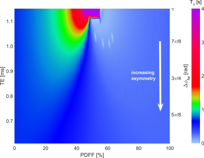
The echo time of balanced steady-state free precession readouts modulates the influence of fat on MOLLI T1 measurements
Ferenc Mozes, Elizabeth Tunnicliffe, Matthew Robson
With the increasing world-wide prevalence of non-alcoholic fatty liver disease it is essential to look for non-invasive diagnostic and monitoring methods, like T1 mapping. It has previously been shown that the presence of fat can artificially prolong liver T1 times measured with modified Look-Locker methods. However, this effect depends on the chosen TR and TE of the readout sequence. Since achievable TR and TE differs for scanner vendors and models, it is important to understand the influence of sequence timings on MOLLI T1 measurements in the presence of fat.
|
|
4976.
 |
83 |
Silent T1-Mapping at 7T Using the Variable Flip Angle Method
Emil Ljungberg, Brian Burns, Peder Larson, Shannon Kolind, Mark Symms, Florian Wiesinger, Gareth Barker
In this work we present quantitative T1-maps obtained using the silent, zero echo-time, RUFIS sequence at 7T. Four flip angles (2,4,8,11)° were acquired in 5 minutes. The obtained T1-maps showed strong contrast between white matter (T1=1.6s) and cortical grey matter (T1=2.3s), in agreement with values in the literature. Reduced contrast was observed in deep grey matter structures, attributed to large B1+-errors in the centre of the brain.
|
|
4977.
 |
84 |
Inversion-recovery MRI based biphasic analysis of porous media: simulations, phantom experiments and in vivo brain study.
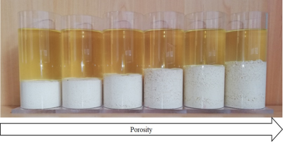
Ledia Lilaj, Jürgen Braun , Thom Fischer, Ingolf Sack, Sebastian Hirsch
A novel technique that combines inversion recovery MRI (IR-MRI) and a biphasic porous tissue model is introduced to quantify in every voxel the porosity, defined as the ratio between the volume of the fluid phase and the total volume of both the fluid and the solid matrix. Simulations revealed precise results over a wide range of values. Porosities of tofu phantoms measured by IR-MRI were in good agreement with the values obtained from reference methods, confirming the stability of our technique.
The same IR-MRI method was then applied to the brains of healthy volunteers providing quantitative maps of porosity.
|
|
4978.
 |
85 |
Oxygen Saturation Dependent Effects on Blood Transverse Relaxation at Low Fields
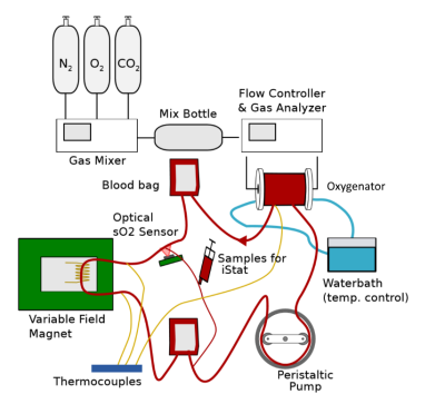
Dion Thomas, Petrik Galvosas, Paul Teal, Graham Wright, Freya Harrison, Max Berry, Yu-Chieh Tzeng, Sergei Obruchkov
The change in T2 due to the oxygen saturation sO2 in blood has been well characterised at high fields, and has been successfully applied for in-vivo oximetry measurements. The effect is known to increase with B0 field strength, but there are few studies at low field. In this work, the relationship between the T2 relaxation rate and blood oxygenation sO2 has been characterised at a range of magnetic fields below 1 Tesla to determine whether changes in oxygenation can be practically observed and accurately quantified at lower fields.
|
|
4979.
 |
86 |
Blood oxygenation measurements in single vessels: lineshape measurements of the water signal
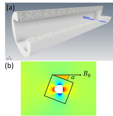
Lukas Buschle, Christian Ziener, Volker Sturm, Patrick Vogel, Ke Zhang, Martin Bendszus, Sabine Heiland, Mirko Pham, Heinz-Peter Schlemmer, Felix Kurz, Thomas Kampf
In this work, the lineshape around a vessel inside a cubic voxel is analytically analyzed in dependence on the orientation of the voxel according to the external magnetic field. Results are validated with phantom measurements and in vivo measurements, that both agree very well with the developed theory. The analytical model therefore allows a determination of the oxygen extraction fraction from single voxel measurements around macroscopic vessels.
|
|
4980.
 |
87 |
Reproducibility of Simultaneous in vivo Blood T1 and T2 Imaging Method
Jialu Zhang, Dingxin Wang, Xiaotong Zhang, Lynn Eberly, Gregory Metzger, Donald Dengel, David Tupper, Anne Murray, Xiufeng Li
The longitudinal and transverse relaxation time constants of blood vary across subjects, developmental stages, physiological states or specific diseases. We implemented a fast method for simultaneous in vivo measurements of blood T1 and T2. Although such an approach has been successfully demonstrated, its repeatability or robustness has not been assessed. We performed a two-session study using our fast in vivo blood T1 and T2 imaging method, and the study results are reported in the following.
|
|
4981.
 |
88 |
Physiologically Accurate Simulations of Endogenous Susceptibility-Based Contrast in Cancer Reveal the Importance of Intravascular Oxygen Variations on Transverse Relaxation
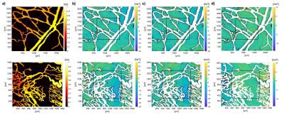
Artur Hahn, Janaka Senarathna, Volker Sturm, Sabine Heiland, Martin Bendszus, Felix Kurz, Arvind Pathak
The heterogeneous nature of tumor vasculature and hemodynamics make it challenging to model transverse relaxation in such tissues. Here we imaged the intravascular oxygen saturation in healthy abdominal wall and breast tumor xenografts in mice using intrinsic optical signal (IOS) imaging, and used these quantitative data in realistic simulations of transverse relaxation. We found that the inclusion of de factooxygen distributions recapitulated the heterogeneity of tumor transverse relaxation rates in contrast to the traditional approach of assuming constant oxygen distribution for the tumor microvascular bed. These findings have important implications for BOLD MRI of tumors.
|
|
4982
 |
89 |
On the influence of two coexisting species of susceptibility-producing structures on the R2’ relaxation rate: the static dephasing regime and diffusion effects
Video Permission Withheld
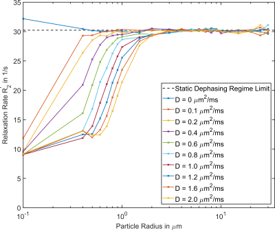
Julian Emmerich, Sina Straub
In this work, we examine the effect of two different species of susceptibility producing-structures within one voxel to deduce whether the relaxation rate is proportional to the sum of the absolute product between volume fraction and susceptibility value. Furthermore, the effect of diffusion on the R2’ relaxation rate beyond the static dephasing regime is analyzed.
|
|
4983.
 |
90 |
Calculation of molar relaxivity and concentration map of Gd-DTPA map using quantitative parameter map before and after injection for brain metastasis
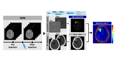
Yuki Matsumoto, Masafumi Harada, Yuki Kanazawa, Takashi Abe, Maki Otomo, Yo Taniguchi, Masaharu Ono, Yoshitaka Bito
R1 map and susceptibility maps before and after injection of Gd-DTPA were calculated using quantitative parameter map technique. Then, concentration map (CM) of the Gd-DTPA was calculated using the susceptibility maps. A linear regression between CM and R1post - R1pre map was performed. The comparison between the CM and R1pre-R1post map each of the metastasis demonstrated a strong correlation (y = 4.0966 - 0.46535, R=0.95) and relaxivity map r1 was empirically calculated from the linear regression. A slope of the linear regression means the relaxivity in metastasis and it approximately matched the relaxivity of the Gd-DTPA in plasma at 3T.
|
|
4984.
 |
91 |
Kidney stone discrimination using ultrashort TE MRI
Johannes Fischer, Agazi Tesfai, Ute Ludwig, Philippe Müller, Arkadiusz Miernik, Michael Bock
Kidney stone disease (urolithiasis) is not only very painful, but can also pose serious health risks, when the fragmentation of infected kidney stones releases bacteria, that may cause post-operative sepsis. In this work we show the ability of Magnetic Resonance Imaging (MRI) to discriminate between common types of kidney stones using relative signal intensity and T2* relaxation times.
|
|
4985.
|
92 |
Insights from the Configuration Model theory accelerate Bloch simulations for dictionary-based T2 mapping
Marco Hauke, Dominik Weidlich, Carl Ganter, Axel Haase, Dimitrios Karampinos
Muscle water T2 has been proposed as an imaging biomarker of disease activity in neuromuscular diseases. 2D multi-echo spin-echo sequences have been used for muscle T2 mapping with known limitations including the sensitivity to transmit B1 inhomogeneities. Confounding effects on the T2 quantification can be removed by matching the experimental signal with pre-simulated theoretical signal decay curves obtained by Bloch simulations. However, up to now it is unclear what discretization over the slice profile is sufficient to determine a meaningful dictionary. The purpose of this work was to utilize the configuration model in order to determine a minimal number of z-location necessary for the simulation and applies the technique for T2 mapping of in vivo thigh musculature data.
|
|
4986.
 |
93 |
Characterising the temporal evolution of fixation in human post mortem brain via linear relaxometry modelling – a marker of cross-linking?
Siawoosh Mohammadi, Sebastian Papazoglou, Herbert Mushumba, Mohammad Ashtarayeh, Klaus Püschel, Gunther Helms, Martina Callaghan, Nikolaus Weiskopf, Tobias Streubel
MRI-based biophysical models are typically validated by comparison to ex-vivo histology of fixed tissue. The fixation process itself and the accompanied autolysis processes strongly modify tissue composition, and lead to MR signal changes, making the validation of biophysical models for in vivo MRI particularly challenging. To better understand the temporal evolution of the fixation process within the whole brain and its influence on MRI parameters, we monitor the temporal evolution of the fixation process of a whole human post-mortem brain using the linear relaxometry model across 15 time-points comprised of one unfixed, in-situ MRI scan and 14 ex-vivo MRI scans at different stages of the fixation process (days 1-93).
|
|
4987
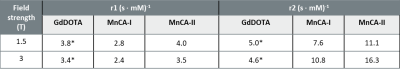 |
94 |
Manganese-based macrocyclic chelates as a potential novel MRI contrast agent – relaxometry & imaging
Video Permission Withheld
Wibeke Nordhøy, Robin Bugge, Tuva Hope, Deirdre Cassidy, Brian Bales, Atle Bjørnerud
In this study, novel Manganese-based contrast agents (MnCAs) were evaluated by relaxometry techniques (NMR and phantom imaging) and tested in naïve pigs using a contrast-enhanced MRA protocol and compared to the well-established agent GdDOTA. Despite some differences in T1 and T2 relaxation performance, both MnCAs and GdDOTA provided strong vascular T1-enhancement in naïve pig imaging thus providing initial evidence that MnCAs could be utilized for a clinical application.
|
|
4988.
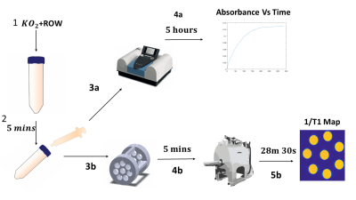 |
95 |
Measuring the Relaxivity of the Superoxide Radical
Martin MacKinnon, Bruce Berkowitz, Yen-Yu Ian Shih
The excessive production of reactive oxygen species (ROS), is commonly thought to be a pathogenic factor in a range of neurodegerative diseases, psychological conditions and in the etiology of aging. Traditionally, ROS have been thought undetectable in-vivo, due to their short half-life and low concentrations in living tissue. The paramagnetism of ROS may provide a means of encoding oxidative stress into MRI data. To investigate how different concentrations of ROS contribute to MRI signals, the T1 relaxivity of ROS must be determined. Using a novel method to detect ROS in-vivo, QUEST-MRI, we show that the relaxivity of the superoxide radical is in the range between 0.135-0.509 LmM-1s-1 - similar to nitroxides used as contrast agents to detect ROS in EPR.
|
|
4989.
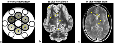 |
96 |
Analysis of magnetization transfer (MT) influence on quantitative mapping of T2 relaxation time
Dvir Radunsky, Tamar Blumenfeld-Katzir, Osnat Volovyk, Tal Assaf, Daniel Barazany, Galia Tsarfaty, Noam Ben-Eliezer
Quantification of T2 values is valuable for a broad range of pathologies, yet, highly challenging due to the contamination of rapid multi-echo spin-echo (MESE) protocols by stimulated and indirect echoes. Bloch-simulations based methods, such as the echo modulation curve (EMC) algorithm, take these signal fluctuations into account and produce accurate, precise, and reproducible T2 values. This work provides detailed analysis of magnetization transfer (MT) effect on MESE signal and on the ensuing T2values, investigating different protocol settings, and using three models: in vitro urea phantom, ex vivo horse brain and in vivo human brain.
|
|
4990.
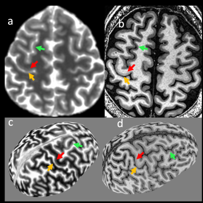 |
97 |
Exploring human cortical microstructure using magnetic resonance fingerprinting at 3T
Irene Wang , Rasim Boyacioglu, Mark Griswold, Stephen Jones, Dan Ma
Human cortical areas are typically differentiated by functions with varying cyto- and myelo- architectures. Since quantitative MR tissue properties, such as T1, T2 and susceptibility, reflect underlying molecular compositions and micro-environment of tissues, multiple quantitative imaging methods have been used to investigate human cortical microstructure at 7T. Here, we used the FSL image analysis tool to process multi-parametric quantitative maps and demonstrated that high resolution multi-parametric maps acquired from a single MR Fingerprinting (MRF) scan at 3T can reveal similar patterns of cyto-architectural information that are typically identified at 7T.
|
|
4991.
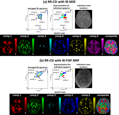 |
98 |
Multidimensional T1 Relaxation -T2 Relaxation Correlation Spectroscopic Imaging with a Magnetic Resonance Fingerprinting Acquisition
Daeun Kim, Bo Zhao, Lawrence Wald, Justin Haldar
T1 Relaxation-T2 Relaxation Correlation Spectroscopic Imaging (RR-CSI) is a novel multidimensional imaging approach that estimates a 2D T1-T2 correlation spectrum at every spatial location, and enables spatial mapping of sub-voxel tissue compartments with very good compartmental resolving capabilities. While RR-CSI was previously demonstrated using data acquired with an inversion recovery multi-echo spin-echo sequence, it can also accommodate data acquired with other encoding schemes. In this work, we investigate an inversion recovery FISP MR fingerprinting acquisition scheme for RR-CSI. Results show, both theoretically and empirically using in vivo human data, that fingerprinting is a viable alternative.
|
|
4992.
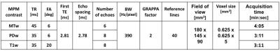 |
99 |
High-resolution quantitative maps of magnetisation transfer, R1 and R2* of the cervical spinal cord in clinically feasible acquisition time using vendor-provided sequences
Daniel Papp, Alex Smith, Romina Mariano, Stuart Clare
Here, we present a fast multi-parameter quantitative MRI protocol to estimate R1, R2* and MT saturation in the cervical spinal cord, acquired using only vendor-supplied sequences, in clinically acceptable acquisition times (~11 minutes total). The proposed protocol can be easily adapted to clinical scanners, and requires only stock sequences and freely available post-processing tools, and is faster than a comparable protocol required for PSR mapping in a qMT framework.
|
|
4993.
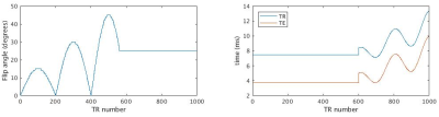 |
100 |
Simultaneous Concentration Quantification of SPIO and ProHance using bSSFP MR Fingerprinting
Ollie Marriott, Chris Bowen, James Rioux, Kimberley Brewer
Superparamagnetic Iron Oxide (SPIO) contrast agents are used extensively in molecular imaging studies as a tool to evaluate various cell types. Enabling simultaneous use of multiple contrast agents would greatly improve molecular imaging studies. We utilize a bSSFP MR fingerprinting sequence, combined with an extension of the concentration-dependent linear relationship, to show that concentrations of SPIO and a second contrast agent (ProHance) can be simultaneously quantified.
|
|
| Top |
Novel Developments in CEST
Digital Poster
Contrast Mechanisms
Thursday, 16 May 2019
| Exhibition Hall |
14:45 - 15:45 |
| |
|
Computer # |
|
4994.
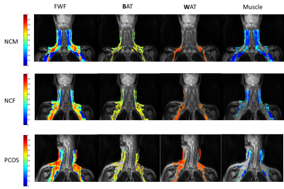 |
101 |
Characterization of brown adipose tissue in PCOS patients by Z-Spectrum Imaging (ZSI)
Li LI, Alessandro Scotti, Jicheng Fang, Chong Wee Liew, Nael Khayyat, WenZhen Zhu, Kejia Cai
Z-spectrum imaging (ZSI) was used to characterize brown adipose tissue (BAT) features in polycystic ovary syndrome (PCOS) patients compared to healthy subjects. Fat-water fraction (FWF) was derived from the fitting of the direct saturation of water and fat in ZSI. BAT segmentation allowed the measurement of BAT-specific FWF and area. PCOS group showed increased FWF and reduced BAT area compared to controls, likely due to reduced BAT cells activity and lipid accumulation. ZSI can therefore be used to study BAT characteristics in vivo in metabolic disorders.
|
|
4995.
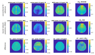 |
102 |
Can a neural network predict B0 maps from uncorrected CEST-spectra?
Felix Glang, Anagha Deshmane, Florian Martin, Kai Herz, Klaus Scheffler, Moritz Zaiss
Analysis of chemical exchange saturation transfer (CEST) effects suffers from B0 inhomogeneity. Common correction methods involve computationally expensive algorithms or even additional measurements. Here we demonstrate that deep neural networks are able to predict B0 maps from raw Z-spectra by training the networks with measured B0 maps. Moreover, we show that CEST contrast parameters representing amide, amine and NOE resonance peaks can be directly predicted from uncorrected Z-spectra in a fast single step. This provides a shortcut to conventional evaluation procedures and will be useful to guide nonlinear model fitting.
|
|
4996.
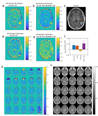 |
103 |
Artifacts in dynamic CEST MRI due to motion and field shifts – implications for glucoCEST MRI at 3T
Moritz Zaiss, Kai Herz, Anagha Deshmane, Mina Kim, Xavier Golay, Tobias Lindig, Benjamin Bender, Ulrike Ernemann, Klaus Scheffler
Dynamic glucose enhanced imaging yields expected CEST effects that are rather small in tissue especially at clinical field strengths (<2 %). Small movements during the dynamic CEST measurement together with a subtraction-based evaluation can lead to pseudo CEST effects of the same order of magnitude. We studied these effects by virtual difference images of a basline scan that were altered by the rigid body transformations and B0 shifts. Minor motion (0.6 mm translations) and B0 artifacts (7 Hz shift) can lead to pseudo effects in the order of 1% in dynamic CEST imaging, despite no glucose was injected at all.
|
|
4997
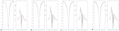 |
104 |
A new EPI multislice evaluation for APT-CEST
Video Permission Withheld
Jan Schüre, Manoj Shrestha, Ralf Deichmann, Elke Hattingen, Marlies Wagner, Ulrich Pilatus
To investigate APT-CEST feasibility as pH-sensitive contrast, a multislice CEST EPI sequence was evaluated. The proposed sequence allows continuous saturation during EPI measurement, leaving sufficient time to capture robust CEST data by multiple repetitions. The saturation scheme was optimized in a phantom study and with healthy volunteers. With the availability of 3D CEST data a subsequent registration of APT-CEST contrast images to 31P-MRSI can be achieved for evaluating pH-related changes in APT-CEST contrast. This is demonstrated in a study of a patient with glioblastoma.
|
|
4998.
|
105 |
Characterization of brain metabolites using CEST and machine learning
Nirbhay Yadav, Akansha Sehgal, Peter van Zijl
In vivo CEST MRI data can include contributions from a vast array of metabolites, mobile proteins and peptides, and immobile macromolecules amongst others. Detecting which components are present in any given dataset is a major challenge. Here, as a first start to address the problem, we have used a machine learning approach to classify a CEST dataset acquired from brain metabolite phantoms. The classifier was successful in all cases and was shown to be robust to a moderate level of noise. The results demonstrate this is a promising technique that could potentially quantify molecular contributions in vivo.
|
|
4999.
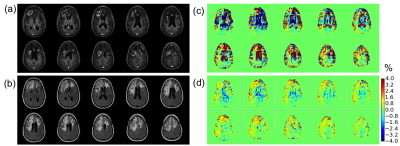 |
106 |
Dynamic Glucose Enhanced Imaging at 3T: Effects of Physiological Changes and Motion
Xiang Xu, Anina Seidemo, Akansha Sehgal, Nirbhay Yadav, Moritz Zaiss, Linda Knutsson, Peter van Zijl
Dynamic glucose enhanced (DGE) MRI has shown potential for imaging D-glucose delivery and brain uptake at fields of 7T and higher. Here we evaluate some issues involved with translating DGE MRI to the clinical field strength of 3T. Due to the reduced effect size subject motion becomes more confounding than at 7T, possibly producing artifacts in terms of dynamic signal changes that are beyond the magnitude of the actual effect size. On the other hand, physiological changes such as ventricular swelling and vascular dilatation may appear as motion to the motion correction procedure, possibly leading to unintended overcorrection.
|
|
5000.
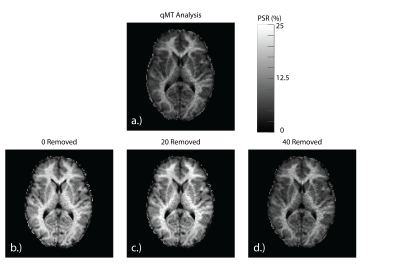 |
107 |
Does the MT Effect Bias CEST Effects? Quantifying CEST in the Presence of MT
Alex Smith, Kevin Ray, Martin Craig, Seth Smith, Michael Chappell
Chemical exchange saturation transfer (CEST) is an MRI technique sensitive to the presence of low-concentration solute protons exchanging with water. However, magnetization transfer (MT) effects due to large macromolecules also exchange with water, and they can bias potential CEST effects if they are not correctly modelled. This study demonstrates that simultaneously modelling CEST and MT may yield incorrect estimates of CEST effects if an insufficient model is utilised. To prevent this, the MT effect should first be quantified outside the CEST domain and subsequently applied to CEST analyses.
|
|
5001.
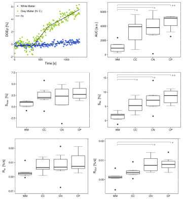 |
108 |
Quantification of brain perfusion using dynamic glucose-enhanced MRI
Charlotte Debus, Patrick Schuenke, Ralf Floca, Myriam Keymling, Amir Abdollahi, Alexander Radbruch, Peter Bachert, Mark Ladd, Heinz-Peter Schlemmer, Daniel Paech
Dynamic glucose-enhanced (DGE) MRI was analyzed to derive quantitative parameters related to tissue perfusion, microvasculature and glucose uptake in the human brain. Adiabatically prepared T1ρ-weighted DGE-MRI was performed on seven healthy volunteers and one glioblastoma patient with a 7T scanner after administration of D-glucose. DGEρ time curves were investigated in different morphological structures of the healthy brain and in tumor tissue by extraction of semi-quantitative parameters and pharmacokinetic modelling using the extended Tofts model. Results show that application of semi-quantitative and pharmacokinetic modeling approaches for quantification of DGE-MRI is feasible in both healthy humans and brain tumor patients.
|
|
5002.
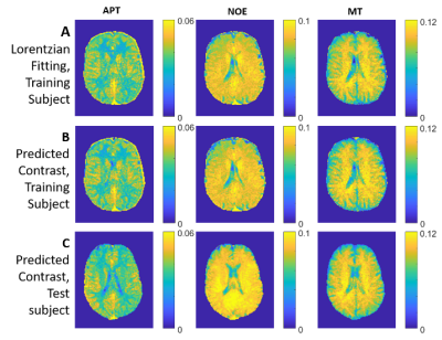 |
109 |
Machine Learning accelerates and stabilizes selective CEST fitting at 3T
Anagha Deshmane, Moritz Zaiss, Kai Herz, Benjamin Bender, Tobias Lindig, Klaus Scheffler
Multi-Lorentzian analysis of chemical exchange saturation transfer (CEST) Z-spectra by non-linear least squares (NLLS) fitting is common at ultra-high field strengths but particularly challenging at clinical field strengths due to broad, coalesced peaks and low SNR. Here we demonstrate that a neural network (NN) trained on just 3 slices of a single subject can robustly predict CEST Lorentzian pool parameters in other subjects, in the presence of motion, and in a brain tumor patient, with a 95 % reduction in computing time, allowing for quick estimation of NLLS initial conditions or initial online reconstruction of spectrally selective CEST contrasts.
|
|
5003.
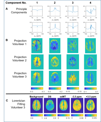 |
110 |
Model-free generation of CEST contrast using principal components of Z-spectra at 3 T
Anagha Deshmane, Moritz Zaiss, Mark Schuppert, Kai Herz, Klaus Scheffler
Fitting of spectrally selective CEST contrasts requires models with limiting assumptions. Snapshot CEST allows us to densely sample the Z-spectrum with rapid volumetric imaging within a clinically feasible scan time. With over 60k spectra available per subject, statistically-driven analysis methods are now possible. Here we demonstrate that principle component analysis can be used for model-free analysis of spectral features. Projection of Z-spectra onto principle components from a group of healthy subjects provides several relevant contrasts which reveal anatomical detail and correlate with Gadolinium uptake signatures in a brain tumor patient.
|
|
5004.
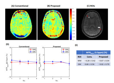 |
111 |
Fast 3D Whole-Brain Steady-State CEST at 3T Using MR Multitasking
Pei Han, Zhengwei Zhou, Anthony Christodoulou, Sen Ma, Nan Wang, Debiao Li
We propose a fast 3D whole-brain steady-state chemical exchange saturation transfer (CEST) method using MR Multitasking. Exploiting the correlation between images throughout the space, time and frequency dimensions, the low-rank tensor framework shows possibility for further acceleration of steady-state CEST, so that a whole-brain Z-spectrum acquisition can be done within 5 min.
|
|
5005.
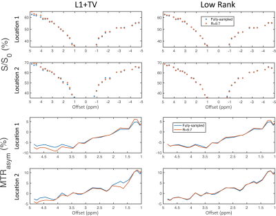 |
112 |
Low Rank Compressed Sensing Accelerated CEST Imaging
Zhipeng Cao, Zhongliang Zu, Kristin O'Grady, Jun Ma, William Grissom, Seth Smith, John Gore
A method to accelerate chemical exchange saturation transfer (CEST) imaging is presented by reconstructing highly undersampled z-spectral image series using low rank compressed sensing. Results show x10 acceleration with an eight channel receiver with human brain imaging at 7T.
|
|
5006.
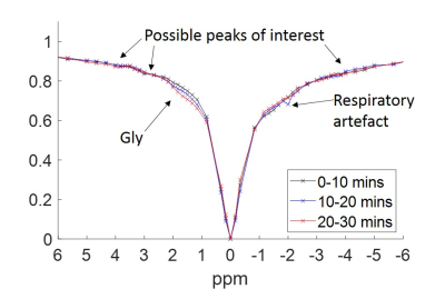 |
113 |
The 7T z-spectrum from the human liver in-vivo: observing the effects of a meal
Andrew Carradus, Emma Doran, Olivier Mougin, Christopher Mirfin, Hans Hoogduin, Stephen Bawden, Penny Gowland
In this work we acquired the first z-spectrum from the human liver in vivo at 7T. Glycogen and NOE peaks were observed, and their evolution over time was monitored after fasting and after a meal. Both the glycogen peak and the NOE peak at -1.7ppm were observed to increase 2-4 hours after a high carbohydrate meal.
|
|
5007.
 |
114 |
A comparison of dynamic and static B0 mapping approaches for correction of CEST MRI at 7T
Esau Poblador Rodriguez, Philipp Moser, Barbara Dymerska, Simon Robinson, Benjamin Schmitt, Andre van der Kouwe, Stephan Gruber, Siegfried Trattnig, Wolfgang Bogner
In contrast to established static B0 correction approaches which assume an invariant static field during a CEST MRI acquisition, we propose three methods which can track and compensate temporal B0 fluctuations by shifting each Z-spectral point separately before MTRasym analysis. We show the benefit of the proposed dynamic methods in comparison to three established static approaches by assessing their performance in the absence/presence of an induced frequency drift. In addition, we investigate the reliability and reproducibility of CEST MRI at ultra-high field (7T) by evaluating the drift’s impact on the B0-corrected MTRasym maps in the brains of five healthy volunteers.
|
|
5008.
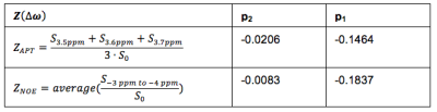 |
115 |
Axillary Chemical Exchange Saturation Transfer (CEST) MRI Contrast is Consistent with Secondary Breast Cancer Treatment-Related Lymphedema
Rachelle Crescenzi, Paula Donahue, Manus Donahue
Breast cancer treatment-related lymphedema (BCRL) is a chronic condition with 30% two-year incidence in cancer survivors treated with lymph node dissection. Changes in the tissue microenvironment indicate edema rich in macromolecular proteins. We hypothesize that chemical exchange saturation transfer (CEST) MRI, after accounting for transmit field (B1) heterogeneity and longitudinal (T1) relaxation time variation, will be sensitive to affected tissues in patients with BCRL. We report that after performing appropriate correction procedures in the upper extremities, it is possible to detect disease-specific CEST contrast in the affected and contralateral arms of BCRL patients.
|
|
5009.
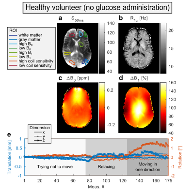 |
116 |
Acquisition protocol for glucoCESL MRI in the human brain at 7T with reduced motion-induced artifacts
Philip Boyd, Johannes Breitling, Ferdinand Zimmermann, Andreas Korzowski, Moritz Zaiss, Kai Herz, Patrick Schuenke, Nina Weinfurtner, Heinz-Peter Schlemmer, Mark Ladd, Peter Bachert, Daniel Paech, Steffen Goerke
In this study, an acquisition protocol for glucoCESL MRI examinations in the human brain is presented, that was optimized for suppression of motion-induced artifacts. This was achieved by using a combination of (i) a 3D imaging readout, which enabled co-registration of the acquired images over the course of time, and (ii) a ΔR1ρ contrast based on quantitative R1ρ maps instead of R1ρ-weighted images. The presented acquisition protocol improves the applicability of glucose-weighted MRI for examinations in humans where motion is present. Feasibility was verified by examination in a first patient with glioblastoma.
|
|
5010.
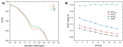 |
117 |
Investigating Gadolinium Depositions in Glycosaminoglycans using CEST MRI - Why T1 Corrections are Important
Patrick Schuenke, Patrick Werner, Matthias Taupitz, Leif Schröder
In this study, we investigated the CEST effect of model solutions composed of gadolinium-based contrast agents (GdCA), heparin as model substance for glycosaminoglycans (GAGs) and ZnCl2 with the aim to provide direct evidence of Gd3+-GAG complex formations. We performed time resolved CEST and T1 relaxation time measurements and quantified the CEST effects using different CEST metrics, including MTRasym and AREX. Our results demonstrate the necessity of T1 correction in CEST MRI in such a case where T1 conditions are affected, too.
|
|
5011.
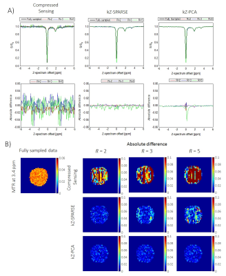 |
118 |
Accelerating CEST MRI by Exploiting Sparsity in the Z-Spectrum Domain
Grzegorz Kwiatkowski, Sebastian Kozerke
Chemical Exchange Saturation Transfer (CEST) is an emerging modality offering an enhanced sensitivity for the detection of dilute metabolites with exchangeable protons. To provide quantitative analysis, an acquisition of multiple images per Z-spectrum is required, leading to long acquisition times in practice. In this report we present a novel approach for rapid acquisition of CEST MRI that exploits sparsity in the Z-spectrum domain. Based on ex-vivo and in-vivo data, an acceleration factor of up to R=5 is shown, without significant loss in data accuracy.
|
|
5012.
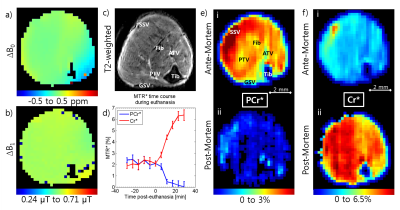 |
119 |
Phosphocreatine CEST (PCrCEST) Detects Phosphocreatine and pH Changes in the Muscle at 15.2T
Julius Chung, Tao Jin, Jung Hee Lee, Seong-Gi Kim
Phosphocreatine plays a major role in the metabolic upkeep of tissues such as the muscles and the heart. Chemical Exchange-sensitive MRI has been shown to have sensitivity to phosphocreatine. We examine the sensitivity of Phosphocreatine CEST (PCrCEST) to changes in phosphocreatine levels and pH via PCrCEST imaging in the mouse hindlimb while inducing euthanasia and present high resolution PCrCEST maps in the ante- and post-mortem.
|
|
5013.
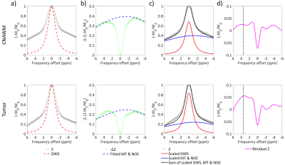 |
120 |
Improved tumor characterization with direct saturation corrected (DISC) amide proton transfer (APT) MRI in glioma patients
Yin Wu, Yinsheng Chen, Jie Liu, Cong Li, Xiaofei Lv, Zhongping Chen, Phillip Sun, Hairong Zheng
Recently, a direct saturation correction (DISC) method has proved to improve APT quantification in pulsed-RF CEST imaging. In this study, performance of tumor characterization was compared among routine asymmetry analysis (MTRasym), three-point offset (APT3pts) and the DISC methods in glioma patients at 3T. Although all methods revealed significant APT elevation in tumor region compared to that in the contralateral normal appearing white matter, the DISC method exhibited substantially higher contrast-to-noise ratio (2.16±0.90) between the two regions than that of MTRasym (1.78±1.08) and APT3pts (0.87±0.57), demonstrating its superiority in improved sensitivity for tumor characterization.
|
|
5014.
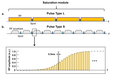 |
121 |
Quantifying the Effects of Pulsed Saturation Transfer at 1.5T and 3T
Rachel Chan, Sten Myrehaug, Greg Stanisz, Arjun Sahgal, Angus Lau
The aim of this study was to investigate the feasibility of CEST imaging at 1.5T and to compare the results to 3T experiments. CEST and MT parameters were quantified in phantoms and in the healthy brain at both fields. A pulsed saturation scheme was used to overcome the single RF amplifier duty cycle limitations of the 1.5T clinical scanner. Parameters were estimated using a Bloch-McConnell two-pool simulation that incorporated the extended phase graph formalism. The new methods demonstrated promise for enabling broader application of CEST MRI for field strengths below 3T.
|
|
5015.
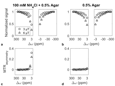 |
122 |
Inaccuracy in Estimated CEST Contrast After Extrapolation from Two-Pool Quantitative MT Fitting
Wilfred Lam, Ryan Oglesby, Greg Stanisz
A recently introduced technique has been used to remove the semisolid macromolecular magnetization transfer (MT) contribution from Z-spectra in order to isolate the contrast from chemical exchange saturation transfer (CEST) and the relayed nuclear Overhauser effect (rNOE). This is usually performed by first fitting Z-spectra acquired with high amplitude RF saturation with a two-pool quantitative MT model and then extrapolating an MT-only Z-spectrum to a lower saturation B1 and subtracting from Z-spectra acquired with the same B1. We aim to show that this two-step pipeline introduces inaccuracies into the extrapolation and describe them.
|
|
5016.
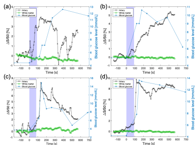 |
123 |
Comparison between glucoCEST at 3T and Blood Glucose Sampling in Humans
Anina Seidemo, Anna Rydhög, Ronnie Wirestam, Xiang Xu, Akansha Ashvani Sehgal, Yi Zhang, Frederik Testud, Pia Sundgren, Peter van Zijl, Linda Knutsson
Dynamic glucose-enhanced (DGE) MRI uses chemical exchange saturation transfer (CEST) to retrieve information about microcirculation using D-glucose as a contrast agent.We performed glucoCEST imaging in four healthy volunteers at 3T and compared arterial input functions (AIFs) and DGE signal in white matter to measured venous blood glucose levels after glucose infusion. An increase in DGE signal following the glucose infusion was observed in cerebral arteries, but not in white matter. We observed a similarity in shape of the AIF and the blood glucose curve, and that a higher blood glucose level corresponds to a higher DGE signal.
|
|
5017.
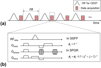 |
124 |
Fully Steady-State Chemical Exchange Saturation Transfer (CEST) imaging for Amide Proton Transfer (APT) at 3T MRI
Byungjai Kim, Seohee So, Namho Jeong, Hyunwook Park
A steady-state chemical exchange saturation transfer (SS-CEST) MRI, which is based on the standard macromolecule transfer (MT) imaging method, has been developed for fast scanning 1,2. In this study, we proposed a SS-CEST imaging combined with steady-state free precession (SSFP) which is so-called fully SS-CEST imaging. The fully SS-CEST method was compared with a SS-CEST combined with spoiled gradient echo (SPGR) through numerical simulations and MRI experiments. The results show that the fully SS-CEST method can provide higher amide proton transfer (APT) contrast than the SS-CEST with SGPR.
|
|
5018.
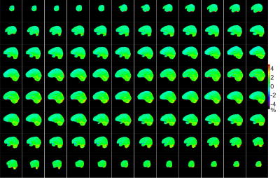 |
125 |
Isotropic Whole-brain CEST Imaging with Fast SPACE Readout
Yi Zhang, Ruibin Liu, Yi Sun, Dan Wu
For Chemical Exchange Saturation Transfer (CEST) imaging to be adopted as a routine clinical sequence, its spatial coverage and acquisition speed need ideally to be comparable to the current anatomical MRI sequences. To the best of our knowledge, whole-brain CEST imaging has only been demonstrated with 3D EPI readout, which, however, is vulnerable to susceptibility artifacts. Here, we propose a novel whole-brain isotropic-resolution CEST sequence utilizing the fast SPACE readout. The SPACE CEST sequence enables whole-brain 2.79mm isotropic CEST imaging in 5min without susceptibility artifacts, and should facilitate the translation of CEST MRI as a clinical routine sequence.
|
|
| Top |
Perfusion & Permeability
Digital Poster
Contrast Mechanisms
Thursday, 16 May 2019
| Exhibition Hall |
14:45 - 15:45 |
| |
|
Computer # |
|
5019.
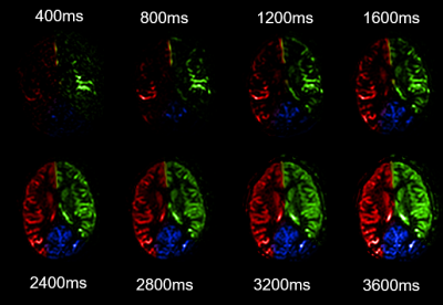 |
126 |
Time-resolved super-selective Arterial Spin Labeling using a ternary matrix based labeling approach
Thomas Lindner, Olav Jansen, Michael Helle
This study presents a novel approach to simultaneously acquire vessel-selective and time-resolved perfusion images combining a ternary encoding matrix approach with self-controlled super-selective Arterial Spin Labeling.
|
|
5020.
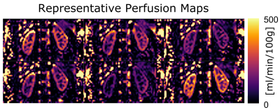 |
127 |
Investigating Variability Sources in Kidney Perfusion Measurements with Pulsed ASL: A Phantom and In Vivo Pilot Study
Jorge Chacon-Caldera, Lisa-Maria Hatz, Anja Kruse, Tanja Uhrig, Fabian Zimmer, Simon Hubertus, Ingo Hermann, Lothar Schad, Frank Zöllner
In this work, the variations of a 3D GRASE sequence with a fast pulsed ASL protocol at 3T were tested in isolation using a phantom and in vivo measurements. In vivo, 25 single scans in breath hold with a single volunteer were obtained in one session, thus reducing breathing motion and registration uncertainties as well as shimming, inter-volunteer and hydration stage variabilities. In phantom, we found a perfusion of 94.5 ± 5.4 ml/min/100g and in vivo, we found perfusion values in the expected physiological range with high standard deviation inter-scans but high correlation between kidneys. This suggests physiological instead of signal-to-noise related variations which would yield random inter-kidney variations.
|
|
5021.
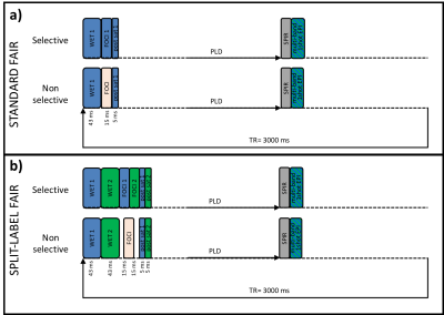 |
128 |
A Split-Label Design for Simultaneous Measurements of Perfusion in Distant Slices by Pulsed Arterial Spin Labeling
Celine Baligand, Lydiane Hirschler, Thom Veeger, Suzanne Franklin, Matthias van Osch, Hermien Kan
Most Arterial-Spin-Labeling (ASL-)MRI in exercised muscle employ single-slice acquisitions. However, fiber-type and oxidative capacity vary along the length of healthy muscles. Therefore, multi-slice acquisitions are desirable. Multi-slice pulsed ASL coverage is limited because the label is created proximally from the stack of slices. In muscle, this implies long transit times to the most distal slice due to slow flow. We propose a split-label design adaptation of FAIR that allows for sufficient labeling for distant slices. We validated our approach in the brain to take advantage of the high resting-state perfusion, and applied it in the lower leg muscle after exercise.
|
|
5022.
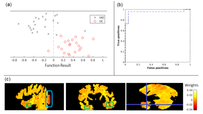 |
129 |
A Pipeline for ASL Quantification and Analysis using Inter-regional Differences and Machine Learning: Application to Young Onset Alzheimer’s Disease
Jack Highton, Enrico De Vita, Jonathan Schott, David Thomas
Arterial Spin Labelling (ASL) is an MRI method to measure cerebral blood flow with potential to assist early dementia diagnosis. Here, ASL data acquired from patients with young onset Alzheimer’s disease (AD) was analysed, using both a novel region based statistical approach and voxel based machine learning. This is the first study to analyse ASL data from patients with Posterior Cortical Atrophy using machine learning. Both approaches are shown to identify regions known to be affected by AD. Inter-region analysis suggests the parietal lobe is the most useful benchmark region, to separate region specific hypoperfusion from global perfusion changes.
|
|
5023.
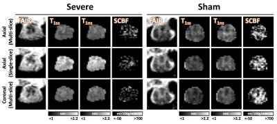 |
130 |
Quantifying Blood Flow of Rat Spinal Cord Injury Using in vivo Flow-sensitive Alternating Inversion Recovery (FAIR)
Seongtaek Lee, Natasha Wilkins, Brian Schmit, Shekar Kurpad, Matthew Budde
Perfusion weighted MRI has been widely used as a non-invasive MR biomarker in brain imaging but its application to spinal cord imaging has been limited due to the inherent difficulties. In this study, we evaluated flow-sensitive alternating inversion recovery to quantify spinal cord blood flow (SCBF) in rat spinal cord with varying severities of contusion injury. A trend of decreasing SCBF was observed with greater injury severity, suggesting that arterial spin labeling may be useful as a reliable non-invasive indicator of spinal cord traumatic injury. Furthermore, T1 values demonstrated greater sensitivity to injury severity and functional outcomes.
|
|
5024.
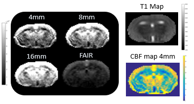 |
131 |
Quantification of Multiple Boli Arterial Spin Labelling in Mice and Rats
Samantha Paterson, Camille Graff, Antoine Vallatos, William Holmes
A need for high SNR quantitative ASL has driven the quantification of mbASL, a high SNR ASL sequence that uses adiabatic pulses to label multiple boli of arterial water. The sequence has a hybrid PASL & CASL nature with a modified Buxton kinetic model used to describe this hybrid-like nature. We have shown that experimental results mirror theoretical predictions with signal distribution changing with labelling slice thickness. High SNR mbASL images in mice and rats with significantly higher signal than the standard FAIR sequence have been produced and CBF images and values acquired that agree with the literature.
|
|
5025.
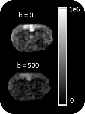 |
132 |
Imaging Blood Brain Barrier Permeability in a Human African Trypanosomiasis Mouse Model using Diffusion Weighted Multiple Boli Arterial Spin Labelling
Samantha Paterson, Antoine Vallatos, Jean Rodgers, William Holmes
Human African Trypanosomiasis is a parasitic disease that causes progressive blood brain barrier breakdown. We have developed a non-invasive high SNR ASL technique (mbASL) combined with bipolar diffusion gradients to determine the ratio of intravascular to extravascular signal from the brain. The ratio of this signal will change in a mouse brain infected with HAT due to the barrier breakdown. We have imaged this changing brain along with producing CBF maps, thus using a novel method in imaging a HAT infected mouse.
|
|
5026.
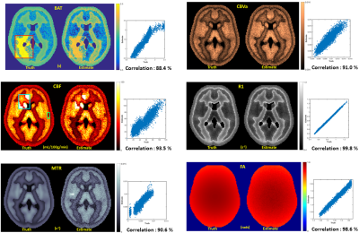 |
133 |
Optimizing MRF-ASL Scan Design towards Precise Quantification of Hemodynamic Properties in Cerebrovascular Disorders
Anish Lahiri, Jeffrey Fessler, Luis Hernandez-Garcia
We investigate an optimization method to make fast, precise quantification of hemodynamic and tissue properties from an MRF ASL scan more robust to their respective feasible ranges, particularly when conditions pertaining to cerebrovascular disorders are included in consideration. We further validate our methods on synthetic and healthy human subject data.
|
|
5027.
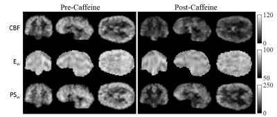 |
134 |
Effect of Caffeine on Blood-Brain Barrier Water Permeability Measured with Intrinsic Diffusivity Encoding of Arterial Labeled Spins (IDEALS)
Kenneth Wengler, Kwan Chen, Mark Schweitzer, Xiang He
Caffeine is a commonly used neurostimulator that also produces cerebral vasoconstriction by antagonizing adenosine receptors. Previous studies demonstrate that acute caffeine intake leads to significant cerebral blood flow (CBF) reduction but have not investigated the effect on blood-brain barrier (BBB) water permeability. Here we provide an initial investigation into the effect of caffeine on BBB water permeability parameters, water extraction fraction (Ew) and permeability surface area product (PSw), using the recently developed Intrinsic Diffusivity Encoding of Arterial Labeled Spins (IDEALS). Significant reductions in CBF, Ew, and PSw were observed after administration of 200 mg caffeine.
|
|
5028.
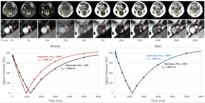 |
135 |
In vivo quantification of arterial blood longitudinal relaxation time during graded hyperoxia at 3T using an intermittent cuff occlusion paradigm
Erin Englund, Ana Rodriguez-Soto, Felix Wehrli
Breathing hyperoxic gas results in dissolved oxygen in blood plasma, shortening T1 of arterial blood (T1a). Quantification of T1a is difficult due to blood flow between the inversion and acquisition. Here, this problem is overcome using an intermittent cuff occlusion protocol to suspend flow in the femoral artery and vein. An inversion recovery-prepared bSSFP sequence was used to measure T1a during normoxia (FiO2=21% O2) and graded hyperoxia (FiO2~33%, 48%, 55%, 100% O2) in ten healthy subjects at 3T. During normoxia, T1a was 1819±142ms, which was shortened to 1522±100ms during maximal hyperoxia, and the relationship between T1a and PETO2 was linear.
|
|
5029.
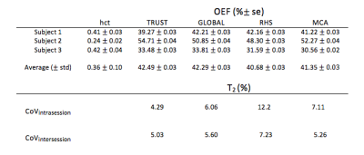 |
136 |
Reproducibility of Selective Localised T2-Relaxation-Under-Spin-Tagging (SL-TRUST) for Regional Cerebral Oxygen Extraction Fraction
Caitlin O'Brien, Thomas Okell, Peter Jezzard
In this study, we offer preliminary results of a reproducibility study on a novel sequence: Selective Localised TRUST (SL-TRUST). SL-TRUST is an MR acquisition method for acquiring spatially specific cerebral tissue oxygen extraction fraction (OEF) values through measurements of venous blood T2 in the superior sagittal sinus. Three subjects underwent four scan sessions over a seven day period and the inter/intra session variability of SL-TRUST was evaluated. The resulting venous blood T2 and tissue OEF values from spatially specific regions (a single hemisphere, and a 70x80x80mm tissue region) are compared to whole brain measures obtained using TRUST.
|
|
5030.
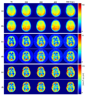 |
137 |
The spiral trajectory correction effect on arterial spin labeling acquired with high-slew-rate gradient on a compact 3T scanner
Daehun Kang, Uten Yarach, Myung-Ho In, Erin Gray, Nolan Meyer, Joshua Trzasko, Yunhong Shu, Matt Bernstein, John Huston
Proper design of the arterial spin-labeled (ASL) readout trajectory can minimize signal loss, reduce artifacts, and consequently improve the quality of ASL-derived perfusion maps. High performance gradients can improve MR image quality in spiral acquisition to reduce susceptibility and off-resonance effect. However, the eddy current and the system delay can degrade image quality by causing image rotation and blurring effects. In this work, the image artifact was corrected with a truer trajectory measured by a dynamic field camera. The effects of the trajectory correction on ASL images are investigated on a low-cryogen, compact 3T MRI system.
|
|
5031.
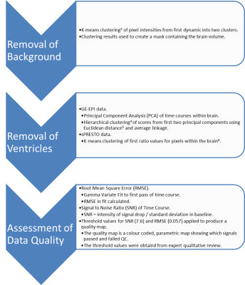 |
138 |
An Algorithm for the Automated Quality Assessment and Perfusion Biomarker Determination of Multicentre Dynamic Susceptibility Contrast (DSC-) MRI
Stephen Powell, Stephanie Withey, Yu Sun, Lesley Macpherson, Laurence Abernathy, Barry Pizer, Richard Grundy, Simon Bailey, Dipayan Mitra, Dorothee Auer, Shivaram Avula, Theodoros N. Arvanitis, Andrew Peet
Dynamic Susceptibility Contrast (DSC-) MRI estimates biomarkers, such as cerebral blood volume (CBV). However, data quality varies between centres and quality control (QC) is carried out by qualitative review, which is time-consuming and subjective. An automated QC pipeline was developed and tested on 34 patient data sets. The pipeline analysed four slices from each patient, producing SNR, RMSE, relative CBV (rCBV), and quality maps for each slice, which were used to quantify QC. Average values for each parameter were produced for each centre, protocol and field strength, showing variability in data quality and providing a basis for multi-centre protocol optimisation.
|
|
5032.
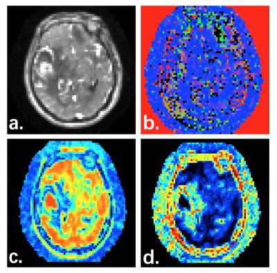 |
139 |
Dynamic Perfusion Tensor Imaging
Presentation Not Submitted
Jing Wang, Yang Fan
DCE-MRIprovides a method to continuously measure the spatial and temporal characteristics of local tissue perfusion. This study points out an interesting feature of DCE-MRI: the voxel wised correlation can be encoded in 26 directions, allowing for the measurement of perfusion tensor. We demonstrate this new method, dynamic perfusion tensor imaging (dPTI), facilitates the reconstruction of the local perfusion field, characterized by a perfusion tensor, from which can be derived quantities related to the structure of the local perfusion field, such as the mean perfusion and perfusion anisotropy.
|
|
5033.
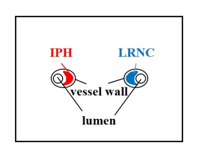 |
140 |
The Effect of the Injection Dose, Rate and Concentration on Carotid Dynamic Contrast-Enhanced MRI: a Simulation Study
Yajie Wang, Yishi Wang, Haikun Qi, Xian Liu, Hua Guo, Huijun Chen
Dynamic contrast-enhanced MR imaging (DCE-MRI) is an effective tool to quantify inflammation in carotid atherosclerotic plaque, while little is known on the effect of the injection protocol on carotid DCE-MRI. In this study, the effect of the contrast injection protocol, including injection dose and effective injection rate (decided by the injection rate and concentration) on the pharmacokinetic parameters estimation in carotid DCE-MRI were investigated. The results indicated that high injection dose (~0.1mmol/kg) with relative low effective injection rate (~0.5 mmol/s, effective injection rate = rate (ml/s) × concentration (mol/L)) was recommended for the simulated bright-blood DCE protocol.
|
|
5034.
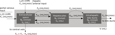 |
141 |
Repeatability of hepatocellular uptake and efflux in the rat liver: A comparison of Gadoxetate DCE-MRI models
Sirisha Tadimalla, Claudia Green, Denise Steinmann, Sascha Koehler, Hans-Paul Juretschke, Iina Laitinen, John Waterton, Paul Hockings, Catherine Hines, Gunnar Schütz, Steven Sourbron
A variety of Gadoxetate DCE-MRI models have been proposed to derive hepatocyte uptake and efflux rates in the rat, but it is unclear which provides most reliable measurements. Here, we compare four models in terms of their test-retest repeatability on 9 rats measured in 3 sites. Results indicate that a two-compartment high-flow model, assuming negligible sinusoidal backflux and a fixed population-based extracellular volume fraction, provides most repeatable measures of hepatocellular function in the healthy rat.
|
|
5035.
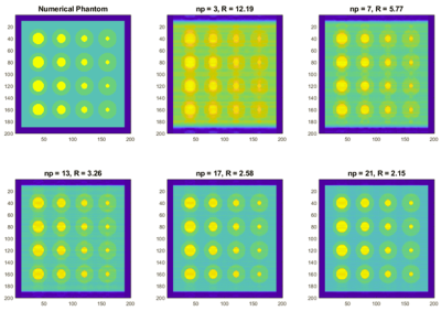 |
142 |
Pharmacokinetic Parameter Accuracy Correlates with Image Quality Metrics in Flexible Temporal Resolution DCE-MRI Simulations
Allister Mason, Nathan Murtha, James Rioux, Sharon Clarke, Chris Bowen, Steven Beyea
The choice of imaging parameters in dynamic contrast enhanced (DCE) MRI, such as temporal resolution, can affect the recovered parameter when using quantitative pharmacokinetic (PK) models, such as Ktrans in the Tofts model. We propose objective image quality metrics (IQMs) as a tool to guide this choice to maximize PK parameter accuracy. DCE MRI simulations were performed on a numerical phantom with user defined PK parameters. IQMs were calculated using the numerical phantom as a reference and references generated from the simulated data. In both cases, a strong correlation between the PK parameter error and IQM score is found.
|
|
5036.
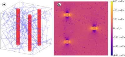 |
143 |
The role of perivascular spaces in white matter dynamic susceptibility contrast MRI
Jonathan Doucette, Enedino Hernández-Torres, Christian Kames, Alexander Rauscher
Using vascular parameters obtained from dynamic susceptibility contrast MRI, the gradient echo (GRE) and spin echo (SE) dynamic susceptibility contrast (DSC) induced changes in ΔR(∗)2 were
simulated at 3T in order to investigate the effects of tissue orientation and perivascular spaces (PVS). We found that the orientation dependence of both ΔR2 and ΔR∗2 are
amplified by PVS, though ΔR2 is
far more sensitive to PVS.
|
|
5037.
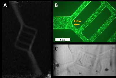 |
144 |
A 3D-Printed Physiologically-Faithful Perfusion Phantom that Recapitulates Microvasculature Structure for Quantitative Experimental Validation of Fluid Transport
John Morgan, Thanh Nguyen, Pascal Spincemaille, Yi Wang
Recapitulation of microvascular structure, function and perfusion in vitro can enable studies of vascular biology, provide a model for diseases such as ischemic stroke or tumor angiogenesis and enable quantitative evaluation of physiologic blood or lymph perfusion. Here we describe the initial design and deployment of a first-generation, self-contained 3D-printed, physiologically-faithful, microfluidic perfusion phantom to form explicit, hierarchically-branching, microvascular structure encapsulated in a type I collagen matrix in vitro, with pump-driven perfusion easily visible via phase-contrast MRI (Fig. 1). The phantom flexibly supports creation of user-defined vessel network geometries with human vascular cells and allows experimental validation of blood flow, i.e., via constitutive equations for convective and diffusive transport that quantitatively relate the flux of tracers from time-resolved images to transport field quantities. Thus, the largely qualitative and unmeasurable global arterial input assumption in the traditional Kety’s method can be replaced with measurable and reproducible MRI experimental data, formulated as quantitative transport mapping (QTM). Preliminary data demonstrate that the QTM phantom is promising for characterizing actual blood transport in vitro in healthy and pathological contexts.
|
|
5038.
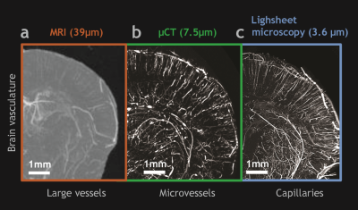 |
145 |
A novel trimodality vascular contrast agent for “image-based systems biology” applications
Akanksha Bhargava, Priyanka Kushwaha , Ryan Riddle, Manisha Aggarwal, Arvind Pathak
Preclinical vascular imaging has been instrumental in advancing our understanding of the role of blood vessels in health and disease. However, currently there are no vascular contrast agents that are simultaneously visible with all three primary imaging modalities, i.e. MRI, CT and optical imaging. Consequently, this poses a hurdle when integrating multiscale, multimodality vascular imaging data for systems biology applications. Therefore, we developed a new vascular contrast agent that delivers trimodal imaging capabilities with MRI, CT and optical imaging for preclinical systems biology investigations. We demonstrate its compatibility with different types of imaging contrasts that allow trimodality, multiscale imaging of healthy (e.g. murine brain, leg, kidney) and pathological microvasculature (e.g. cancer xenografts).
|
|
5039.
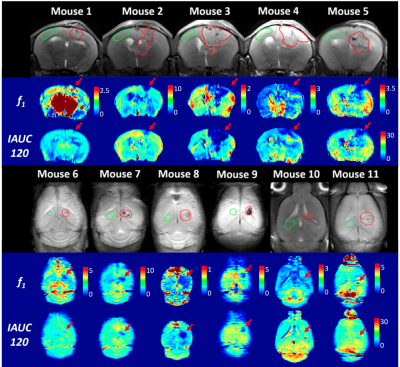 |
146 |
Using D2O As a Diffusible Tracer in Characterizing Tumor Properties on Mice
Pei-Lun Yu, Ming Cheng, Xiayu Lyu, Sheng-Min Huang, Kung-Chu Ho, Fu-Nien Wang
As a diffusible tracer, D2O was employed as a negative contrast agent in investigating tumor perfusion in this study. Results show that the transfer constant derived from D2O perfusion is able to characterize tumor flow properties. Furthermore, we also demonstrated that the initial area under curve of D2O perfusion has potential in detecting the flow difference between tumor and normal tissue, suggesting the feasibility of semi-quantitative indices for D2O perfusion.
|
|
5040.
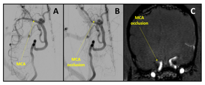 |
147 |
Predicting the rate of stroke evolution in canines using MR-derived time-to-peak perfusion maps
Robert King, Matthew Gounis, Mohammed Salman Shazeeb
Mechanical thrombectomy for the treatment of ischemic stroke shows high rates of recanalization; however, some patients still have poor clinical outcome. The canine large vessel occlusion model has been developed to better understand new treatments. This model has a drawback of inconsistent rates of stroke growth. Here, MRI perfusion based time-to-peak maps were used to predict the rate of infarct growth as validated by ADC-derived maps. Classification of canines into either fast or slow evolvers was reliably shown with this method of analysis, allowing for a better understanding of new therapeutics and potentially for better patient selection for thrombectomy.
|
|
5041.
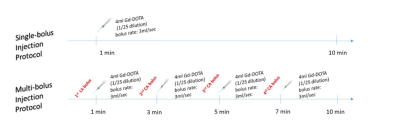 |
148 |
Using multi-bolus injection protocol to improve the robustness of DCE-MRI: application in rabbit liver
Sofia Veneti, Daniel BALVAY, Gwennhäel AUTRET, Charles CUENOD
Quantitative DCE-MRI suffers from limitations in the liver due to its dual input (hepatic artery, portal vein). Single- and multi-bolus (four) injection protocols were performed in rabbits on a 4.7T small-animal MRI. The Hepatic Perfusion Index (HPI), total hepatic blood flow (F), and distribution volume (Vd) were studied using a computer simulation and evaluated by the Coefficient of Variation (CV) and the 95% Confidence Interval (CI) factors. Statistical tests were performed for 1000 iterations with Mann-Whitney test and p<0.002 statistically significant. Multi-bolus injection protocol strongly improve the DCE precision in the liver particularly for HPI, which is a critical parameter.
|
|
5042.
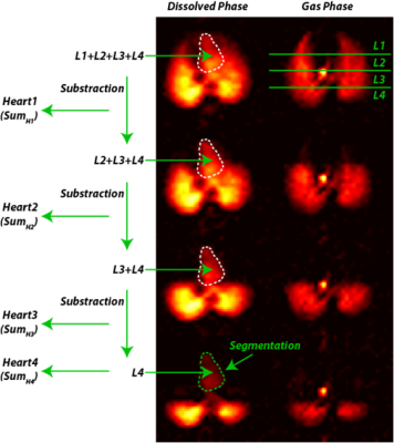 |
149 |
Measuring Regional Gas Transport in Injured Rabbit Lungs Using Hyperpolarized Xenon
Yi Xin, Kai Ruppert, Maurizio Cereda, Faraz Amzajerdian, Hooman Hamedania, Mehrdad Pourfathi, Sarmad Siddiqui, Ian Duncan, Luis Loza, Tahmina Achekzai, Federico Sertic, Ryan Baron, Harrilla Profka, Stephen Kadlecek, Rahim Rizi
Hyperpolarized 129-Xenon MRI measures the regional content of tracer gas in the lungs; it can also differentiate between Xenon contained in the gas phase (GP) and in the dissolved phase (DP), allowing us to characterize regional gas diffusivity and uptake in the pulmonary capillary blood in addition to capturing parameters of alveolar aeration. By measuring absorbed Xenon signal in the left heart and aorta shortly after inhalation, it is theoretically possible to study the next step of gas transfer by measuring the gas that reaches the arterial blood. In this study, we explore the regional gas transport of injured rabbit lungs in two different states of recruitment.
|
|
5043
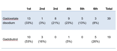 |
150 |
Transient respiratory motion artifacts in multiple arterial phases of contrast-enhanced dynamic MR imaging of the abdomen: a comparison using gadoxetate disodium and gadobutrol.
Video Permission Withheld
Masahiro Tanabe, Mayumi Higashi, Keisuke Miyoshi, Sei Nakao, Etsushi Iida, Matakazu Furukawa, Katsuyoshi Ito
This study compared the occurrence of transient respiratory motion artifacts (TRMA) in multiple arterial phases of contrast-enhanced MR imaging using a rapid acquisition technique with high temporal and spatial resolution between gadoxetate disodium and gadobutrol. This study showed that the frequency of TRMA after third arterial phase was significantly higher in patients using gadoxetate disodium than in patients using gadobutrol. In multiple arterial phase dynamic MR imaging, the frequency of TRMA in gadoxetate disodium increased, compared with gadobutrol, caused by the intolerable respiratory suspension after third arterial phase, possibly due to contrast agent-related effect.
|
|
| Top |
Electromagnetic Tissue Mapping
Digital Poster
Contrast Mechanisms
Thursday, 16 May 2019
| Exhibition Hall |
14:45 - 15:45 |
| |
|
Computer # |
|
5044.
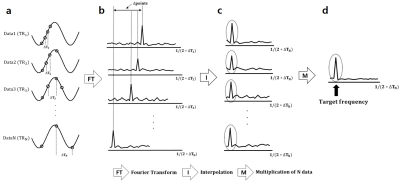 |
151 |
Multiple TR Approach for Direct Detection of Fast Oscillating Magnetic Fields
Je-Seok Ham, Hyun-Soo Lee, Sung-Hong Park
Due to weak amplitude and fast oscillation, it is still controversial whether neuronal activities can be directly detected through MR imaging. In this study, we propose a novel method, multiple-TR approach, which utilized 1) multi-phase acquisition and 2) frequency spectrum multiplication for detecting weak and fast oscillating magnetic fields. We demonstrated with phantom experiments that SNR at the stimulation frequency on the spectrum was remarkably enhanced with the higher number of TRs under almost the same scan time, amplifying oscillation frequency component while suppressing systematic noises. This proposed approach will increase possibility of directly detecting neural oscillations in vivo.
|
|
5045.
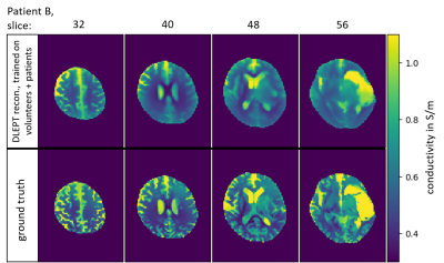 |
152 |
Deep learning brain conductivity mapping using a patch-based 3D U-net
Nils Hampe, Ulrich Katscher, Cornelius van den Berg, Khin Tha, Stefano Mandija
Conventional Electrical Properties Tomography (EPT) suffers from reconstruction artifacts related to assumptions necessary for solving the equations analytically. To circumvent the necessity for these assumptions, in this study a deep learning approach is utilized to approximate the analytically unsolvable equations. For this purpose, a 3D convolutional neural network was trained on simulations and in-vivo data from healthy volunteers and cancer patients. Results demonstrate the potential of this method, as noise-free conductivity maps were obtained without anatomic apriori information in less than 1:30 min per reconstruction.
|
|
5046.
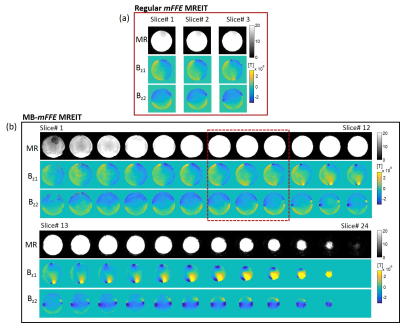 |
153 |
Fast MREIT acquisition using Multi-Band and SENSE Techniques
Munish Chauhan, Michael Schär, Sulagna Sahu, Thomas Mareci, Rosalind Sadleir
Recent studies demonstrated the first current density and conductivity tensor images of human heads during transcranial electrical stimulation (tES), using MREIT techniques. These techniques allowed imaging of three 5-mm slices in a sequence lasting 12 minutes, a typical therapeutic tES administration time. To obtain whole brain coverage, it is important to accelerate existing MREIT sequences. In this study, we combined our existing Gradient-Echo (mFFE) MREIT acquisition methods with parallel imaging. We found the acquisition speed of image data reflecting current distributions in tES was improved 8 times by using Multi-Band (MB) imaging, while maintaining good image quality.
|
|
5047.
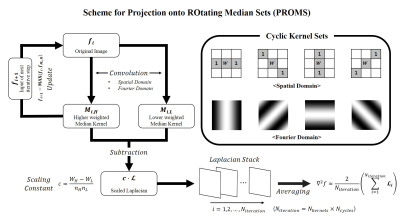 |
154 |
Discrete Laplacian Estimation Using Projection onto ROtating Median Sets (PROMS) for MR Electrical Property Tomography
Han-Jae Chung, Jong-Min Kim, You-Jin Jeong, Jae-Won Yoo, Jeong-Hee Kim, Chulhyun Lee, Chang-Hyun Oh
The novel contrast mechanism, MREPT, is still difficult to use in the clinical field. This is because the reconstruction of MREPT is performed under many assumptions, which lead to limitations for solving the governing equations, and is highly noise sensitive characteristics to noise in practical use. In this work, we propose a new discrete Laplacian estimation method using Median kernel sets, which has fast and noise-robust features, and show its performance by using in-vivo experiments and EM simulation. For validation of the proposed method, three healthy volunteers were scanned on a clinical MRI system with identical imaging parameters.
|
|
5048.
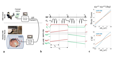 |
155 |
Does RF spoiling enhance human in-vivo brain MR Current Density Imaging (MRCDI)?
Cihan Göksu, Lars G. Hanson, Hartwig Siebner, Philipp Ehses, Klaus Scheffler, Axel Thielscher
MRCDI is an emerging modality for non-invasive measurement of weak currents in the human brain, which is important in several neuroscientific applications. It is based on current-induced field measurements and requires high sensitivity to the extrinsic field changes. Measurement sensitivity can be compromised by irrelevant field changes caused by physiological variation. Here, we compare the performance of the so far most sensitive MRCDI method based on steady-state free precession free induction decay (SSFP-FID) with its RF-spoiled counterpart fast low angle shot (FLASH). No significant sensitivity differences were observed in slices covering the upper part of the brain, but SSFP-FID had ~20% lower noise floors in lower slices. For the relevant acquisition parameters, FLASH exhibits no remarkable image quality enhancements in 2D.
|
|
5049
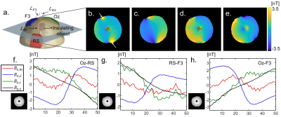 |
156 |
Compensation of lead-wire magnetic field contributions in MREIT experiment using image segmentation: a phantom study
Video Permission Withheld
Saurav Zaman Khan Sajib, Munish Chauhan, Guita Banan, Sulagna Sahu, Luning Wang, Thomas Mareci, Rosalind Sadleir
For the measurement of current-induced phase using MRI, the effect of stray magnetic fields caused by the current carrying wire on images must be minimized prior to reconstruction of the current density. In this study, we report a method which can effectively remove the effect of lead wire magnetic fields interference during MREIT measurements. Results from phantom experiments and numerical simulations demonstrate the feasibility of the method, which can be used to correct for lead effects when measuring current density in human transcranial direct current stimulation (tDCS) measurements using magnetic resonance electrical impedance tomography (MREIT).
|
|
5050.
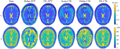 |
157 |
Improving Tissue Electrical Properties Reconstructions by Exploiting the Benefits of Combining Deep Learning-EPT and 3D Contrast Source Inversion-EPT
Reijer Leijsen, Cornelis van den Berg, Rob Remis, Andrew Webb, Stefano Mandija
We propose a two-step approach to EPT reconstruction where we use the results from a deep-learning approach as the initial estimate for a 3D contrast-source inversion algorithm. The combination of these two methods builds upon the strengths of each. Results using an anatomically accurate head model with and without an artificially inserted tumour show that CSI-EPT improves DL-EPT reconstructions in structures that are not present in the training set, while DL-EPT used as an initial guess for CSI-EPT leads to improved accuracy and convergence.
|
|
5051.
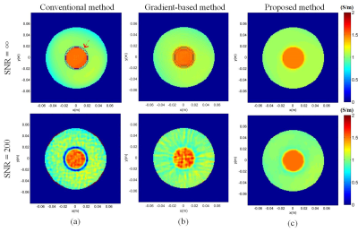 |
158 |
A dual constraints-based approach to electrical conductivity imaging using MR phase
Xiangdong Sun, Lijun Lu, Yingjie Mei, Xiaoyun Liu, Wufan Chen
Electrical conductivity imaging of tissue can potentially provide electrical property information of tissues. Here, we proposed a dual constraints based electrical conductivity imaging method to reconstruct the distribution of conductivity. The proposed method transforms the partial differential equation into an optimization problem with total variation and wavelet transform regularization terms. To evaluate the performance of the proposed method numerical simulations and phantom experiments were implemented at 3T. The results showed that the proposed method improves the accuracy of the conductivity estimate and suppress the effect of noise. The proposed method is expected to be applied clinically in the future.
|
|
5052.
 |
159 |
Inverse problem approach to cr-MREPT
Yusuf Ider, Celik Boga
The convection-reaction partial differential equation (PDE) based algorithm for Magnetic Resonance Electrical Properties Tomograpy, cr-MREPT, does not suffer from internal boundary artifacts but has the Low Convective Field (LCF) artifact. The cr-MREPT PDE is rearranged such that H1+ is the unknown variable and the coefficients depend on the EPs. The EPs are iteratively adjusted to minimize the difference between calculated and measured H1+. The method can be applied to a Region-of-Interest without considering the whole object. Conductivity reconstructions for simulation objects and also for an agar phantom demonstrate that the proposed method does not suffer from boundary and LCF artifacts.
|
|
5053.
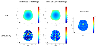 |
160 |
Estimation of transceive phase via LORE-GN algorithm and its use in MREPT
Safa Ozdemir, Yusuf Ider
Balanced steady state free precession (bSSFP) is a widely used MR sequence since it has high speed, high SNR, motion insensitivity and automatic eddy current compensation. Besides all these advantages, bSSFP sequence is susceptible B0 inhomogeneity and banding artifact occurs in certain off-frequency regions. In this paper, one of the correction methods, LORE-GN, is utilized to obtain transceive phase free from the distortions originating from B0 inhomogeneity. As an application, acquired transceive phase maps are used to obtain conductivity maps.
|
|
5054.
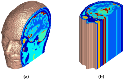 |
161 |
Limitations of 2-D Field Structure Assumptions in Electrical Properties Tomography and its 3-D CSI-EPT Solution
Patrick Fuchs, Reijer Leijsen, Rob Remis
CSI-EPT was originally implemented in a two-dimensional formulation and has since been extended to 3-D to allow for volumetric reconstructions without any assumptions on the field structures. Since the 3-D method is computationally much more complex than its 2-D counterpart, here we investigate the 2-D assumption and its requirements. Unfortunately the 2-D assumption breaks down when the object in consideration is not sufficiently longitudinally invariant, even if the fields can still be considered E-polarised. Therefore, to achieve accurate and robust reconstructions of EPs in a practical or clinical setting the 3-D CSI-EPT method is a recommended starting point.
|
|
5055.
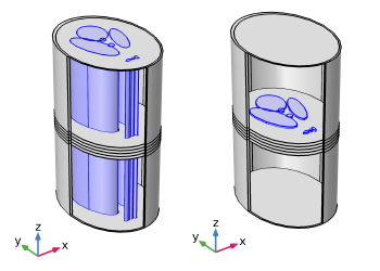 |
162 |
An Explicit EPT Reconstruction Method Based on the Dbar Equation Incorporating Longitudinal Magnetic Field Variations
Motofumi Fushimi, Takaaki Nara
This paper presents a novel explicit reconstruction method for magnetic resonance-based electrical properties tomography (EPT). We derive the dbar equation for the electric field that incorporates the variation of the magnetic field along the longitudinal axis. This new dbar equation allows an explicit reconstruction of the electrical properties even when the magnetic field varies along that axis. We also propose an iterative reconstruction procedure that solves the dbar equation by the Cauchy–Pompeiu formula and updates EP values in each step. The proposed method correctly reconstructed EPs even in the case that the magnetic field and EPs vary along the longitudinal direction.
|
|
5056.
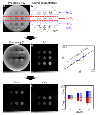 |
163 |
Validation of magnetic susceptibility source separation: Monte Carlo simulation and phantom experiment
Presentation Not Submitted
Jingu Lee, Hyeong-geol Shin, Hyunsung Eun, Dongmyung Shin, Jongho Lee
In this study, the recently proposed magnetic susceptibility source separation method, which separates the paramagnetic susceptibility source from the diamagnetic susceptibility source, was validated using Monte-Carlo simulation and phantom experiment. The results demonstrate that the method successfully separates the paramagnetic and diamagnetic susceptibility sources in both simulation and experiment.
|
|
5057.
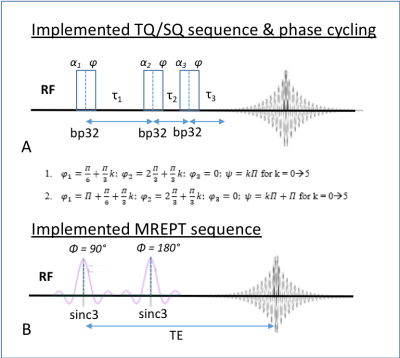 |
164 |
Investigating the Relationship Between Conductivity and Bound Sodium Fractions at 21.1 T
Ghoncheh Amouzandeh, Nastaren Abad, Jens Rosenberg, Samuel Grant
This study investigates the relation between electrical conductivity evaluated using MR Electrical Properties Tomography (EPT), sodium concentrations and their mobility. In particular, the project seeks to determine if conductivity values calculated from EPT differentiate between bound and free sodium measured by triple quantum (TQ) coherence selection. TQ and EPT acquisitions were evaluated over a range of sodium concentration and with different binding conditions to provide insight into the sources of tissue conductivity changes. The correlation between electrical conductivity and ionic content can provide more in depth understanding of how sodium ions are changing in pathological conditions.
|
|
5058.
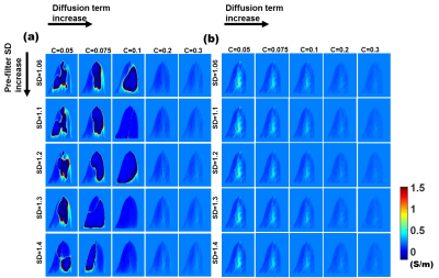 |
165 |
Filter design for Breast Conductivity imaging Using phase-based gradient EPT (gEPT)
Jun-Hyeong Kim, Jaewook Shin, Soo-Yeon Kim, Dong-Hyun Kim
EPT has a potential for immediate clinical use since it does not require additional hardware. However, there are various problems when applying clinical practice. In the case of breast image, to overcome SNR, a reconstruction method called magnitude-weighted polynomial fitting was applied. Recently, a new phase-based technique called ‘gradient-based EPT’ (pgEPT) was proposed. This method removes the boundary artifact by adding a gradient of phase and reduce noise by adding diffusion term. But, it doesn’t work properly for low SNR. In this study, an improved pgEPT for breast conductivity by replacing the Laplacian term with polynomial fitting is proposed.
|
|
5059.
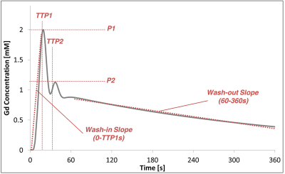 |
166 |
Optimal temporal resolution for accurate AIF measurement and error-constrained pharmacokinetic modelling of DCE data
Silvin Knight, James Meaney, Andrew Fagan
A highly-controlled and validated phantom-based method was used to investigate the effects of acquisition temporal resolution (Tres) on the arterial input function (AIF) measurement accuracy and precision for DCE-MRI. The propagation of these AIF measurement errors into errors in pharmacokinetic modelling parameter values could thus also be investigated. Guideline Tres values which can be used to constrain errors in Ktrans, kep and ve within defined limits (e.g. <5%, <10% etc) are presented.
|
|
5060
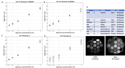 |
167 |
Dynamic susceptibility contrast MRI phantom for validation of clinical perfusion imaging
Video Permission Withheld
Slavka Carnicka, Katy Keenan , Stephen Russek, Nikki Rentz, Karl Stupic, Brandon Hancock, John Kirsch, Matthias van Osch , Bradley Erickson, Yunhong Shu, Chad Quarles, Ashley Stokes, Yuxiang Zhou, Edward Jackson, Alex Antolak, Vicky Liao, Alexander Lin , Elizabeth Mirowski, Michael Boss, Nancy Obuchowski, Ona Wu
Dynamic susceptibility contrast MR imaging (DSC MRI) is a very promising quantitative imaging technique used increasingly as both a diagnostic and research tool. This technique quantifies susceptibility-induced (R2*) signal loss to assess tissue perfusion (blood supply) and viability. Development of reference phantoms is crucial to determine the in vitro accuracy, test-retest repeatability, and inter-platform reproducibility of ?R2* quantification protocols. Hence, we developed a static DSC phantom suitable for simple and reliable evaluation of acquisition methods to assess susceptibility changes across multiple scanners and time. We also finalized acquisition protocols and developed software to analyze the DSC phantom data.
|
|
5061.
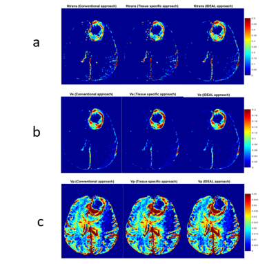 |
168 |
To evaluate the effect of different initial guess selection approaches on quantitative analysis of DCE-MRI data of brain tumor patients
Dinil Sasi, Sameer Manickam, Rakshit Dadarwal, Ayan Debnath, Snekha Thakran, Rakesh K Gupta, Anup Singh
Quantitative analysis of dynamic-contrast-enhanced(DCE)-MRI data using various tracer kinetic models is widely used in cancer diagnosis and follow-up. In general, voxelwise model fitting using nonlinear-least-square method requires a long processing time depending upon image-resolution, data noise, choice of initial guess, model type and computer-platform. In this study, we proposed a tissue specific initial guess selection approach, for the voxel wise fitting using nonlinear–least-square method, which substantially reduced computation-time without compromising accuracy of parameters compared to regular global initial guess approach. It also performed better than recently proposed Image-Downsampling-Expedited-Adaptive-Least-squares fitting approach. Parallel-processing was also implemented to further reduce the time
|
|
5062.
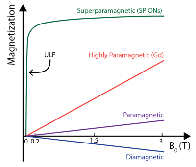 |
169 |
Susceptibility Contrast at Ultra-low Magnetic field with Superparamagnetic Nanoparticles
David Waddington, Thomas Boele, Richard Maschmeyer, Zdenka Kuncic, Matthew Rosen
MRI scanners operating at ultra-low fields (ULF) promise to reduce the cost and expand the clinical accessibility of MRI. Here, we use an ULF (6.5 mT) MRI scanner and an efficient balanced steady-state free precession MRI protocol to image superparamagnetic iron oxide nanoparticles (SPIONS) in solution. We observe strong susceptibility effects due to the highly-magnetized state of SPIONs even at ULF. These susceptibility effects enable the most sensitive imaging of a contrast agent at ULF that we are aware of. These results will broaden the clinical applications of ULF MRI, and have implications for drug tracking and delivery in nanotheranostics.
|
|
5063.
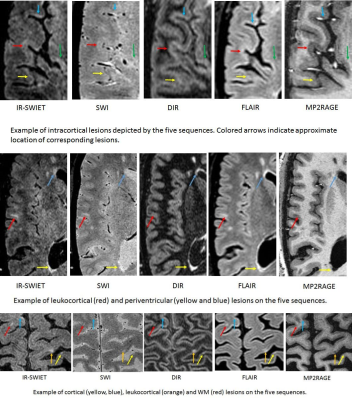 |
170 |
Inversion Recovery Susceptibility Weighted Imaging with Enhanced T2 Weighting (IR-SWIET): Application to Multiple Sclerosis (MS) Lesions
Neville Gai, Govind Nair, Erin Beck, Daniel Reich
Susceptibility weighted imaging provides important information regarding iron deposition and lesions in multiple sclerosis. However, CSF appears hyperintense on SWI images and can interfere with cortical lesion detection. Cortical lesions are associated with disability and disability progression in MS patients. Here a new 3D sequence (IR-SWIET), which suppresses CSF while maintaining T2 and T2* contrast of SWI is designed and evaluated. IR-SWIET was compared with four other commonly used sequences. CNR analysis in 30 lesions from MS patients showed that the sequence provided superior lesion depiction compared with SWI and compared well with 3D-DIR, MP2RAGE, and FLAIR.
|
|
5064.
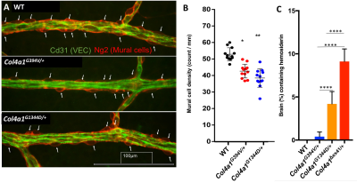 |
171 |
Identification of mutation-dependent heterogeneity in murine models of cerebral small vessel disease using susceptibility weighted imaging at 14.1 Tesla
Brice Tiret, Genki Hayashi, Cassandre Labelle-Dumais, Douglas Gould, Myriam Chaumeil
Cerebral small vessel diseases (cSVDs) are a poorly understood group of conditions that manifest as intracerebral hemorrhage, microinfarcts, microbleeds and white matter lesions. Here, we used innovative mouse models that recapitulate the clinical spectrum of human cSVDs to investigate whether T2-weighted (T2-w) and Susceptibility Weighted Imaging (SWI) at 14.1Tesla could be used to differentiate between disease subtypes. Our results show that SWI could differentiate between models, with the number of SWI-detected lesions in line with both disease severity and hemosiderin staining. High-field SWI thus demonstrate high potential to non-invasively monitor cSVD evolution and response to therapies in those models.
|
|
5065.
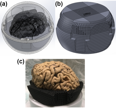 |
172 |
3D-Printed whole-brain holder for multiple orientation magnetic susceptibility measurements and precise dissection
Kwok-Shing Chan, Jeroen Mollink, Renaud Hedouin, Norbert Hermesdorf, Anne-Marie van Cappellen van Walsum, José Marques
In this study, we present a whole-brain holder for ex vivo experiments which allows rotating the sample inside a conventional head coil (while ensuring no deformation occurs) and provides guidance for precise correspondence between the MR data and excised tissue. We demonstrate some of these features with two experiments aimed at validating magnetic susceptibility measurements using MRI, where small 5mm cube samples located in different slices through the whole brain can be excised with great precision.
|
|
5066.
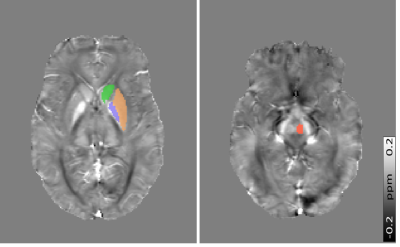 |
173 |
MRI Susceptibility Mapping Suggests Elevated Brain Iron in Sickle Cell Anaemia
Russell Murdoch, Jamie Kawadler, Fenella Kirkham, Karin Shmueli
Sickle Cell Anaemia (SCA) is a genetic condition characterized by haemolytic anaemia, cerebral vasculopathy and cognitive impairment. The effect of SCA on brain iron concentrations has not been extensively studied. Brain iron is important in cognitive function and iron overload may accelerate neurodegeneration. Here, susceptibility mapping (QSM) was used to compare brain tissue susceptibility values in 86 SCA patients and 25 healthy controls. Elevated susceptibility was found in the red nucleus of the SCA group versus controls, suggesting increased iron accumulation. In SCA subjects there was no significant effect of silent cerebral infarcts or anaemia severity on brain susceptibility values.
|
|
5067.
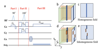 |
174 |
B0 Field Map estimation with View Line sequence
Shuang Liu, Wenqi Qiu, Gabriel Maggiora, Akihiro Kuwahata, Moriaki Kusakabe, Pablo Irarrazaval, Masaki Sekino
In order to visualize the concentration and distribution of magnetic nanoparticles in pre-clinical research of magnetic nanoparticles-based sentinel lymph node biopsy, Quantitative Susceptibility Mapping(QSM) is a promising quantification tool in MRI. However, the strong magnetic field generated by high concentrations of magnetic particles causes the failure of conventional B0 map used in QSM. A novel spin echo-based View Line sequence with radius basic function interpolation-based reconstruction method has been proposed to obtain the B0 map for high concentrations of magnetic nanoparticles. This method has been verified by MRI data and simulation data for a range of iron in Resovist from 41 ug to 654 ug. Comparison of the reconstructed field map and theoretical field map provides normalized root mean square of 0.085±0.054 for
the line field map, and 0.065±0.024 for
the interpolated field map.
|
|
5068.
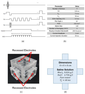 |
175 |
Effect of Intense Utilization of Gradients in Magnetic Resonance Current Density Imaging and its Removal
Mehdi Sadighi, Hasan Hüseyin Eroglu, B. Murat Eyüboglu
Intense utilization of gradients causes spatial and temporal variations of the main magnetic field which are consistent with resistive heating of the magnet structures. Since MR phase measurements are sensitive to the errors related to the B0B0 inhomogeneities
correction strategies are required. Here, it is shown that field variations due to the temperature change of MR equipment in Magnetic Resonance Current Density Imaging (MRCDI) using Induced Current Nonlinear Encoding-Spoiled Multi Gradient Echo (ICNE-SPMGE) pulse sequence introduces a bias to noise distribution of magnetic flux density (BzBz)
measurements. Removing this bias reduces the measured noise standard deviation of the BzBz measurements.
|
|
Back to Program-at-a-Glance |  Back to Top Back to Top |
| The International Society for Magnetic Resonance in Medicine is accredited by the Accreditation Council for Continuing Medical Education to provide continuing medical education for physicians. |
