|
MRS to CEST & What Is In-Between
Combined Educational & Scientific Session
ORGANIZERS: Lucio Frydman, Elena Vinogradov
Monday, 13 May 2019
| Room 510A-D |
08:15 - 10:15 |
Moderators: Gopal Varma, Christoph Juchem |
Skill Level: Intermediate
Session Number: M-03
Overview
MRS and CEST provide two complementary windows to detect and quantify molecular species in their native tissue environments. Few tools provide a comparable degree of information at near no cost of invasiveness. Still, the methods provide different, often complementary but sometimes disagreeing, pictures on the molecular status. The present session seeks to lay out the bases of both molecular imaging approaches, their unique strengths and weaknesses, and provide a state-of-the-art outlook on the new opportunities that each of them provides for furthering biological understanding and clinical diagnosis.
Target Audience
Basic researchers, biologists, clinicians, molecular imaging trainees, and experts.
Educational Objectives
As a result of attending this course, participants should be able to:
- Identify the strengths and weaknesses of MRS and CEST for imaging molecules in vivo: sensitivity, resolution, imaging compatibility;
- Describe the main applications of each of these methods for biological research and clinical applications, including their ability to quantify various parameters (concentrations, exchange rates, pH); and
- Recognize the origin of conflicting pictures arising from both sets of tools when.
08:15
|
|
MRS in Tissue: What Are We Measuring?

Anke Henning
|
08:35
|
0047.
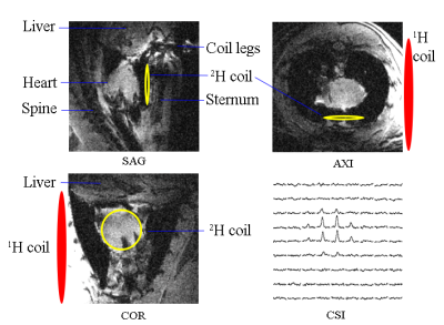 |
Dynamic Deuterium MRS Imaging for Studying Rat Heart Energy Metabolism in vivo – Initial Experience
Huan Li, Xiao-Hong Zhu, Wei Zhu, Byeong-Yeul Lee, Hannes Wiesner, Yi Zhang, Tao Wang, Wei Chen
Assessment of myocardial energy metabolism is crucial for understanding heart function and viable myocardium after myocardial infarction. Based on our recently developed in vivo Deuterium (2H) MR spectroscopic (DMRS) approach, we further exploited the DMRS and imaging (DMRSI) methods for dynamic measurement of the myocardial energy metabolism in rat heart at 16.4 T. This work demonstrates the feasibility of in vivo DMRS for assessing myocardial energy metabolisms, and its potential to directly image the viable myocardium in hearts under conditions such as myocardial infarction.
|
08:47
 |
0046.
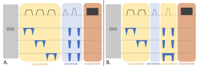 |
Novel methods to record MR spectra in human brain without suppressing or exciting the water signal to investigate exchange-sensitive protons.
Martyna Dziadosz, Wolfgang Bogner, André Döring, Roland Kreis
Non-water-suppressed MR spectroscopy in the form of metabolite-cycled MRS and longitudinal-relaxation-enhanced MRS (also termed non-water-excitation, NWE) has come into focus when studying compounds with exchanging protons (observed in the downfield region) or compounds like NAD+ that seem to be affected by cross relaxation from water. Here, we strive to implement new NWE techniques that can be used at 3T and allow for very short TE and observation of fast exchanging protons. 2D I-CSE, a combination of 2D ISIS and slice-selection with chemical shift selective excitation, fulfills this profile and first human brain applications show large signal contributions from exchanging protons.
|
08:59
|
 |
CEST in Tissue: What Are We Measuring?
Daniel Gochberg
In CEST, specificity is the key issue.
|
09:19
 |
0048.
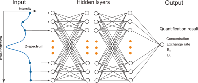 |
High-resolution phosphocreatine mapping of human skeletal muscle by artificial neural network-based chemical exchange saturation transfer MRI at 3T
Lin Chen, Michael Schär, Kannie Chan, Jianpan Huang, Zhiliang Wei, Hanzhang Lu, Qin Qin, Robert Weiss, Peter van Zijl, Jiadi Xu
The creatine kinase reaction provides energy for cells by reversibly regenerating adenosine triphosphate from a phosphocreatine pool. Functional impairment of this system is observed in many neurodegenerative and muscle diseases. In this study, we developed a high-resolution phosphocreatine (PCr) mapping approach that can be used on standard magnetic resonance clinical scanners by specifically detecting PCr via the water proton signal using a chemical exchange saturation transfer (CEST) method. An artificial neural network was employed to achieve absolute quantification of PCr concentration. Such a mapping method offers a non-invasive, rapid imaging tool to quantify abnormalities in PCr content and distribution in musculoskeletal diseases.
|
09:31
|
0049
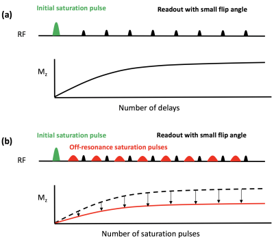 |
Translating pH-sensitive PROgressive saturation for Quantifying Exchange using Saturation Times (PRO-QUEST) MRI to a 3T Clinical Scanner
Video Permission Withheld
Mina Kim, Marco Battiston, Eleni Demetriou, Aaron Kujawa, Torben Schneider, Vincent Evans, Sachi Okuchi, David Atkinson, Claudia Wheeler-Kingshott, Xavier Golay
In this work, a recently developed method called PRO-QUEST (PROgressive saturation for Quantifying Exchange using Saturation Times) is translated to a 3T clinical scanner for assessing pH-sensitive indices in phantoms and a healthy volunteer. Our results demonstrate that quantification of pH sensitive indices using PRO-QUEST is feasible at 3T within clinically acceptable acquisition times. Our initial findings suggest that PRO-QUEST has the potential to provide a new biomarker to study neurological disorders associated with brain tissue acidosis.
|
09:43
|
|
CEST & MRS: Biochemistry (Breast Cancer)
Kristine Glunde
CEST & MRS: Biochemistry (Breast Cancer)
|
10:03
|
0050.
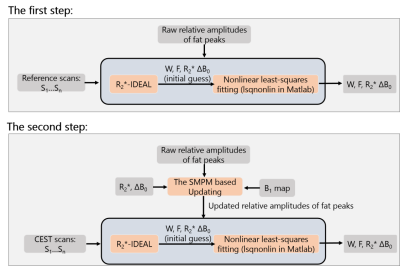 |
Self-adapting Multi-peak Water-fat Separation for Removing Lipid Artifacts in Breast Chemical Exchange Saturation Transfer (CEST) Imaging
Yu Zhao, Jianqi Li
Chemical exchange saturation transfer (CEST) MRI show potential for breast lesion characterization. However, artifacts caused by strong lipid signals hinder its widespread application. To remove the artifacts, water-fat separation based on multipoint Dixon acquisition is used to obtain water-only images. Considering that RF pulses with various frequency offset in CEST preparation saturate each fat peak at different level, relative amplitudes of fat peaks are updated for building fat signal model by the numerical simulation. Based on this self-adapting multi-peak model (SMPM), a method combining nonlinear least-squares fitting and R2 *-IDEAL is used to perform the water-fat reconstruction. Phantom and in vivo breast experiments demonstrate that the proposed method successfully removes lipid artifacts.
|
10:15
|
|
Adjournment |
|
