|
Multimodal fMRI: From Animal to Human
Combined Educational & Scientific Session
ORGANIZERS: Benedikt Poser, Susan Francis, Richard Buxton, Hanzhang Lu
Monday, 13 May 2019
| Room 710B |
08:15 - 10:15 |
Moderators: Xin Yu, Richard Buxton |
Skill Level: Intermediate to Advanced
Session Number: M-04
Overview
An active research area is the combination of innovative fMRI techniques with other state of the art neuro-techniques to provide a deeper probe of brain function in both health and disease, with applications extending from animal models to human studies. This course will provide an overview and sampling of current work in this area, including novel MR imaging and analysis methods and novel combinations of fMRI with current and emerging neuro-techniques, such as genetic tools, optical imaging and electrophysiological recordings. Clinical applications of these methods will be described.
Target Audience
Anyone interested in how electrophysiology relates to BOLD fMRI.
Educational Objectives
As a result of attending this course, participants should be able to:
-Identify the complementary aspects of current neuroimaging and neuro-measurement techniques;
-Describe how multimodal methods are combined in an experimental design to provide a deeper probe of brain function.
08:15
|
|
Multi-Modal Imaging: From Animal Models to Human

Catie Chang
Integrating fMRI with complementary neurophysiological measurements can provide a more comprehensive understanding of brain function, and help to clarify the neural basis of BOLD fMRI signals. Here, we will discuss multi-modal imaging studies of the human brain using simultaneous EEG-fMRI, along with studies in animal models, which allow for more direct, invasive monitoring and manipulation of neural circuits. This talk will also briefly discuss technical challenges and methodology involved in acquiring and analyzing simultaneous fMRI-electrophysiological data.
|
08:45
|
0051.
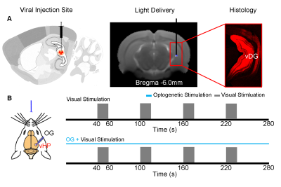 |
Optogenetic fMRI reveals ventral hippocampal modulatory effects on large-scale visual processing
Eddie Wong, Alex Leong, Celia M. Dong, Anthea To, Ed Wu
Hippocampus has been traditionally associated to learning, memory, navigation and emotional behaviors. However, little is known regarding whether and how it influences the processing of large-scale visual sensory information. In this study, we combined optogenetic stimulation and visual fMRI to investigate the influence of the ventral hippocampus on visual processing across the central visual pathway, including superior colliculus, lateral geniculate nucleus and visual cortex. Our optogenetic fMRI results reveal for the first time the differential influences of high and low frequency ventral hippocampal activities on visual processing along the central visual pathway.
|
08:57
|
0052.
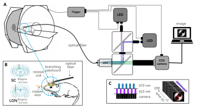 |
Neurovascular coupling in task and resting state using simultaneous calcium fiber photometry and fMRI in rats
Chuanjun Tong, Jiankun Dai, Yanqiu Feng, Zhifeng Liang
Neurovascular coupling is the foundation of functional brain imaging. We developed a dual site, dual color simultaneous GCaMP6-based fiber photometry and fMRI recording system in rats, to simultaneously record calcium and BOLD signals. Our results revealed the strong couplings in the task condition, and much weaker but still significant coupling in the resting state. We also showed that in the resting state such coupling was susceptible to different preprocessing steps. Our results provided a novel perspective on neurovascular coupling in task and resting state conditions.
|
09:09
 |
0053.
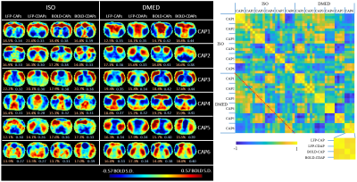 |
LFP-triggered Co-activation Patterns Show That the Relationship between LFP and BOLD Is Driven by a Few Distinct Events
Xiaodi Zhang, Wen-Ju Pan, Shella Keilholz
To gather electrophysiological evidence of time-varying functional networks, we developed a new method to analyze simultaneous fMRI and LFP data, which averages the fMRI frames at LFP power higher or lower than a threshold. The results not only show that the correlation between LFP power and BOLD is driven by a few distinct instead of a continuous interaction, but also suggests that the non-stationary resting state networks found in fMRI studies represent the time-varying behavior of LFPs.
|
09:21
|
|
Multi-modal Imaging in Epilepsy Research
Steven Stufflebeam
|
09:51
|
0054.
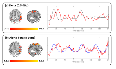 |
Exploration of the spatial and temporal characteristics of sensorimotor neural activity in the preterm human brain with simultaneous EEG-fMRI
Tanya Poppe, Kimberly Whitehead, Sofia Dall'Orso, Camilla O'Keefe, Jakki Brandon, Katy Vecchiato, Rui Pedro A G Teixeira, Felipe Godinez, Anthony Price, A David Edwards, Lorenzo Fabrizi, Tomoki Arichi
In the developing animal brain, different patterns of neural activity have distinct roles in the establishment of brain networks at different scales. Although studies suggest that the human preterm period is a crucial time for establishing brain connectivity, the role of different frequencies of neural activity has not been studied. We therefore used simultaneous EEG-fMRI and a robotic somatosensory stimulus to study the temporal and spatial characteristics of evoked neural activity in a group of preterm infants. Specific types of neural activity were associated with different BOLD responses, suggesting that these methods offer new insights into developing brain activity.
|
10:03
|
0055.
 |
Identifying focal thalamic activity underlying sleep and wake states through EEG-fMRI at 7 Tesla
Laura Lewis, Giorgio Bonmassar, Kawin Setsompop, Robert Stickgold, Bruce Rosen, Jonathan Polimeni
The thalamus plays an important role in regulating brain states, but remains poorly understood due to the technical challenges in imaging small brain structures with simultaneous electrophysiology. We implemented simultaneous fast fMRI and EEG at 7 Tesla to achieve high-SNR imaging of thalamic dynamics during human sleep. We found that we could detect selective activity within a focal set of thalamic nuclei that preceded the moment of awakening. These results identify potential network mechanisms engaged in regulating brain states, and demonstrate the potential for multimodal 7T imaging to identify new roles for deep brain structures in regulating cortical function and cognition.
|
10:15
|
|
Adjournment |
|
