Educational Course
Cardiac Microstructure Back to Program-at-a-Glance Back to Program-at-a-Glance
|
|
Cardiac Microstructure
Weekday Course
ORGANIZERS: Jennifer Keegan, Bruno Quesson
Monday, 13 May 2019
| Room 512A-H |
13:45 - 15:45 |
Moderators: Daniel Ennis, Christopher Nguyen |
Skill Level: Basic to Advanced
Session Number: M-05
Overview
Diffusion tensor imaging is an established neuroimaging technique that uses anisotropic diffusion of water to investigate white matter organisation in the brain and how it changes with aging and pathology. Over recent years, these techniques have been further developed to study the microstructure of the heart where they can provide information relating to the alignment and integrity of cardiomyocytes and potentially provide new insights into the mechanisms of heart disease.
This session describes the importance of cardiac microstructure, how diffusion tensor imaging may be used to probe that structure, and what clinically useful information it may extract.
Target Audience
This session targets scientists and clinicians interested in understanding why imaging cardiac microstructure is important and how diffusion tensor imaging can be used to probe that structure.
Educational Objectives
As a result of attending this course, participants should be able to:
- Describe the microstructure of the myocardium;
- Recognize state-of-the-art diffusion tensor imaging (DTI) techniques for probing cardiac microstructure;
- Recognize current clinical studies; and
- Identify key technical developments to allow further clinical application.
13:45
|
|
Understanding Cardiac Microstructure

Alistair Young
Detailed information can now be obtained on the microstructural architecture of the heart from MRI data. Changes in microstructure in disease have significant impacts on cardiac performance. This talk will give course participants an overview of our current understanding of the relationships between cardiac microstructure and cardiac function. Recent clinical applications will be reviewed and areas of productive future research will be highlighted.
|
14:15
|
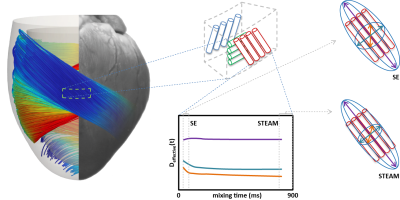 |
Cardiac DTI: Acquisition Techniques
Christian Stoeck
The two most commonly used approaches to overcome motion induced signal dephasing in cardiac diffusion weighted imaging rely on stimulated echo acquisition mode and motion compensated spin echo diffusion weighted imaging. Both sequences employ different strategies to generate diffusion contrast. In recent in-vivo imaging as well as Monte Carlo simulations it has become apparent that the quantitative parameters such as mean diffusivity and fractional anisotropy substantially differ between the two imaging approaches. The aim of this educational is to explain motion compensation strategies and discuss the resulting differences in measurements.
|
14:45
|
 |
Cardiac DTI: Processing Techniques
Elizabeth Tunnicliffe
This talk will outline how the diffusion tensor is calculated from the diffusion weighted images. Tensor-derived invariants (mean diffusivity and fractional anisotropy) as well as cardiac-specific directional parameters will be discussed.
|
15:15
|
|
Cardiac DTI: Current & Future Clinical Applications
Dudley Pennell
Only one tool exists to perform in-vivo, human non-invasive assessment of the myocardium at the microstructural level, namely diffusion tensor (DT) cardiac magnetic resonance (CMR). DT-CMR quantifies water diffusion in the myocardium, which is constrained by the myocardial micro-architecture. This talk examines potential clinical applications of DT-CMR.
|
15:40
|
|
Self Assessment Module (SAM) |
15:45
|
|
Adjournment |
|
Back to Program-at-a-Glance |  Back to Top Back to Top |
| The International Society for Magnetic Resonance in Medicine is accredited by the Accreditation Council for Continuing Medical Education to provide continuing medical education for physicians. |
