|
Mapping the Liver
Combined Educational & Scientific Session
ORGANIZERS: Utaroh Motosugi, Mustafa Shadi Bashir
Monday, 13 May 2019
| Room 510A-D |
13:45 - 15:45 |
Moderators: Bachir Taouli, Scott Reeder |
Skill Level: Basic to Advanced
Session Number: M-07
Overview
Through rapid technical development, it is now possible to obtain quantitative maps of a variety of MR-related properties of the liver. Many of these correspond directly with important physical properties of the liver, such as R2* mapping for iron quantification and proton density fat fraction mapping for fat quantification. During this session, both technical developments and clinical applications in quantitative mapping in the liver will be presented.
Target Audience
MDs and PhD scientists interested in quantitative MRI techniques applied to the liver.
Educational Objectives
As a result of attending this course, participants should be able to:
- Identify emerging quantitative mapping techniques applied to the liver; and
- Discuss current and likely future clinical applications of quantitative mapping techniques in the liver.
13:45
|
|
Technical Development

Diego Hernando
This presentation will describe MRI-based techniques for quantification of fat and iron deposition in the liver. Specifically, this presentation will cover: 1) the relevant MR contrast mechanisms related to the presence of fat and iron, 2) the types of pulse sequences used to probe these contrast mechanisms, 3) the main challenges for quantification of fat and iron, 4) the technical solutions to these challenges, and 5) the state of the art of development and validation of MRI-based techniques for quantification of fat and iron deposition in the liver.
|
14:15
|
|
Clinical Applications
Jeong Min Lee
New advances in liver MRI including T1-,T2*- and T1 rho mapping techniques, proton density fat fraction (PDFF) and elastography techniques may enable diagnosis of unseen pathologies by conventional techniques in the liver. In addition, Gd-EOB-DTPA can enable assessment of liver function by using postcontrast hepatobiliary phase or T1 reduction rate (normally above 60%). In the near future, MR fingerprinting may enable single slice acquisition and easy implementation of multiparametric MRI and follow-up of patients. These noninvasive imaging techniques serve an alternative or complimentary role to invasive liver biopsy. Mapping techniques of the liver, for fat, iron, and fibrosis quantitative imaging is increasingly being used in clinical practice, and may soon become standard of care.
|
14:45
|
0157.
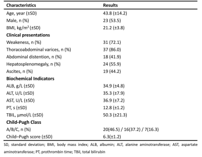 |
Evaluating the clinical efficacy of magnetic resonance elastography in patients with Budd-Chiari syndrome
Peng Xu, Kai Xu, Weiqiang Dou, Yong Shen
Budd-Chiari syndrome (BCS) is a rare disorder with an obstruction of the hepatic venous outflow tract. Previous studies reported that liver stiffness (LS) was associated with liver function in some chronic liver diseases. In this study, magnetic resonance elastography was for the first time applied to investigate the LS levels of BCS patients staged with different Child-Pugh grades. As a result, significant correlation between LS levels and liver function properties was shown in BCS patients. In addition, higher LS levels were found in BCS patients with higher grades and lower LS levels were observed for patients after receiving treatment.
|
14:57
|
0158.
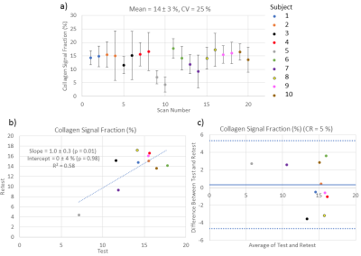 |
Quantification and Repeatability of the Collagen Signal Fraction in the Healthy Liver Using Ultrashort Echo Time (UTE) MRI
Adrienne Siu, Ferenc Mózes, Luca Biasiolli, Matthew Robson
Liver injury can lead to fibrosis, i.e. an accumulation of collagen. Fibrosis is clinically assessed via biopsy. Due to the health risks and unrepresentative sampling associated with biopsy, a non-invasive method of quantifying collagen would be beneficial. Here, an ultrashort echo time (UTE) pulse sequence was employed to quantify the collagen signal fraction in 10 subjects with healthy livers in a test-retest study at 3 T. The collagen signal fraction was repeatable, with a mean of 14 ± 3 %.
|
15:09
|
0159.
 |
Feasibility Study of MRI IDEAL-IQ Sequence in Evaluating Liver Function of Patients with Liver Cirrhosis
Did Not Present
Nan Wang, Ailian Liu, Qinhe Zhang, Lizhi Xie
Liver biopsy is the gold standard for the diagnosis of liver cirrhosis, but because the biopsy is invasive, and liver tissue sampling is insufficient and sampling error (puncture location) exists. IDEAL-IQ sequence can be used to analyze liver fat fraction and iron content (R2*) simultaneously, and it is easy to operate.
|
15:21
|
0160.
 |
Can Intravoxel Incoherent Motion Diffusion-weighted Imaging Be used for Preoperative Assessment of Microvascular Invasion in Hepatocellular Carcinoma ?
Did Not Present
Yi Wei, Hehan Tang, Xiaocheng Wei, Bin Song
Microvascular invasion (MVI) is one of the most important factors for the recurrence of hepatocellular carcinoma (HCC), however, accurate preoperative evaluation of MVI is quietly difficult because of the controversy results caused by the conventional imaging features. Compared with diffusion-weighted imaging (DWI), Intravoxel incoherent motion (IVIM) diffusion-weighted MR imaging could better characterize heterogeneity and irregularity of tissue components, and thus may have the potential to better evaluate MVI. In this study, we prospectively determine the usefulness of IVIM parameters and conventional radiologic features for preoperative prediction of MVI in patients with HCC.
|
15:33
|
0161.
 |
In vivo mapping of liver microstructure using quantitative Temporal Diffusion Spectroscopy Imaging
Xiaoyu Jiang, Junzhong Xu, John Gore
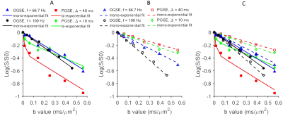
Architectural and morphological changes of hepatocytes (the major parenchymal cells carrying out most of the metabolic functions of the liver) are key diagnostic findings for liver diseases and are associated with important biological events. However, such information can currently only be assessed by liver biopsy. Quantitative temporal diffusion spectroscopy imaging (qTDSI), which uses different modulated gradient waveforms to measure ADC values equivalent to the use of multiple diffusion times (Δ), has been shown to provide accurate, high-resolution maps of cell size in solid tumors. In this study, we demonstrated that qTDSI can map hepatocyte sizes in mice in vivo.
|
15:45
|
|
Adjournment |
|
