Demyelination in the Spinal Cord: MS, NMO & ALS
Oral
Neuro
Tuesday, 14 May 2019
| Room 513D-F |
13:30 - 15:30 |
Moderators: Cornelia Laule, John Port |
13:30
 |
0520.
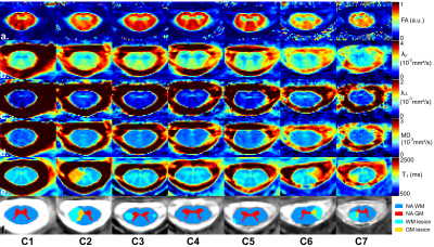 |
High-resolution multiparametric quantitative MRI of the cervical spinal cord at 7T: preliminary results at the early stage of multiple sclerosis

Aurélien Massire, Sarah Demortière, Pierre Lehmann, Henitsoa Rasoanandrianina, Maxime Guye, Bertrand Audoin, Jean Pelletier, Virginie Callot
In this exploratory study, a high-resolution multiparametric quantitative MRI protocol including T1 mapping and diffusion tensor imaging was used to explore the cervical spinal cord of multiple sclerosis patients at 7T, in comparison to healthy controls. These patients were also explored at 3T to investigate the potential benefits of 7T MRI in terms of lesion detection and delineation. The obtained preliminary results showed an improved qualitative anatomical depiction when compared to clinical fields and for the first time an in vivo multiparametric quantitative characterization of the pathological cervical spinal cord in multiple sclerosis at ultra-high field.
|
13:42
|
0521
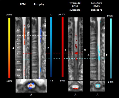 |
Spinal cord atrophy in Neuromyelitis Optica is associated to spinal cord lesions and clinical disability
Video Permission Withheld
Loredana Storelli, Laura Cacciaguerra, Paola Valsasina, Sarlota Mesaros, Jelena Drulovic, Alessandro Meani, Massimo Filippi, Maria A. Rocca
Although spinal cord involvement is one of the major magnetic resonance imaging (MRI) and clinical finding in Neuromyelitis Optica (NMO), there is a lack of quantitative studies in this field. This study quantifies and localizes spinal cord atrophy in 40 NMO patients and assesses its relationship with spinal cord lesions and clinical variables. Differently from what happens in multiple sclerosis, NMO cord atrophy seems related to the occurrence of spinal cord lesions and not to a more diffuse damage. Atrophy measures are strongly correlated with global EDSS and its pyramidal and sensitive subscores, suggesting its clinical relevance.
|
13:54
 |
0522.
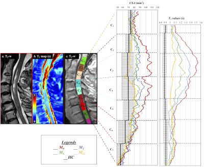 |
An exploratory study to evaluate the potentiality of advanced multi-parametric MRI for longitudinal follow-ups of patients with Neuromyelitis optica spectrum disorder
Henitsoa Rasoanandrianina, Bertrand Audoin, Aurélien Massire, Lauriane Pini, Claire Coste, Maxime Guye, Romain Marignier, Virginie Callot
The temporal pattern of spinal cord (SC) tissue changes in Neuromyelitis optica spectrum disorder, a rare neuro-inflammatory condition, was evaluated for the first time using a multi-parametric MR protocol covering the whole cervical SC (high-resolution T2*-weighted anatomical imaging, diffusion tensor imaging (DTI), 3D-MP2RAGE T1-mapping, conventional and inhomogeneous Magnetization Transfer (MT/ihMT) imaging). Results showed substantial pathological variations of all MR metrics at baseline in agreement with the presence of a multi-level inflammatory lesion and a temporal pattern suggesting progressive recovery to control values, with T1 values being the most sensitive to temporal changes and showing residual tissue destructuration following the myelitis.
|
14:06
|
0523.
 |
Reproducibility of Quantitative Cervical Spinal Cord MRI for Multi-center Clinical Trials in Multiple Sclerosis
Joo-won Kim, Haocheng Cai, Mohamed El Mendili, Peng Sun, Courtney Dula, Christina Alfonso, Sheng-Kwei Song, Matilde Inglese, Robert Naismith, Junqian Xu
Quantitative cervical spinal cord (C-spine) MRI offers promising biomarkers for progressive multiple sclerosis (MS). The intra- and inter-site, short- and long-term reproducibility of these quantitative C-spine MRI measurements is crucial for designing longitudinal or multi-site study. In this two-site pilot study, we evaluated the reproducibility of quantitative C-spine MRI and demonstrate the feasibility of multi-site study with high reproducibility using a harmonized protocol.
|
14:18
|
0524.
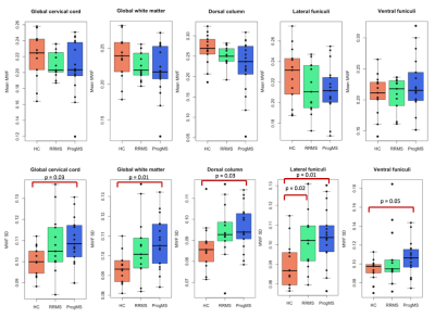 |
Does myelin content heterogeneity in the spinal cord reflect disability in multiple sclerosis?
Lisa Eunyoung Lee, Adam Dvorak, Hanwen Liu, Shawna Abel, Poljanka Johnson, Irene Vavasour, Cornelia Laule, Roger Tam, David Li, Jillian Chan, Robert Carruthers, Anthony Traboulsee, Shannon Kolind
The spinal cord is inherently more difficult to image than brain resulting in fewer MRI studies in multiple sclerosis (MS) patients. We used GRASE-derived myelin water fraction (MWF) mean (myelin content) and standard deviation (SD; myelin content heterogeneity) to better understand MS cervical spinal cord pathology compared to healthy controls. We found significant differences in cervical spinal cord MWF SD between progressive MS, relapsing-remitting MS and healthy controls. Further, MWF SD was correlated with disability measures in progressive MS. Our findings suggest that MWF SD as a measure of myelin abnormality in cervical cord relates to disability in MS.
|
14:30
|
0525.
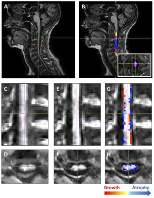 |
Improving longitudinal spinal cord atrophy measurements in multiple sclerosis using the Generalised Boundary Shift Integral
Marcello Moccia, Ferran Prados, Massimo Filippi, Maria Rocca, Paola Valsasina, Wallace Brownlee, Chiara Zecca, Antonio Gallo, Alex Rovira, Achim Gass, Jacqueline Palace, Carsten Lukas, Claudia Gandini Wheeler-Kingshott, Olga Ciccarelli, Frederik Barkhof
Spinal cord atrophy is a clinically-relevant feature of multiple sclerosis (MS), but can be difficult to estimate longitudinally using segmentation-based methods. We applied a fully-automated registration-based technique for spinal cord atrophy measurement (Generalised Boundary Shift Integral-GBSI-) on MS patients (n=282) and controls (n=82), from MAGNIMS and Queen Square cohorts. GBSI provided similar spinal cord atrophy rates, compared with cervical cord cross-sectional area (CSA), but with lower variability and favourable sample size estimates. GBSI performed better than CSA in differentiating cases from controls, and in depicting MS clinical features. GBSI could be used to monitor disease progression and in neuroprotective trials.
|
14:42
|
0526.
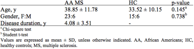 |
Spine atrophy and sensory-motor disability in African Americans with Multiple Sclerosis
Mohamed Mounir El Mendili, Maria Petracca, Amgad Droby, Giacomo Boffa, Swetha Paduri, Christopher Langston, Ilena George, Claire Riley, Jonathan Howard, Sylvia Klineova, Matilde Inglese
African Americans with multiple sclerosis (MS) present a more severe disease course than Caucasians with MS, but the contribution of spinal lesions and cervical spinal cord damage to clinical disability has never been explored. In the present study, we investigated the extent of cervical spinal cord (CSC) damage in AA MS patients compared to age-, sex- and race matched healthy controls. Our study showed that CSC damage in terms of both macroscopic lesions and atrophy significantly impacts motor and sensory performances in AA with MS.
|
14:54
|
0527.
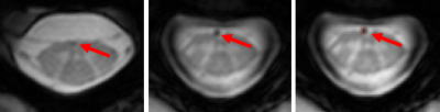 |
7T MRI Shows Enlarged Anterior Vein in the Spinal Cord of Multiple Sclerosis Patients
Atlee Witt, Bonner Kirkland, Bryson Reynolds, Benjamin Conrad, Aashim Bhatia , Seth Smith
T2*-weighted Gradient Echo (T2*FFE) were collected at 7-Tesla in patients with relapsing-remitting multiple sclerosis (MS) and healthy controls for a more detailed image of the spinal cord, specifically the vasculature of the spinal cord. Analyzing the anterior vein may provide insight into the connection between the vasculature and lesion presence in the spinal cord of MS patients. Our results demonstrate significantly enlarged anterior veins in MS patients compared to healthy controls. The anterior vein in the spinal cord has not been studied in great detail prior to this study.
|
15:06
|
0528.
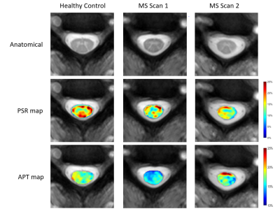 |
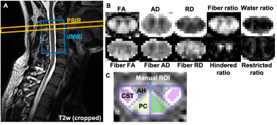
Using Quantitative MT- and CEST-derived Metrics to Evaluate Longitudinal Tissue Changes in the Spinal Cord of Multiple Sclerosis Patients at 3T
Richard Lawless, Quinn Weinberg, Haley Feiler, Sam By, Alex Smith, Francesca Bagnato, Seth Smith
Conventional T1 and T2 weighted MRI are ubiquitously used to diagnose and monitor disease progression in multiple sclerosis, but are only sensitive to later-stage inflammatory lesions and atrophy. Imaging biomarkers sensitive to tissue changes earlier in disease pathology may have significant implications in the diagnosis and prognosis of MS. Quantitative magnetization transfer (qMT) and chemical exchange saturation transfer (CEST) MRI have shown sensitivity to macromolecules and tissue biochemistry, respectively. In this work, we investigate quantitatively derived metrics from qMT and CEST as potential biomarkers for pathological changes which precede lesion formation.
|
15:18
|
0529.
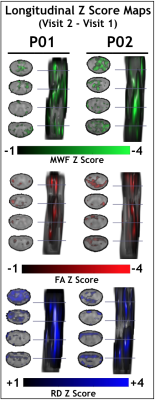 |
Quantitative MRI with spatiotemporal normalization can detect longitudinal changes in primary lateral sclerosis cervical spinal cord
Adam Dvorak, Poljanka Johnson, Hanwen Liu, Emil Ljungberg, Irene Vavasour, John Kramer, Alex MacKay, Cornelia Laule, Hannah Briemberg, Neil Cashman, Shannon Kolind
Analysis of longitudinal quantitative MRI data can be confounded by a variety of factors. We use spatiotemporal normalization and a symmetric diffeomorphic normalization algorithm to compare quantitative MRI metrics in a patient-specific halfway space. Utilizing these methods, myelin water imaging and diffusion tensor imaging were able to detect changes in primary lateral sclerosis (PLS) spinal cord similar to those found in the faster-progressing amyotrophic lateral sclerosis (ALS). Future studies could employ spatiotemporal normalization with more subjects and imaging timepoints to investigate the use of longitudinal MWI and DTI as a biomarker for motor neuron disease progression.
|
|
