Brain Tumor: CEST, MRS, & Diffusion MRI
Oral
Neuro
Wednesday, 15 May 2019
| Room 512A-H |
13:30 - 15:30 |
Moderators: Olusola Adeleke, Dariya Malyarenko |
13:30
|
0851.
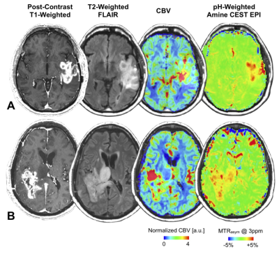 |
Association between tumor acidity and hypervascularity within human gliomas using pH-weighted amine chemical exchange saturation transfer echoplanar imaging (CEST-EPI) and dynamic susceptibility contrast (DSC) perfusion MRI at 3T

Yulin Wang, Jingwen Yao, Ararat Chakhoyan, Catalina Raymond, Noriko Salamon, Linda Liau, Phioanh Nghiemphu, Albert Lai, Whitney Pope, Timothy Cloughesy, Benjamin Ellingson
In the current study, we employed a fast pH-weighted molecular MRI technique using amine chemical exchange saturation transfer echoplanar imaging (CEST-EPI) and compared to dynamic susceptibility contrast (DSC) perfusion MRI, in order to examine the association between tumor acidity and vascularity in 82 patients with histologically confirmed gliomas. We observed colocalized regions of altered vascularity and acidity in tumors within individual patients, and significant positive correlation between median magnetization transfer ratio asymmetry (MTRasym) at 3ppm and relative cerebral blood volume (rCBV) within T2 hyperintense lesions. But areas of contrast enhancement were more complex and did not show a strong, predictable relationship.
|
13:42
|
0852.
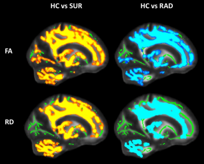 |
Evaluating white matter microstructure in childhood brain tumour survivors: A combined DTI and MTR approach.
Logan Richard, Jovanka Skocic, Ashley Ferkul, Elizabeth Cox, Suzanne Laughlin, Eric Bouffet, Donald Mabbott
Diffusion tensor imaging has been utilized to study the impact of cancer treatment on white matter microstructure in paediatric brain tumour survivors. We utilized magnetization transfer imaging (MTI), which provides more specific information on myelin, along with DTI, to determine if treatment for paediatric brain tumours has specific or non-specific impacts on white matter structure. When compared to their healthy counterparts, children treated for brain tumours exhibit decreased anisotropy and increased diffusion metrics without any significant differences in MT. This suggests treatment may impact fiber organization rather than myelin structure in this patient population.
|
13:54
 |
0853.
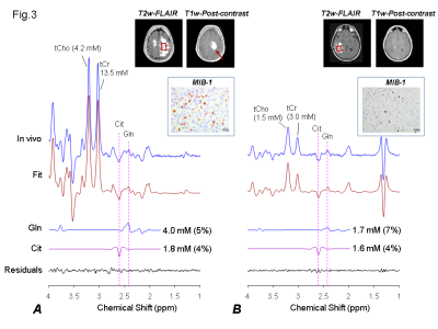 |
Glutamine Mediated Citrate Elevation- A ‘Metabolic-Rearrangement’ for Proliferation in High Grade Tumors: In vivo MRS Studies at 3T
Vivek Tiwari, Zhongxu An, Sandeep Ganji, Elena Daoud, Kimmo Hatanpaa, Michael Levy, Toral Patel, Elizabeth Maher, Edward Pan, Bruce Mickey, Changho Choi
Although substantial progress has been made in deciphering genetic and histological events in cancers, metabolic rearrangements that provide building blocks to elicit uncontrolled proliferation in cancers is still not understood. Nucleotide and lipids are the basic units needed for cell proliferation and membrane synthesis. Our in vivo MRS studies at 3 T in glioma patients indicate that tumors rearrange glutamine metabolism to produce citrate for increased lipid biosynthesis for membrane formation, and nucleotide for cell-multiplication.
|
14:06
|
0854.
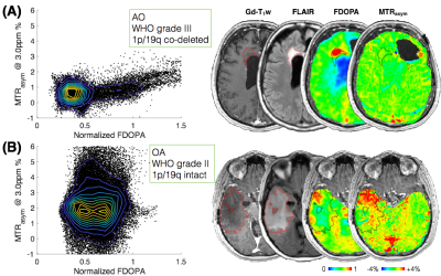 |
Low extracellular acidification in 1p/19q co-deleted gliomas confirmed using pH-weighted amine CEST-EPI, T2 relaxometry, and amino acid PET
Jingwen Yao, Ararat Chakhoyan, Catalina Raymond, Noriko Salamon, Linda Liau, Phioanh Nghiemphu, Albert Lai, Whitney Pope, Timothy Cloughesy, Benjamin Ellingson
The 1p/19q co-deletion in gliomas is associated with better response to therapies and better patient prognosis. In this study, we demonstrate that 1p/19q co-deleted gliomas are less acidic than gliomas with intact 1p/19q using a combination of pH-sensitive amine CEST-EPI, T2 relaxometry, and 18F-FDOPA PET. Results suggest amine CEST-EPI may serve as a quick non-invasive imaging biomarker for identifying 1p/19q co-deleted tumors. Our results also support the hypothesis that the better prognosis and higher sensitivity to treatment of 1p/19q co-deleted gliomas may be related to less acidity in tumor microenvironment.
|
14:18
|
0855.
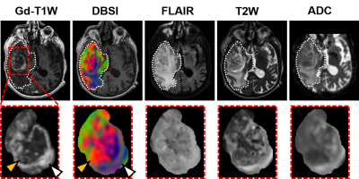 |
Noninvasive Assessment of Tumor Histopathology in Glioblastoma Specimen and Patient
Zezhong Ye, Joshua Lin, Richard Price, Peng Sun, Sam Gary, Jeffrey Viox, Chunyu Song, Ajit George, Jie Zhan, Ruimeng Yang, Sonika Dahiya, Albert Kim, Sheng-Kwei Song
Glioblastoma (GBM) is the most frequent malignant brain tumor in adults, accounting for approximately 45-50% of all primary malignant brain tumors. To circumvent the shortcomings of current clinical MRI techniques, we applied modified diffusion basis spectrum imaging (DBSI) to accurately detect pathology in GBM. In this study, we demonstrate modified-DBSI efficacy in detecting different histopathological structures in ex vivo and in vivo scans of glioblastoma.
|
14:30
|
0856.
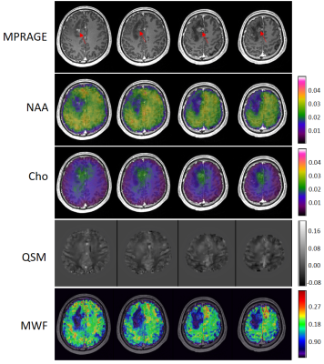 |
Multimodal Imaging of Brain Tumors Using High-Resolution 1H-MRSI Without Water Suppression
Jun Liu, Yudu Li, Tianyao Wang, Tianxiao Zhang, Ziyu Meng, Ke Xue, Rong Guo, Yibo Zhao, Yiping Du, Qun Chen, Zhi-Pei Liang, Yao Li
The characterization and grading of brain tumors are challenging using conventional MRI technology because of its poor specificity. MRSI has been recognized as a powerful tool for mapping the metabolic fingerprints of tumors. However, existing MRSI methods have poor spatial resolution which limits their practical applications in brain tumor imaging. In this paper, we investigate the use of a high-resolution MRSI technique for multimodal imaging of brain tumors. We demonstrate the capability of performing simultaneous mapping of brain metabolites (at 2.0×2.4×2.0 mm3 nominal spatial resolution), MWF and QSM using our new MRSI technology. This capability enables us to capture metabolic changes of small brain tumors (<10mm3).
|
14:42
 |
0857.
 |
GLINT: GlucoCEST in neoplastic tumors at 3 T – preliminary application in glioma patients using optimized preparation, imaging, and post-processing
Kai Herz, Tobias Lindig, Anagha Deshmane, Benjamin Bender, Xavier Golay, Ulrike Ernemann, Klaus Scheffler, Moritz Zaiss
Dynamic glucoCEST at clinical field strengths is very challenging due to the low effect size. Here, we present a saturation, imaging and post-processing protocol for minimizing possible artifacts to detect dynamic CEST effects reliably, and demonstrate the application in two glioblastoma patients at 3 T.
|
14:54
|
0858.
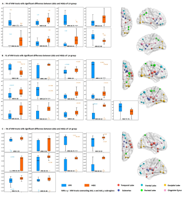 |
Hemispheric Specification of Remote Effect of Cerebral Glioma on White Matter Connectivity
Siqi Cai, Chunxiang Jiang, Xiaojing Long, Zhifeng Shi, Liang Chen, Kai Wang, Hairong Zheng, Xin Liu, Lijuan Zhang
Biological aggressiveness of glioma extends beyond the radiological territory. Based on the atlas based method and diffusion metrics derived from diffusion tensor imaging, the remote effect of cerebral gliomas on the white matter connectivity was analyzed at the global level. LGGs and HGGs showed difference in the diffusion metrics in the contralesional brain regions with hemispheric and anatomical specifications, which possibly underlie the functional neuroplasticity confounding the aggressiveness assessment and management of glioma.
|
15:06
|
0859.
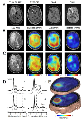 |
Deuterium Metabolic Imaging (DMI) of glucose metabolism in patients with brain tumors.
Henk De Feyter, Zachary Corbin, Isabel Prado, Robert Fulbright, Douglas Rothman, Robin de Graaf
Deuterium Metabolic Imaging (DMI) is a novel approach providing high 3D spatial resolution metabolic data from both animal models and human subjects. DMI relies on 2H MRSI in combination with administration of 2H-labeled substrates. Here we describe the first experiences with using DMI to map steady state metabolism of orally administered [6,6’-2H2]-glucose in patients diagnosed with a brain tumor. DMI revealed striking image contrast based on regional differences of glucose metabolism, with high-grade tumor lesions depicting metabolite labeling with consistently high lactate production and low glucose oxidation, a metabolic phenotype known as the Warburg effect.
|
15:18
|
0860.
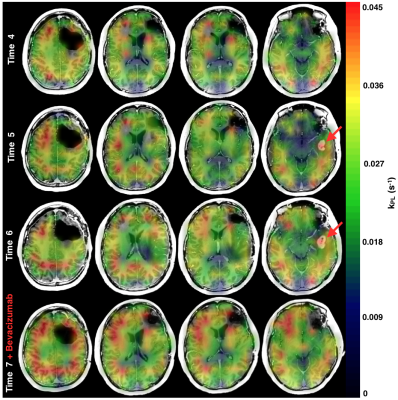 |
Serial characterization of HP [1-13C]pyruvate metabolism in the brains of patients with glioma and healthy controls
Adam Autry, Jeremy Gordon, Hsin-Yu Chen, Daniele Mammoli, Marisa Lafontaine, Javier Villanueva-Meyer, Susan Chang, Duan Xu, Peder Larson, Daniel Vigneron, Sarah Nelson, Yan Li
Serial dynamic hyperpolarized [1-13C]pyruvate imaging was performed on 3 patients undergoing treatment for recurrent brain tumors and 2 healthy controls using a frequency-specific EPI sequence (20 total scans). To evaluate metabolism within normal-appearing white matter (NAWM), rate constants for pyruvate-to-lactate (kPL) and pyruvate-to-bicarbonate (kPB) conversion were kinetically modeled. Healthy control data provided reference rate constants in NAWM and demonstrated replicabililty of test-retest type scans across hardware platforms. Serial patient data also showed similar, replicable data with standard-of-care treatment, as well as evidence that kPL, NAWM is increased 148-290% following administration of anti-angiogenic agent Bevacizumab, which promotes vascular normalization.
|
|
