Brain Tumor Vascular
Oral
Neuro
Tuesday, 14 May 2019
| Room 511BCEF |
08:15 - 10:15 |
Moderators: Laura Bell, Mary Kate Manhard |
08:15
|
0390.
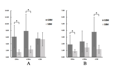 |
To discriminate glioblastoma from solitary brain metastasis with inflow-based vascular-space-occupancy (iVASO): comparison with dynamic susceptibility contrast MR imaging

Xiaodan Li, Danni Wang, Shukun Liao, Liuji Guo, Yingjie Mei, Xiang Xiao, Xiaomin Liu, Jun Hua, Jay Pillai, Yikai Xu, Yuankui Wu
Accurate differentiation between GBM and SBM is of vital importance clinically. DSC-MRI cannot differentiate them accurately using measures derived from tumoral regions. Moreover, DSC-MRI requires gadolinium contrast agent administration. Inflow-based vascular-space-occupancy (iVASO) is a novel perfusion technique without the need for exogenous contrast agents. In this study, the capability of iVASO in differentiating GBM and SBM was investigated and compared with that of DSC-MRI. The results showed that iVASO-based measures within tumoral regions could differentiate them accurately. This suggests that iVASO may be used as an alternative for perfusion study on brain tumor.
|
08:27
 |
0391.
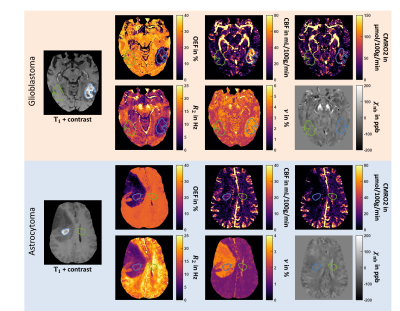 |
MRI-based Oxygen Extraction Fraction and Cerebral Metabolic Rate of Oxygen Mapping in High-Grade Glioma Using a Combined Quantitative Susceptibility Mapping and Quantitative Blood Oxygenation Level-Dependent Approach
Simon Hubertus, Sebastian Thomas, Junghun Cho, Shun Zhang, Ilhami Kovanlikaya, Yi Wang, Lothar R. Schad
MRI-based oxygenation mapping would be beneficial for treatment planning of high-grade gliomas. We used dynamic contrast-enhanced imaging and a combined quantitative susceptibility mapping and blood oxygenation level-dependent approach to quantify the oxygen extraction fraction (OEF) and cerebral metabolic rate of oxygen (CMRO2) in 6 patients with glioblastoma multiforme and 2 with anaplastic astrocytoma. Robust reconstruction of physiologically meaningful uniform OEF maps in healthy tissue was achieved and OEF was significantly lower in the tumor compared to the contralateral side. Blood flow was significantly higher in the tumor only for glioblastoma multiforme. CMRO2 showed no significant differences.
|
08:39
|
0392.
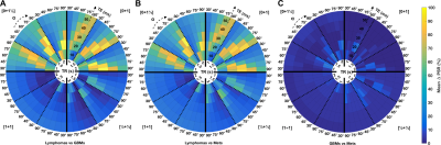 |
Investigating the Influence of DSC-MRI Acquisition Methods on the Clinical Application of Percentage Signal Recovery in Brain Tumors
Natenael Semmineh, Laura Bell, Ashley Stokes, Ethan Mathew, Matthew Lee, Jerrold Boxerman, C. Chad Quarles
Percentage signal recovery (PSR) derived from DSC-MRI has been shown to exhibit unique properties between enhancing malignant lesions, due to T1 and T2* contrast agent leakage effects. Currently there is no consensus on the choice of acquisition protocols to maximize the effectiveness of PSR to differentiate between tumor types. Using a validated DSC-MRI digital reference object we characterize the influence of preload dosing schemes and pulse sequence parameters on PSR. The goal of this study is to leverage the digital reference object to identify the optimal combination of parameters that provide effective PSR difference between GBM, metastasis, and lymphoma.
|
08:51
|
0393
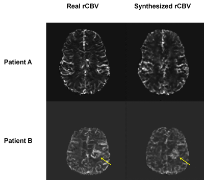 |
Synthesizing rCBV maps from DCE-MRI of brain tumors using conditional adversarial networks
Video Permission Withheld
Jeremiah Sanders, Henry Chen, Jason Johnson, Donald Schomer, Jingfei Ma, Ho-Ling Liu
In this work we investigate conditional adversarial networks for synthesizing relative cerebral blood volume (rCBV) maps from dynamic contrast enhanced (DCE)-MRI. A network based on the pix2pix framework is trained to map DCE-MRI to rCBV maps using rCBV maps generated from dynamic susceptibility contrast (DSC)-MRI in the same patient cohort. The results demonstrate the feasibility of synthesizing realistic rCBV maps from DCE images, potentially improving MR perfusion imaging of the brain using a single contrast injection.
|
09:03
|
0394.
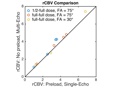 |
Evaluation of Single Bolus, Multi-Echo Dynamic Susceptibility Contrast Protocols in Brain Tumor Patients
Ashley Stokes, Ashley Nespodzany, Lea Alhilali, Leland Hu, C. Chad Quarles, Leslie Baxter
Relative cerebral blood volume (rCBV) obtained from dynamic susceptibility contrast (DSC) MRI is adversely impacted by contrast agent leakage in brain tumors. Using a digital reference object, we previously demonstrated that multi-echo DSC-MRI protocols provide advantages in terms of contrast agent dosing, pulse sequence flexibility, and rCBV accuracy. The purpose of this study is to assess the in-vivo performance of multi-echo acquisitions in patients with brain tumors. Multi-echo rCBV obtained without a preload is compared to the standard single-echo rCBV obtained with preload. In addition, two contrast agent doses and two flip angles are compared for the multi-echo acquisition.
|
09:15
|
0395.
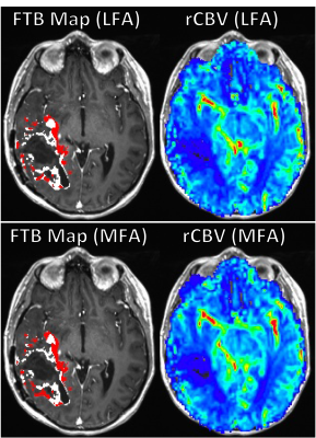 |
Evaluation of Fractional Tumor Burden (FTB) fidelity using a no-preload, low-flip angle dynamic susceptibility contrast MRI acquisition scheme
Melissa Prah, Leland Hu, Jerrold Boxerman, C. Chad Quarles, Jennifer Connelly, Kathleen Schmainda
This study examines the fidelity of a no pre-load, low flip-angle (LFA) dynamic susceptibility contrast MRI acquisition approach in the calculation of Fractional Tumor Burden (FTB) maps, which have shown promise as a predictive biomarker in glioblastoma patients. The LFA approach was recently identified as providing similar accuracy to the standard mid-range flip-angle approach with preload. FTB was found to have robust quantitative and spatial agreement between LFA and MFA approaches. The results of this study bode well for increased adoption of FTB as a biomarker amenable to both the standard and newer LFA approach.
|
09:27
|
0396
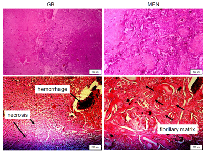 |
Detection of fluid/solid tissue behavior of neurotumors by magnetic resonance elastography
Video Permission Withheld
Kaspar Streitberger, Ledia Lilaj, Felix Schrank, Jürgen Braun, Josef Käs, Karl Hoffmann, Martin Reiss-Zimmermann, Ingolf Sack
It is known that glioblastoma (GB) display high heterogeneity and porosity (higher water content) than meningioma (MEN) suggesting a higher fluidity of GB than MEN. However, we will demonstrate by MR elastography (MRE) in patients and phantoms that the concept of fluidity cannot be naively transferred to brain tumors. Instead, the macroscopic viscosity-fluidity behavior of GB and MEN can be understood MRE of materials that comprise various amounts of water including materials with fibrillary architecture such as tofu. Similar to tofu, viscosity and fluidity in GB and MEN seem to reduce with increasing water content, indicating an ‘anomalous’ viscosity-fluidity behavior.
|
09:39
|
0397.
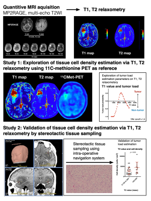 |
T1, T2 relaxometry for tissue cell density quantification in glioma imaging: Exploration study via 11C-methionine PET and its validation via stereotactic tissue sampling
Manabu Kinoshita, Masato Uchikoshi, Souichiro Tateishi, Shohei Miyazaki, Mio Sakai, Tomohiko Ozaki, Katsunori Asai, Yuya Fujita, Takahiro Matsuhashi, Yonehiro Kanemura, Eku Shimosegawa, Jun Hatazawa, Shinichi Nakatsuka, Haruhiko Kishima, Katsuyuki Nakanishi
In this study, the authors attempted to use T1, T2 relaxometry for predicting tumor cell density within the brain in glioma patients. The study was conducted in two stages, first as an exploratory study comparing T1, T2 relaxometry and 11C-methinonie PET images, and second as a validation study using intraoperative stereo-tactically obtained tissues. The authors were able to identify a range of T1 and T2 relaxation time indicative of high cell density, which finding was confirmed by stereotactic tissue sampling. This technique was further able to create predictive tumor cell density map by T1, T2 relaxometry alone.
|
09:51
|
0398.
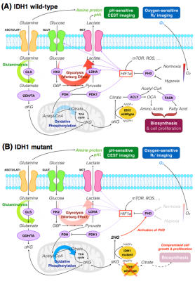 |
Differentiation of IDH1 mutant and wild-type gliomas based on metabolic signatures obtained from pH- and oxygen-sensitive molecular MRI
Jingwen Yao, Ararat Chakhoyan, Catalina Raymond, Noriko Salamon, Linda Liau, William Yong, Phioanh Nghiemphu, Albert Lai, Whitney Pope, Timothy Cloughesy, Benjamin Ellingson
In the current study, we have demonstrated that pH- and oxygen-sensitive amine CEST-SAGE-EPI (chemical exchange saturation transfer spin-and-gradient-echo echoplanar imaging) is a clinically feasible, powerful imaging technique for distinguishing between IDH1-mutant and wild-type gliomas. Results suggest that IDH1 mutation is associated with lower MTRasym at 3.0ppm and lower R2’, implying lower acidity and vascular hypoxia. We hypothesize that 2-HG produced by IDH1-mutant activates PHD and the degradation of HIF1α, subsequently preventing a metabolic shift from oxidative phosphorylation to glycolysis. This is supported by our histological findings of loss of correlation between levels of hypoxia and HIF1α tissue expression in IDH1 mutants.
|
10:03
|
0399.
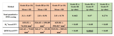 |
Quantitative intra-tumoral-susceptibility-signal (ITSS) vasculature volume (IVV) using QSM vs R2* approach for Glioma Grading
Rupsa Bhattacharjee, Jaladhar Neelavalli, Mamta Gupta, Snekha Thakran, Dinil Sasi, Rakesh Kumar Gupta, Anup Singh
Susceptibility-weighted imaging (SWI) improves the diagnostic accuracy by detecting intra-tumoral-susceptibility-signal-intensities (ITSS). Existing semi-quantitative methods are observer-dependent which manually counts intra-tumoral-susceptibility-signal-intensities (ITSS); a combination of haemorrhage and vasculature. Recent reported study uses the R2*-relaxivity-maps of ITSS, automatically removes haemorrhages from ITSS based on high- R2*-relaxivity of hemorrhage and calculate ITSS-vasculature-volume(IVV) within glioma. Current study evaluates role of QSM for segmentation of ITSS into hemorrhage and vasculature; compute QSM-based-IVV and compare the results with R2*-based-IVV for glioma-grading. High-correlation between both these methods is found, QSM-based-IVV can significantly differentiate between grade-II-vs-III and grade-II-vs-IV. For grade-III-vs-IV R2*-based-IVV scores better.
|
|
