Neurodegeneration (Other than AD)
Oral
Neuro
Wednesday, 15 May 2019
| Room 512A-H |
15:45 - 17:45 |
Moderators: Thijs Dhollander, Daniela Prayer |
15:45
|
0956.
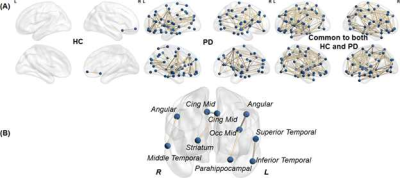 |
Unique Structural Backbone Topological Organization in Early Stage Parkinson’s Disease

Virendra Mishra, Karthik Sreenivasan, Xiaowei Zhuang, Zhengshi Yang, Dietmar Cordes, Zoltan Mari, David Eidelberg, Ryan Walsh
This study shows the presence of a distinctive anatomical backbone network in early Parkinson’s Disease (PD) involving cortical and subcortical regions that are known to be involved in various stages of PD, including early PD. Impaired network segregation and rich-club measures were further found within this backbone network in PD which were associated with disease duration. We hope that by identifying a set of core abnormalities in PD structural connectivity, this will permit improved ability to understand structural-network related disease progression and also to understand changes in these core structural connectivity measures with disease-modifying therapies as they emerge.
|
15:57
|
0957.
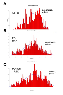 |
Braak model investigation in Parkinson disease using multimodal MRI biomarkers
Nadya Pyatigorskaya, Lydia Yahia-cherif, Romain Valabregue, Claire Ewenczyk, Rahul Gaurav, Marie Mongin, Cecile Gallea, Fatma Gargouri, Eric Bardinet, Marie Vidailhet, Stephane Lehericy
The aim of this study was to test using multimodal MRI biomarkers if the pattern of neurodegeneration in the brain of patients with Parkinson disease (PD) fits the model of disease progression proposed by Braak and al. (2003). Partial Least Squares Path-Modeling of multi-modal imaging data was in-line with the Braak pathology model confirming the gradual lesions in PD brain. While substantia nigra damage was observed in all PD patients, Braak’s model of disease progression was better followed by PD patients with idiopathic rapid eye movement sleep behavior disorders (RBD) than without RBD as demonstrated by model goodness of fit.
|
16:09
|
0958.
 |
The alteration of white matter tract of patients with Parkinson's disease combined with the changes in tract-connected gray matter : a longitudinal report
Shi-Ming Wang, Sung-han Lin, Chih-Chien Tsai, Yi-Hsin Weng, Yao-Liang Chen, Xiang-An Zhao, Jiun-Jie Wang
Parkinson's disease (PD) is a progressive nervous system disorder as a result from the loss of cell in basal ganglia. Fixel-Based Analysis can qualify the fibre density and fibre-bundle cross-section in the nerve fibre. Further, gray matter degeneration was determined by using ROI-based Analysis to investigate Mean Diffusivity (MD) changes. Therefore, the current study proposed to investigate the structural connectivity of atrophy cortical regions in the PD patients with axonal injury over a period of 2 years.
|
16:21
|
0959.
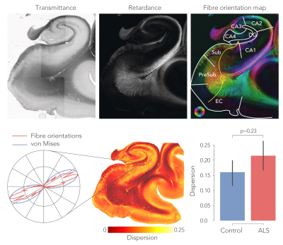 |
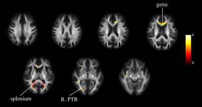
White matter changes in the perforant path in patients with amyotrophic lateral sclerosis
Jeroen Mollink, Marlies Hiemstra, Istvan Huszar, Mark Jenkinson, Karla Miller, Olaf Ansorge, Menuka Pallebage-Gamarallage, Anne-Marie van Cappellen van Walsum
Patients with amyotrophic lateral sclerosis (ALS) and frontotemporal dementia (FTD) share clinical overlap in terms of cognitive decline and are both characterised by the deposition of pathological TDP-43 inclusions in the brain. Here, we hypothesize that white matter degeneration of the perforant path in the hippocampus is a key feature of ALS patients developing FTD-like symptoms. Using diffusion MRI, polarized light imaging (PLI) and immunohistochemical (IHC) analysis we analysed white matter in the perforant path. dMRI and PLI measures suggest white matter degeneration in this pathway; however, densitometric analysis of IHC did not support this interpretation.
|
16:33
 |
0960.
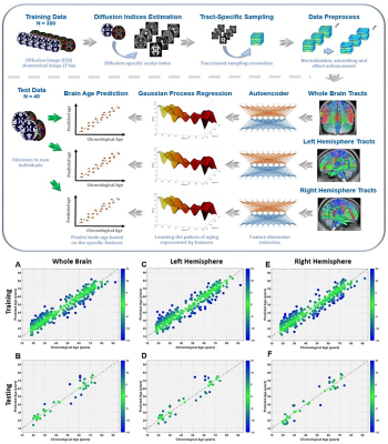 |
Advanced brain aging in patients with right mesial temporal lobe epilepsy: A machine learning approach based on white matter tract integrity
Chang-Le Chen, Yao-Chia Shih, Horng-Huei Liou, Yung-Chin Hsu, Fa-Hsuan Lin, Wen-Yih Tseng
It is unclear whether left and/or right side lesions of mesial temporal lobe epilepsy (MTLE) exhibit different degrees of brain aging. Therefore, we developed machine-learning-based brain age models to quantify the brain aging of patients with unilateral MTLE and of healthy controls. The significantly overestimated brain age was found in the right but not left MTLE patients. Also, the degree of overestimated brain age was correlated with the clinical factors. Moreover, the right uncinate fasciculus was the most contributing feature to the overestimated brain age. This study uncovered the underpinning of advanced brain aging in right MTLE patients.
|
16:45
|
0961.
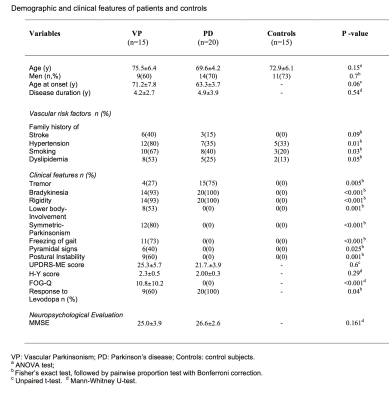 |
Microstructural Changes of Normal-Appearing White Matter in Vascular Parkinsonism
Maria Eugenia Caligiuri, Maria Salsone, Andrea Quattrone, Michaela Montilla, Ivan Rektor, Aldo Quattrone
White matter hyperintensities (WMH) play a crucial role in the pathogenesis of Vascular Parkinsonism (VP), a clinical entity characterized by parkinsonism, postural instability, marked gait difficulty and poor levodopa response. However, involvement of normal-appearing white matter (NAWM) in VP still remains unknown. Here we analyzed NAWM microstructure in VP compared to Parkinson’s disease (PD) and controls using MRI and DTI. We found extensive DTI alterations in VP, but not in PD nor in controls. NAWM damage in the genu of corpus callosum correlated with clinical core features such as postural instability, freezing-of-gait and symmetry of parkinsonism.
|
16:57
|
0962.
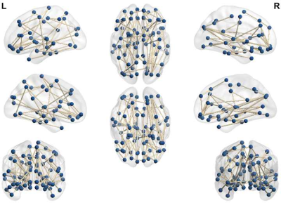 |
Distinctive structural connectivity between cognitively impaired and nonimpaired active professional fighters
Virendra Mishra, Karthik Sreenivasan, Xiaowei Zhuang, Zhengshi Yang, Sarah Banks, Dietmar Cordes, Charles Bernick
Using neuropsychological scores from the Professional Fighters Brain Health Study (PFBHS), this study first identified 70 cognitively impaired active professional fighters, and then matched 70 nonimpaired fighters matched on demographics, and other fighting criteria. Utilizing cortico-cortical connectivity obtained from diffusion MRI (dMRI) dataset, this study identified that there is an overall lower structural connectivity strength in cognitively impaired fighters suggesting that repeated head trauma induces a global brain shift in fighters experiencing cognitive decline.
|
17:09
 |
0963.
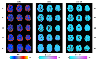 |
Improving strain diagnosis of prion disease by diffusion MRI and biophysical modelling
Marco Palombo, Matteo Figini, Riccardo Pascuzzo, Paola Caroppo, Mattia Verri, Torben Schneider, Claudia Gandini Wheeler-Kingshot, Veronica Redaelli, Daniel Alexander, Hui Zhang, Giorgio Giaccone, Alberto Bizzi
Sporadic Creutzfeldt–Jakob disease (sCJD) is the most common form of prion disease, characterized by five different strains, presenting intracellular vacuoles with different diameter/distribution. Unfortunately, no reliable non-invasive method for strain identification currently exists. Here we provide the first quantitative maps of MR-measured vacuolar diameter/density in five sCJD patients, using multishell diffusion MRI and biophysical modelling. Results show distribution of small and larger vacuoles in the brain lesions of each patient, presumably corresponding to different sCJD strains, and absence of vacuoles in five age-matched healthy controls. If validated, this method would be extremely valuable for non-invasive diagnosis of sCJD strain
|
17:21
|
0964
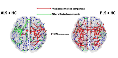 |
Structural and functional organization of the brain connectome in patients with different motor neuron diseases: a multicenter study
Video Permission Withheld
Camilla Cividini, Federica Agosta, Silvia Basaia, Francesca Trojsi, Nilo Riva, Cinzia Femiano, Cristina Moglia, Edoardo G. Spinelli, Maria Rosaria Monsurrò, Yuri Falzone, Andrea Falini, Giancarlo Comi, Adriano Chiò, Gioacchino Tedeschi, Massimo Filippi
We investigated structural and functional brain network topology in amyotrophic (ALS), primary lateral sclerosis (PLS) and progressive muscular atrophy (PMA) patients and in healthy controls (HC), using graph analysis and connectomics. ALS and PLS patients showed widespread microstructural alterations in sensorimotor network, basal ganglia area and prefrontal cortex and posterior brain regions compared to HC, while PMA subjects did not show significant brain damages. All groups had a relatively preserved global and local functional connectome properties compared to each other. Graph analysis and connectomics might represent a powerful approach to understand the pathophysiological process associated with motor neuron diseases.
|
17:33
|
0965.
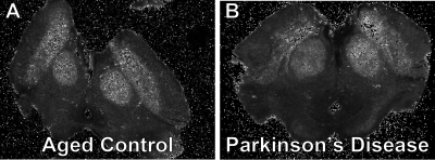 |
Ex vivo Correlations of MRI Parametrics and Histological Measures in Parkinson’s Disease Midbrain
Mark Meadowcroft, Miranda Salvo, Amanda Snyder, Carson Purnell, Jean Copper, Keith Cheng, Xuemei Huang, James Connor, Qing Yang
Parkinson’s disease is neurodegenerative disease characterized by the loss of dopaminergic neurons in the substantia nigra resulting in a range of motor deficits. There is uncertainty in how MRI metrics relate to disease pathology, especially in regard to cellular changes, iron, and the presence of neuromelanin. This work aims to analyzing MRI parametrics on ex vivo PD midbrains to determine how image contrast, relaxation, and susceptibility changes are related to cellularity and integrity in this brain region. Changes in MRI parametrics were found in the substantia nigra of PD subjects in relation to histological markers.
|
|
