Neuropsychiatry & Psychoradiology
Oral
Neuro
Monday, 13 May 2019
| Room 518A-C |
13:45 - 15:45 |
Moderators: Meiyun Wang, Steven Williams |
13:45
 |
0182.
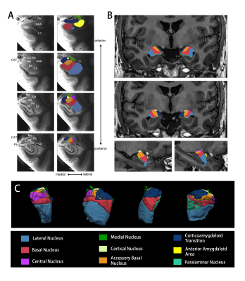 |
Anatomic alterations in amygdala subregions in medication-free patients with obsessive-compulsive disorder

Lianqing Zhang, Xinyu Hu, Hailong Li, Shi Tang, Lu Lu, Xuan Bu, Xiaoxiao Hu, Yingxue Gao, Yanlin Wang, yanchun Yang, John Sweeney, Qiyong Gong, Xiaoqi Huang
Most previous human neuroimaging studies measured the volume of amygdala as a whole, however, the amygdala consists of several functionally distinct subnuclei. Recent advances in structural MR image segmentation technique have made it possible to study amygdala subnuclei volumes with a robust, automatic approach using a Bayesian inference-based atlas building algorithm. Using this algorithm, we for the first time provide a distinctive profile of amygdala subnuclei volume abnormality in a relatively large sample of drug-free obsessive-compulsive disorder patients, and provide an insight that these subnuclei contribute to different aspect of neuropathology in the disorder.
|
13:57
|
0183.
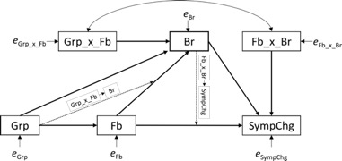 |
Mediators of the real-time fMRI amygdala neurofeedback training effect on PTSD symptom reduction: a whole brain structural equation model mapping analysis
Masaya Misaki, Raquel Phillips, Vadim Zotev, Frank Krueger, Matthew Feldner, Jerzy Bodurka
We determined mediators of the real-time fMRI neurofeedback (rtfMRI-nf) amygdala training inducing PTSD symptom reduction in combat veterans. Thirty-six veterans with PTSD (25 experimental group and 11 control group receiving a feedback from a control region) completed three rtfMRI-nf training sessions in separate days. We employed a novel whole brain structural equation model mapping (SEMM) analysis to identify brain regions that mediated the effects of the rtfMRI-nf procedure on PTSD symptoms. Results revealed that PTSD symptom reduction in the experimental group was mediated by lower activation than the control group in default mode network regions during the neurofeedback training.
|
14:09
|
0184.
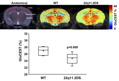 |
Imaging hippocampal glutamate alterations in a 22q11.2 deletion syndrome mouse model of schizophrenia
Puneet Bagga, David Roalf, Hari Hariharan, Anderson Stewart, Douglas Coulter, Raquel Gur, Ravinder Reddy
Schizophrenia (SZ) is a common, severe mental illness caused by neurobiological disturbances in glutamate and dopamine. Glutamate is the major excitatory neurotransmitter in the brain and can be detected using MR spectroscopy and glutamate-weighted chemical exchange saturation transfer (GluCEST) MRI. In this study, we performed high-resolution GluCEST MRI in the 22q11.2 deletion syndrome (22q11.2DS) mouse model of SZ to evaluate glutamatergic alterations in the dorsal and ventral hippocampus. The GluCEST contrast was found to be lower in the hippocampus of 22q11.2DS mice compared to that in the age-matched control mice indicating the lower hippocampal glutamate level in the preclinical model of SZ.
|
14:21
|
0185
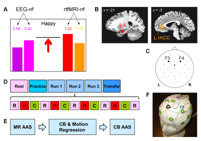 |
Emotion Self-Regulation Training with Simultaneous Real-Time fMRI and EEG Neurofeedback in Major Depression
Video Permission Withheld
Vadim Zotev, Ahmad Mayeli, Masaya Misaki, Jerzy Bodurka
We report a study of emotion self-regulation training in patients with major depressive disorder (MDD) using simultaneous real-time fMRI and EEG neurofeedback (rtfMRI-EEG-nf). Emotion-relevant target nf measures included fMRI activities of the left amygdala and left rACC, and frontal EEG asymmetries in the alpha and high-beta bands. MDD patients successfully learned to upregulate all four measures simultaneously using rtfMRI-EEG-nf during a happy emotion induction task. EEG-fMRI data analyses provided new insights into mechanisms of rtfMRI-EEG-nf. These findings may lead to development of more efficient neurotherapies for MDD.
|
14:33
|
0186.
 |
Discrete Regions of Gray Matter Loss Underlie Major Depressive Disorder: A Replication and Expanded Investigation
Sarah Hellewell, Thomas Welton, Jerome Maller, Matthew Lyon, Mayuresh Korgaonkar, Stephen Koslow, Leanne Williams, Evian Gordon, A Rush, Stuart Grieve
The aims of this study were to construct a biomarker based on anatomical regions of profoundly reduced gray matter volume in subjects with major depressive disorder. Our biomarker successfully discriminated MDD subjects from controls at an accuracy of 73%, suggesting a possible role for network measurements of GM susceptibility in MDD.
|
14:45
|
0187.
 |
Neural Correlates of Rumination in Normal and Major Depressive Disorder: A Brain Network Analysis
Yael Jacob, Laurel Morris, Kuang-Han Huang, Molly Schneider, Gaurav Verma, James Murrough, Priti Balchandani
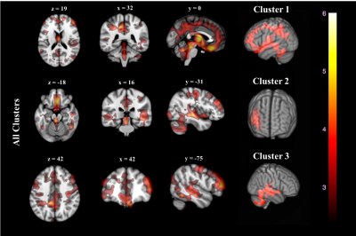
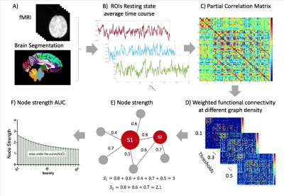
Major depressive disorder (MDD) patients exhibit higher rumination levels; repetitive thinking and focus on negative states. Rumination is known to be associated with brain cortical midline and limbic structures, yet, the underlying brain network topological organization remains unclear. Implementing a graph-theory analysis we tested whether whole brain network connectivity hierarchies during fMRI resting state are associated with rumination. We found a significant correlation between right caudal anterior cingulate (cACC) connectivity strength and subjective rumination tendency. This result emphasize the cACC impact during self-reflective processing, which might serve as biomarker for clinical diagnosis.
|
14:57
|
0188.
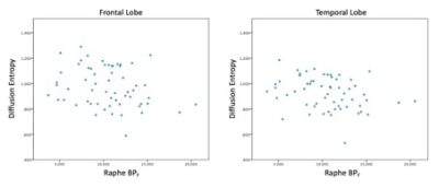 |
PET derived Raphe Nuclei [11C]-CUMI binding potential is associated with MR derived diffusion entropy in the frontal and temporal lobes
Mario Serrano-Sosa, Karl Spuhler, Christine DeLorenzo, Ramin Parsey, Chuan Huang
Using a new DTI processing technique, named diffusion entropy, and dynamic PET data we observed a statistically significant inverse correlation between diffusion entropy and [11C]-CUMI binding potential (BPF) in a dataset with bipolar, major depressive disorder patients and controls. This newfound analysis can give rise to further understanding the pathophysiology that undermines psychiatric diseases and, also, a non-invasive, non-ionizing radiation procedure to estimate [11C]-CUMI BPF.
|
15:09
|
0189.
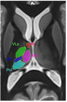 |
Thalamic Substructures in Alcoholism: Volume Deficits and Functional Correlates
Natalie Zahr, Kilian Pohl, Manoj Saranathan
Volumes of 5 thalamic substructures—mediodorsal (MD), anterior (AV), ventral lateral posterior (VLp), ventral posterior lateral (VPl), pulvinar (Pul))—were quantified using a novel automated segmentation algorithm in 40 individuals with Alcohol Use Disorder (AUD) and 60 controls (Con). Multiple regressions on supratentorial-volume-corrected measures revealed that volumes of AV, VLp, VPl, and Pul were smaller with older age, and volumes of AV, VLp, Pul, and MD were smaller in the AUD than Con group. Functional ramifications of thalamic substructures indicated relations between back pain and smaller Pul and VLp volumes, and poor ataxia scores with smaller VPI volumes.
|
15:21
|
0190.
 |
Mapping atypical functional connectome organization and hierarchy in autism spectrum disorders
Seok-Jun Hong, Reinder Vos De Wael, Richard Bethlehem, Sara Lariviere, Casey Paquola, Sofie Valk, Adriana Di Martino, Daniel Margulies, Michael Milham, Jonathan Smallwood, Boris Bernhardt
One paradox of autism is the co-occurrence of deficits in sensory and higher-order socio-cognitive processing. The current work examined whether these phenotypical patterns may relate to abnormal macroscale hierarchy affecting both unimodal and transmodal networks. Combining connectome gradient and stepwise connectivity analysis based on task-free fMRI, we demonstrated atypical connectivity transitions between sensory and higher-order default mode regions in a large cohort of autism individuals. Supervised pattern learning revealed that hierarchical features predicted deficits in social cognition and low-level behavioral symptoms. Our findings provide new evidence for network imbalances in autism, offering a parsimonious reference to consolidate its diverse features.
|
15:33
|
0191.
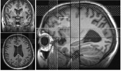 |
Modulation of dorsolateral prefrontal cortex GABA and Glx (glutamate + glutamine) levels following repetitive magnetic stimulation therapy of major depressive disorder
Pallab Bhattacharyya, Murat Altinay , Jian Lin, Amit Anand
Abnormalities of Gamma Amino Butyric Acid (GABA), the major inhibitory neurotransmitter, and Glx (glutamate, major excitatory neurotransmitter + glutamine) have been implicated in the pathology of depression. Modulation of GABA and Glx at left dorsolateral prefrontal cortex, the site of application of repetitive transcranial magnetic stimulation (rTMS) to treat major depressive disorder for patients not adequately responsive to medication treatment, was investigated using MEGA-PRESS spectroscopy. No change in GABA level was observed, while Glx/creatine level increased with rTMS therapy. Patients with higher Glx/Creatine level tended to respond better to rTMS, and the response was inversely correlated with increase in Glx/Creatine.
|
|
