Pediatrics: Fetal & Neonatal
Oral
Neuro
Tuesday, 14 May 2019
| Room 512A-H |
15:45 - 17:45 |
Moderators: Dianna Bardo, Yogesh Rathi |
15:45
|
0628.
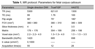 |
The feasibility and clinical value of single-direction diffusion-weighted imaging in the fetal corpus callosum

Cong Sun, Xin Chen, Yufan Chen, Jinxia Zhu, Ruiqin Shan, Guangbin Wang
We investigated the value and feasibility of single-direction diffusion-weighted imaging for the assessment of the fetal corpus callosum in 67 healthy fetuses and 26 fetuses with corpus callosum dysplasia. The results showed that single-direction DWI is significantly superior in optimal visibility and contrast ratio (CR) compared to the conventional HASTE and TrueFISP sequences. This study demonstrated that the application of single-direction DWI is promising in fetal corpus callosum lesions and can be used in routine clinical examination.
|
15:57
 |
0629.
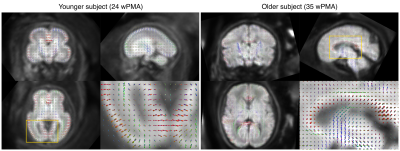 |
Fetal diffusion MRI acquisition and analysis in the developing Human Connectome Project
Daan Christiaens, Lucilio Cordero-Grande, Anthony Price, Jana Hutter, Emer Hughes, Serena Counsell, J-Donald Tournier, Joseph Hajnal
Fetal diffusion MRI (dMRI) can offer unique insight in brain development during the period of rapid, formative growth in the third trimester of pregnancy. The developing Human Connectome Project (dHCP) aims to create a comprehensive mapping of brain connectivity development in a large neonatal and fetal imaging cohort. Here, we design a state-of-the-art acquisition and analysis pipeline for fetal dMRI, including closely integrated dynamic distortion correction and slice-to-volume motion correction. We present results in the first 125 fetal dHCP subjects acquired, demonstrating high quality motion and distortion corrected output in the large majority of subjects and across a broad age range.
|
16:09
|
0630.
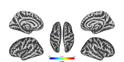 |
In utero exposure to chemotherapy affects cortical neurodevelopment
Jeroen Blommaert, Ahmed Radwan, Charlotte Sleurs, Ron Peeters, Stefan Sunaert, Tineke Vandenbroucke, Gwen Schroyen, Kristel Van Calsteren, Sabine Deprez, Frédéric Amant
One in every 1000-2000 pregnancies is complicated by maternal cancer, for which chemotherapy is increasingly administered during pregnancy. However, only limited knowledge exists on the long-term impact of in utero exposure to cancer therapy. This study investigated the impact of prenatal exposure to chemotherapy, at the age of nine, on cortical development using surface-based morphometry. We found cortical thickness to be significantly lower in the superior part of the left pre-central sulcus of the prenatal-exposed children, compared to controls, whereas the gyrification index was significantly higher in the left post-central sulcus of this group, possibly impacting attentional development.
|
16:21
 |
0631.
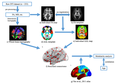 |
Altered development of structural brain networks in young children with prenatal alcohol exposure
Xiangyu Long, Preeti Kar, Ben Gibbard, Catherine Lebel
Prenatal alcohol exposure (PAE) can result in lifelong cognitive and behavioral deficits. Structural brain abnormalities have been shown in older children, but whether they are apparent in younger children is unclear. We investigated structural brain connectivity in 32 children with PAE aged 2-7 years compared to 95 healthy controls using diffusion tensor imaging. Group differences in structural connectivity and correlations with age were examined within and between eight different brain networks. Children with PAE had lower connectivity within and between several networks, but faster development of connectivity than controls, suggesting delayed development.
|
16:33
|
0632.
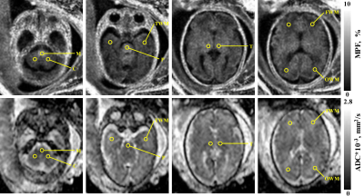 |
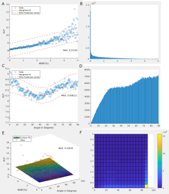
Direct comparison between macromolecular proton fraction and apparent diffusion coefficient as quantitative biomarkers of the human fetal brain maturation
Vasily Yarnykh, Irina Prihod'ko, Andrey Savelov, Alexandra Korostyshevskaya
Apparent diffusion coefficient (ADC) is known as a quantitative biomarker of prenatal brain maturation. Fast macromolecular proton fraction (MPF) mapping is an emerging method for quantitative assessment of myelination that was recently adapted to fetal MRI. This study compared spatiotemporal trajectories of MPF and ADC changes in the brain anatomic structures of 42 fetuses in utero. MPF and ADC demonstrated qualitatively similar but quantitatively different spatiotemporal patterns. MPF appeared more sensitive to changes in the brain structures with known prenatal onset of myelination.
|
16:45
|
0633.
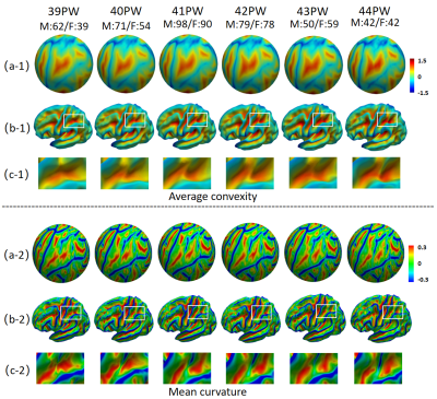 |
Building 4D neonatal cortical surface atlases using Wasserstein barycenter
Zengsi Chen, Zhengwang Wu, Liang Sun, Fan Wang, Li Wang, Weili Lin, John Gilmore, Dinggang Shen, Gang Li
Spatiotemporal (4D) neonatal cortical surface atlases are important tools for understanding the dynamic early brain development. To better preserve the sharpness and clarity of cortical folding patterns on surface atlases, we propose to compute the Wasserstein barycenter under the Wasserstein distance metric, for the construction of 4D neonatal surface atlases at each week from 39 to 44 postmenstrual weeks, based on a large-scale dataset with 764 neonates. Our atlases show sharper and more geometrically-faithful cortical folding patterns than the atlases built by the state-of-the-art method, thus leading to boosted accuracy for spatial normalization and facilitating early brain development studies.
|
16:57
|
0634.
 |
T1 and T2 Weighted Image Segmentation from 1.0T Neonatal MRI
Andrew Melbourne, Guotai Wang, Michael Ebner, Tom Vercauteren, Debra Rosenbaum, Yair Kasirer, Netanel Wasertil, Eli Ben-David, Alona Bin-Nun
In this work we present joint-image segmentation results from a novel 1.0T neonatal-specific MRI scanner. Data from machines such as this represent a new way to observe the growth and development of the preterm brain and can transfer our understanding of neurodevelopment prematurity. Advanced image analysis techniques are required to understand these developmental processes and we show preliminary results showing that good results can be obtained from these data.
|
17:09
|
0635.
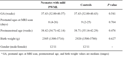 |
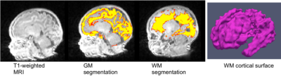
Cortical thickness is a sensitive biomarker for characterizing the gray matter abnormities in neonates with mild white matter injury
Miaomiao Wang, Jing Xia, Jian Chen, Xianjun Li, Congcong Liu, Li Wang, XiaoCheng Wei, Gang Li, Dinggang Shen, Jian Yang
White matter injury is common in neonates. The most common punctate white matter lesions (PWML) can disappear along with time and are easily missed diagnosis, but quantitative measurements may find more subtle alterations. Since DTI is not sensitive to detect the white-matter microstructural changes in mild PWML, alterations in gray-matter (GM) may provide additional knowledge for predicting prognosis. This study aims to quantitatively assess alterations of GM in neonates with mild PWML. Compared with controls, a significant reduction of cortical volume is observed in neonates with mild PWML, and cortical thickness is a sensitive biomarker for characterizing the GM abnormalities.
|
17:21
|
0636.
 |
Myelin Water Imaging and R2* Mapping in Neonates
Alexander Weber, Yuting Zhang, Alexander Rauscher
Understanding sources of R2* in the brains of infants and adults could lead to novel imaging biomarkers that could inform clinicians and researchers about white matter microstructure, and iron and myelin content. These biomarkers could in turn help determine the health, disease state, or developmental progress of white matter tissue, with many potential benefits to diagnosis, treatment, and therapeutic research. As opposed to adults who show a remarkable fit of R2* against fibre orientation, neonates showed very little dependence on myelin or fibre orientation.
|
17:33
|
0637.
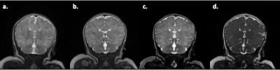 |
Rapid Anatomical Imaging of the Neonatal Brain Using T2-Prepared 3D Steady-State Free Precession
Jinho Park, Catherine Limperopoulos, Zungho Zun
Anatomical imaging of the neonatal brain is typically acquired using T2-weighted imaging based on either 3D fast spin echo (FSE) or 2D single shot FSE (SSFSE). While 3D imaging provides higher signal-to-noise ratio (SNR) and spatial resolution than 2D, its scan time is relatively long.In this study, we developed rapid 3D anatomical imaging for the neonatal brain using 3D steady-state free precession (SSFP) with T2 preparation. Our proposed method demonstrated similar T2 contrast to that of 3D FSE and 2D SSFSE while achieving shorter scan time than 3D FSE and higher through-plane resolution and higher SNR than 2D SSFSE.
|
|
