Traumatic Brain Injury
Oral
Neuro
Monday, 13 May 2019
| Room 513D-F |
08:15 - 10:15 |
Moderators: Brenda Bartnik Olson, Seung Hong Choi |
08:15
|
0076.
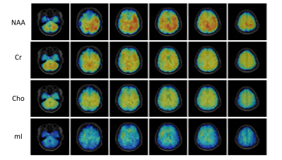 |
Metabolic Imaging of Traumatic Brain Injuries Using Ultrahigh-Resolution 1H-MRSI

Tianyao Wang, Jun Liu, Tianxiao Zhang, Ziyu Meng, Danni Wang, Ke Xue, Yudu Li, Rong Guo, Yibo Zhao, Xin Yu, Zhi-Pei Liang, Yao Li
Traumatic brain injury (TBI) is a significant public health problem that contributes to a large number of injury-induced deaths each year. MRSI has long been recognized as a powerful tool for detection of neurometabolic alterations induced by TBI; however, most existing MRSI studies of TBI are limited by low resolution which severely reduce the detection sensitivity. In this study, we performed MRSI scans on TBI patients using a newly developed ultrahigh-resolution 1H-MRSI technique. Our experimental study yielded very encouraging results and showed that ultrahigh-resolution MRSI can capture neurometabolic alterations induced by TBI effectively.
|
08:27
|
0077.
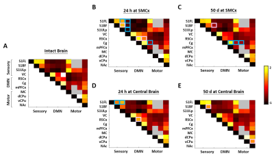 |
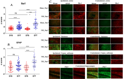
Connectivity Reorganization after Repetitive Mild Traumatic Brain Injury is Impact Site Associated
Yu-Chieh Jill Kao, Chia-Feng Lu, Bao-Yu Hsieh, Cheng-Yu Chen
Different degrees of functional reorganization associated with impact site was unraveled in the acute and chronic phase in the well-controlled animal model of repetitive mTBI. This is the first demonstration of impact site-dependent connectivity alteration without significant parenchymal damage after mTBI in rats.
|
08:39
|
0078.
 |
Free water elimination is necessary to characterize Diffuse Axonal Injury in moderate Traumatic Brain Injury
Jacob Alappatt, Drew Parker, Abdol Aziz Ould Ismail, Junghoon Kim, Ragini Verma
As the prevalence of diffusion MRI for clinical use grows, it is important to address the influence of injury-related extracellular water on the clinical interpretation of diffusion measures in conditions such as traumatic brain injury (TBI). The presence of extracellular free water from edema pollutes the estimation of diffusion measures, leading to flawed conclusions about the microstructure of the white matter. We demonstrate that Fernet, a robust single-shell free-water elimination method, can be used to decouple the effects of extracellular edema and tissue damage, to improve clinical understanding of the effects of injury on underlying white matter structure.
|
08:51
|
0079.
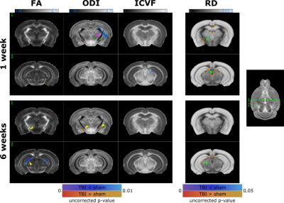 |
White matter changes caused by mild traumatic brain injury in mice
Lisa Gazdzinski, Miranda Mellerup, Tong Wang, John Sled, Brian Nieman, Anne Wheeler
White matter pathology following mild traumatic brain injury (mTBI) is complex and unlikely to be characterized by a single neuroimaging metric. DTI has been used to probe white matter microstructure, but is known to be non-specific. We show that the orientation dispersion index from the NODDI model may be more sensitive to white matter damage following mTBI in a mouse closed-skull impact model. Combined with quantitative susceptibility mapping, our data suggest minimal overt myelin loss, but progressive white matter injury, which may include myelin disruption, up to 6 weeks post-mTBI.
|
09:03
 |
0080.
 |
Exploring static and dynamic functional connectivity differences between cognitively impaired and non-impaired active professional fighters
Xiaowei Zhuang, Virendra Mishra, Yang Zhengshi, Karthik Sreenivasan, Charles Bernick, Dietmar Cordes
Both static and dynamic FC differences between cognitively impaired and non-impaired active fighters were explored using resting-state fMRI. Reduced network strength in anterior default mode network and cerebellar network were observed in impaired fighters, as compared to the non-impaired fighters. Four dynamic FC states were identified with k-means clustering and abnormities in state 2 were observed in impaired fighters. Higher classification accuracy was obtained using dynamic FC matrices as input features to a non-linear classifier, as compared to using static FC matrices as input features, which demonstrates that the time-varying brain activities carry richer cognitive impairment-related information.
|
09:15
|
0081.
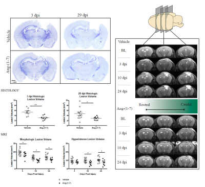 |
In vivo magnetic resonance imaging demonstrates that subcutaneous administration angiotensin-(1-7) is neuroprotective following severe traumatic brain injury in mice
Zachary Janatpour, Asamoah Bosomtwi, Alexandru Korotcov, Andrew Knutsen, Shalini Jaiswal , Nathanael Allison, Aviva Symes, Bernard Dardzinski
The effectiveness of angiotensin-(1-7), a novel peptide derivative of angiotensin II, for the treatment of traumatic brain injury in mice have been investigated using non-invasive MRI techniques. Our results demonstrate that treatment of mice with angiotensin-(1-7) after control cortical impact reduced lesion volume compared to saline-treated mice. The MRI data are validated by histological results. The observed data show for the first time that angiotensin-(1-7) has potential therapeutic use for TBI.
|
09:27
|
0082.
 |
Advanced diffusion-weighted imaging reveals distinct neuropathological processes in concussed youth
Guido Guberman, Jean-Christophe Houde, Isabelle Gagnon, Alain Ptito, Maxime Descoteaux
Because concussions are undetectable by conventional medical imaging, their diagnosis is dependent on symptoms, which can be unreliable. Conventional diffusion-weighted imaging (DWI) can detect abnormalities in concussed individuals, but lack specificity and hence cannot provide information about the underlying neuropathology, especially during the sub-acute stage of concussions where different neuropathologies occur simultaneously. In this study, we used emerging DWI methods to disambiguate the neuropathological basis of a common concussive symptom, memory problems. We found evidence of different white-matter neuropathologies in concussed youth which contributed differently to memory problems. This study is an important step towards developing neuropathologically-informed biomarkers of concussion.
|
09:39
|
0083.
 |
Traumatic Brain Injury fast-forwards Alzheimer's parthenogenesis: A radiological-pathological investigation in a mouse AD model.
Neha Soni, Rodrigo Medeiros, Fatima Nasrallah
Traumatic brain injury (TBI) today is the strongest epigenetic risk factor for Alzheimer's disease (AD) . To date, the underlying mechanisms of these comorbidities are still unclear, which has hindered diagnosis and monitoring. We have investigated the effect of TBI in a PR5 tauopathy model using Diffusion Tensor Imaging and histology. We concluded that neuroinflammation is a key trigger in worsening taupathy accumulation and MRI can detect this cascade of events at an early stage. This study will enhance our understanding, not only of the effect of TBI progression in AD, but the potential of MRI for translational purposes.
|
09:51
|
0084
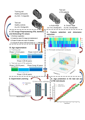 |
Mild traumatic brain injury accelerates progressive brain ageing
Video Permission Withheld
Shuoqiu Gan, Wen Shi, Yingxiang Sun, Lijun Bai, Ming Zhang
To analyze mild traumatic brain injury (mTBI) accelerating brain ageing, we trained a brain age prediction model based on diffuse tensor image (DTI) data by using machine learning method. Conducting a longitudinal observation from acute to chronic stages, we found that mTBI accelerated brain age process from acute stage to chronic stage. This prolonged abnormal brain ageing level could be predicted by information processing speed. In conclusion, mTBI persistently induces brain ageing process deviating from normal trajectory, and this process can be revealed by information processing speed at very early period after injury.
|
10:03
|
0085.
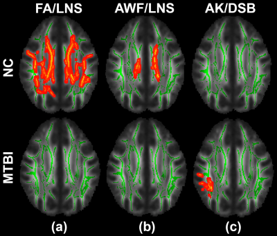 |
Changes In White Matter Microstructure In Relation To Working Memory After Mild Traumatic Brain Injury: Multi-Shell Diffusion MRI Study
Sohae Chung, Els Fieremans, Xiuyuan Wang, Dmitry Novikov, Prin Amorapanth, Steven Flanagan, Joseph Rath, Yvonne Lui
Working memory is a critical cognitive functions affected after mild traumatic brain injury (MTBI). We investigate associations between white matter (WM) microstructure and working memory, using multi-shell diffusion MRI and WAIS-IV subtests. The significant positive correlations observed in normal controls (NC) between tissue microstructure markers (fractional anisotropy (FA) and axonal water fraction (AWF)) with letter-number sequencing (LNS) were not present in MTBI. For MTBI, a significant positive correlation was observed between axial kurtosis (AK) and digit span backward (DSB), not seen in NC. These results show clear differences in the relationship between WM microstructure and working memory performance after injury.
|
|
