Tuesday, 14 May 2019
| Room 511BCEF |
15:45 - 17:45 |
Moderators: Georges El Fakhri, Su Lui |
15:45
 |
0618.
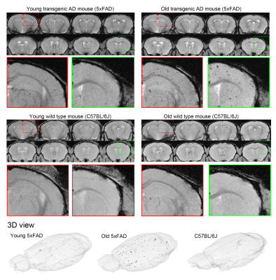 |
In vivo MRI detection of ß-amyloid pathologies at early and late stages of Alzheimer’s disease

Celia M. Dong, Anthea To, Shuai Guo, Kannie W. Y. Chan, Alex T. L. Leong, Ed X. Wu
Early diagnosis of Alzheimer’s disease (AD) is crucial. However, there is a lack of effective diagnostic tools to detect AD at the early stage. Early stage β-amyloid (Aβ) oligomers (AβOs) and late stage Aβ plaques are the pathological hallmarks of AD brains. Recently, we have synthesized a novel curcumin-conjugated magnetic nanoparticle (Cur-MNP) to target Aβ pathologies, visualized in MRI. In this study, we investigated the in vivo feasibility of Cur-MNPs to detect the Aβ pathologies at early and late stages of AD progression, and performed immunohistology to validate the specific targeting of Aβ pathologies.
|
15:57
|
0619.
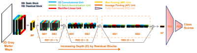 |
Predicting Progression from Mild Cognitive Impairment to Alzheimer’s Disease
Anees Abrol, Zening Fu, Vince Calhoun
This exploratory analysis tests the suitability of deep residual networks to learn neuroanatomical abnormalities from the structural MRI (sMRI) modality, and utility of dynamic (i.e. time-varying) functional connectivity approaches in delineating discriminative functional MRI (fMRI) features to predict progression of individuals with mild cognitive impairment to Alzheimer’s disease. Results demonstrate better than state-of-the-art prediction performance using the structural MRI modality alone. Multimodal prediction performed significantly better than unimodal sMRI or fMRI predictions, thus corroborating the benefits of predicting in the augmented space. Results also corroborate the diagnostic utility of the sMRI and fMRI features used to make the predictions.
|
16:09
 |
0620.
 |
Cerebrovascular reactivity (CVR) as a potential biomarker in vascular cognitive impairment: relationship with cognition, clinical diagnosis, amyloid and tau proteins.
Sandeepa Sur, Zixuan Lin, Yang Li, Sevil Yasar, Paul Rosenberg, Rita Kalyani, Abhay Moghekar, Zheyu Wang, Kaisha Hazel, George Pottanat, Cuimei Xu, Peter van Zijl, Jay Pillai, Peiying Liu, Marilyn Albert, Hanzhang Lu
Is whole brain cerebrovascular reactivity (CVR) associated with cognitive function in subjects with cognitive impairment and normal cognition? In a cross-sectional study of 72 subjects, whole brain CVR as assessed with the BOLD response to a CO2 breathing challenge, was significantly associated with measures of cognitive function. These relationships remained after adjusting for cerebrospinal fluid biomarkers of Alzheimer’s disease pathology and measures of vascular risk. These findings suggest that whole brain CVR may be useful as a biomarker for assessing altered cognition resulting from vascular dysfunction separate from Alzheimer’s disease.
|
16:21
|
0621.
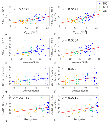 |
Product-Ratios of Metabolite Concentrations as Potential Alzheimer’s Disease Biomarker
Ariane Fillmer, Laura Göschel, Semiha Aydin, Theresa Köbe, Agnes Flöel, Bernd Ittermann
Previous studies have reported changes in the concentrations of several neurometabolites in Alzheimer’s disease (AD). Nevertheless, group differences of these metabolites between healthy controls, mild cognitively impaired, and AD patients remain small. The transition to ultrahigh fields enables the assessment of further metabolites, and some of them, like GABA and glutamate, have been observed to change in AD. In this study, the combination of several metabolite concentrations associated with AD into a product-ratio to serve as a stronger MRS biomarker for Alzheimer’s than individual concentrations of metabolites, and their relationship with volume of brain structures and memory performance is investigated.
|
16:33
|
0622.
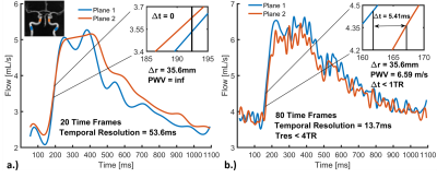 |
Intracranial Pulse Wave Velocity in Alzheimer’s Disease using Flow Encode Split and Low Rank Reconstructed 4D Flow MRI
Leonardo Rivera-Rivera, Sterling Johnson, Chuck Illingworth, Kevin Johnson
Experimental, clinical and epidemiologic evidence shows vascular factors play a role in Alzheimer’s Disease (AD); however, whether AD has a basis in vascular diseases is controversial. Intracranial pulse wave velocity (PWV) is a potential parameter that can probe arterial stiffness changes in AD, but is technically demanding due to requirements of high temporal resolution. In this study, we investigated high temporal resolution 4D flow using 3D radial sampling, low-rank regularization and interleaved encoding splitting for intracranial PWV estimates. Preliminary results from 26 subjects showed an increased intracranial PWV in an AD group when compared to controls.
|
16:45
|
0623.
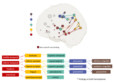 |
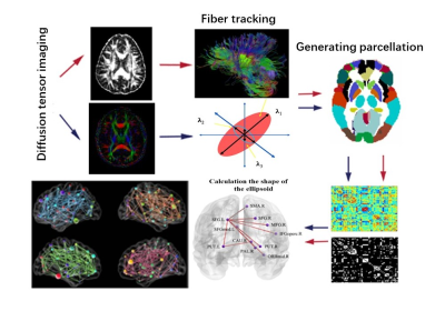
Tau Propagation Pattern Is Suggested Through Associations with Structural Tract Alterations: A Data Driven Approach
Qiuting Wen, Shannon Risacher, Sourajit Mustafi, Jaroslaw Harezlak, Linhui Xie, John West, Martin Farlow, Frederick Unverzagt, Liana Apostolova, Andrew Saykin, Yu-Chien Wu
To test the hypothesis that the propagation of tau is facilitated by microstructural alterations in white-matter tracts, we applied a data driven approach to study the “upstream tau – tract alteration – downstream tau” associations throughout the whole brain using tau positron emission tomography (tau-PET) and diffusion MRI (dMRI). The discovered pathways of spread support the characteristic pattern observed in Braak staging.
|
16:57
|
0624.
 |
Progressive alterations in structural topological properties in T2DM with and without mild cognitive impairment
Ying Xiong, Qiang Zhang, Yang Fan, Wenzhen Zhu
This study aims to investigate the topological organization in T2DM with and without impairment, and characterize its relationships with clinical measurements. Forty T2DM patients were divided into two sub-groups(impaired and normal cognition), together with ten healthy controls, were imaged at a 3T scanner. We found that the T2DM patients with cognitive impairment had decreased global efficiency, local efficiency, but increased shortest path length than those with normal cognition and healthy controls. Decreased nodal properties were also detected. Decreased clustering coefficients correlated with the neuropsychological assessment and disease duration. The structural topological properties research shows potential feasibility in characterizing intrinsic alterations of diabetic encephalopathy.
|
17:09
|
0625.
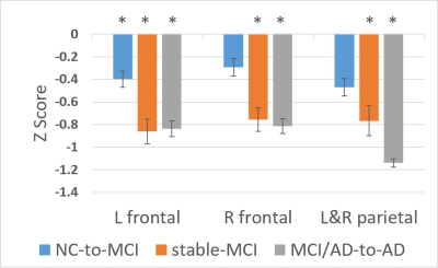 |
White Matter Damage of Alzheimer’s Disease using Magnetization Transfer Imaging
Wenna Duan, Parshant Sehrawat, James Becker, Oscar Lopez, H. Gach, Weiying Dai
The magnetization transfer rate has not been widely used as a biomarker for Alzheimer’s disease (AD). In this study, we measured the magnetization transfer rates in the cardiovascular health study (CHS) cognition study (CHS-CS) cohort at 1.5 T by acquiring T1 maps with and without off-resonance saturation. The magnetization transfer rates were analyzed both cross-sectionally and longitudinally. The longitudinal analysis indicated that damage to white matter regions in the frontal lobe may be associated with AD progression. The study findings demonstrated that the magnetization transfer rate of frontal white matter may be a promising biomarker for AD.
|
17:21
|
0626.
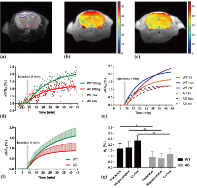 |
Dynamic glucoCEST MRI detects regional changes in cerebral glucose uptake in Alzheimer’s disease at 3T
Jianpan Huang, Jiadi Xu, Celia Dong, Lin Chen, Xiongqi Han, Ed Wu, Peter van Zijl, Kannie Chan
Alzheimer’s disease (AD) is affecting over 50 million people globally. Altered glucose uptake is an early hallmark in AD. Here, we apply glucoCEST, in particular, dynamic glucose enhanced (DGE) MRI to study regional glucose uptake in AD at 3T. First, we optimize saturation parameters, and then we apply these parameters to study glucose uptake in AD mice. Moreover, we use a piecewise exponential fitting to extract specific changes related to glucose uptake and utilization. Results showed a global and regional decrease in cerebral glucose uptake in AD mice compared to WT, which could be an effective mean for early diagnosis.
|
17:33
|
0627.
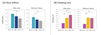 |
Magnetic Resonance Elastography of the Hippocampal Subfields in Alzheimer’s Disease
Lucy Hiscox, Curtis Johnson, Matthew McGarry, Helen Marshall, Edwin van Beek, Neil Roberts, John Starr
Magnetic resonance elastography (MRE) of the hippocampus has shown promise as an imaging biomarker for Alzheimer’s disease (AD). In this work, we report that MRE of the hippocampal subfields are consistent with the known cytoarchitecture. We also found softening of the hippocampal subfields in patients with AD compared to healthy controls with a more pronounced reduction in the subiculum and CA1 compared to the global hippocampus. The dentate-gyrus/CA3 was also more viscoelastic in AD (indicating greater elasticity and less viscosity), whereas no viscoelastic effect was detected globally. MRE of the subfields may provide additional information regarding hippocampal integrity in disease.
|
|
