Monday, 13 May 2019
| Room 510A-D |
16:00 - 18:00 |
Moderators: Moran Artzi |
16:00
|
0287.
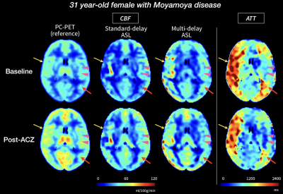 |
Arterial Spin Labeling Underestimates CBF in Regions with Fast Arrival Times: a Simultaneous [15O] PET/MRI Study with Acetazolamide Challenge

Yosuke Ishii, Thoralf Thamn, Jia Guo, Mohammad Khalighi, Mirwais Wardak, Dawn Holley, Harsh Gandhi, Jun Park, Bin Shen, Gary Steinberg, Frederick Chin, Greg Zaharchuk, Audrey Fan
In this study, we investigated the effects of arterial transit time (ATT) reduction and the quantitative accuracy of standard and multi-delay arterial spin labeling (ASL) MRI on healthy controls and Moyamoya patients using acetazolamide (ACZ) by the voxel-wise parametric testing. Administration of ACZ shortened ATT and extended the region where cerebral blood flow (CBF) was underestimated by standard ASL and by multi-delay ASL compared to simultaneous [15O]-water PET reference. Consideration of short ATT is critical for accurate ASL measurements of CBF in the deep gray matter and for quantification of cerebrovascular reactivity after a vasodilation challenge that decreases ATT.
|
16:12
|
0288.
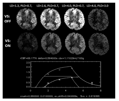 |
ASL signal model for simultaneously measuring CBF and CBV based on ASL imaging for characterizing hemodynamic perfusion state in normal subjects and patients with moyamoya disease.
Hirohiko Kimura, Yoshifumi Higashino, Shota Ishida, Naoyuki Takei, Yasuhiro Fujiwara, Masayuki Kanamoto, Nobuyuki Kosaka, Hiroyuki Kabasawa
Not only cerebral blood flow (CBF) but also cerebral blood volume (CBV) plays an important role for the maintenance of cerebral blood perfusion. We hypothesized that the ASL signal difference caused by vessel suppression (VS) scheme should be dependent on arterial CBV fraction of total ASL signal. In this study, we introduced modified two-compartments model based on ASL signal with and without VS, so that we can calculate arterial volume fraction of total ASL signal. The objective of this study is to demonstrate the feasibility of arterial CBV map as well as CBF based on ASL imaging.
|
16:24
|
0289.
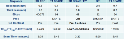 |
The Quantification of Feuromxytol Uptake on the Post Enhance in Intracranial Atherosclerotic Plaque by using a 3D high resolution Black Blood(BB) Multiple Echo(ME) T2* Imaging Technique
Seong-Eun Kim, J Scott McNally, Matthew Alexander, Dennis Parker, Bradley Bolster Jr, Gerald S Treiman, Adam de Havenon
Post-gadoliniun enhancement(PGE) in ICAD may be related to endothelial dysfunction or breakdown or secondary to plaque inflammation. Delayed ferumoxytol imaging allows intravascular clearance with retention in the macrophages present in vulnerable atherosclerotic plaque. A 3D BB ME T2* Imaging technique allows the quantitative ferumoxytol imaging on delayed scans by measuring T2* in intracranial atherosclerotic plaque. We performed 3D BB T2* sequences on ten patients with ICAD and measured T2* changes between baseline and 72 hour after ferumoxytol injection. The iron nanoparticle uptake in symptomatic ICAD presented in this work may provide important mechanistic implications for the pathophysiology of PGE.
|
16:36
|
0290.
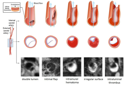 |
High Risk Characteristics of Cervicocranial Artery Dissection Associated with Ischemic Stroke: A Head and Neck Combined Vessel Wall MR Study
ye wu, fang wu, qi yang
The aim of this study was to investigate high risk characteristics of dissected artery using VWMRI. A total of 114 Patients with CeAD were prospectively recruited and 139 dissected vessels were analyzed. Dissected arteries in the stroke group showed a significantly higher prevalence of irregular surface, intraluminal thrombus and severe stenosis (>70%) compared with that of the non-stroke group. Logistic regression analysis showed that the presence of irregular surface and intraluminal thrombus were independently associated with ischemic stroke in CeAD. Our results provide insights into the vascular pathophysiology of symptomatic CeAD and may reveal important predictor of stroke in CeAD.
|
16:48
 |
0291.
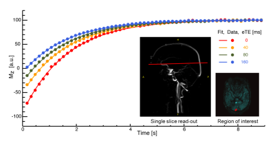 |
Cerebral hypometabolism measured with intravascular T2-prepared tissue relaxation with inversion recovery (T2-TRIR) and pCASL in adults with sickle cell disease
Lena Vaclavu, Esben Thade Petersen, Henri Mutsaerts, Jan Petr, Charles Majoie, John Wood, Ed VanBavel, Bart Biemond, Aart Nederveen
Cerebral metabolic rate of oxygen (CMRO2) quantifies the amount of oxygen consumed by the brain, and relies on continuous delivery of nutrients and oxygen via cerebral blood flow (CBF). In sickle cell disease (SCD), CBF is elevated to compensate for chronic anaemia. This study investigates CMRO2 in adults with SCD using T2-prepared tissue relaxation with inversion recovery (T2-TRIR). CBF increased after acetazolamide-induced vasodilation in both groups but CMRO2 reduced even further in SCD patients while it remained stable in controls. Our results suggest that cerebral shunting is exacerbated by high flow conditions.
|
17:00
|
0292.
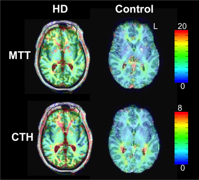 |
Mean Transit Time and Capillary Transit Time Heterogeneity at the Basal Ganglia of Patients with Huntington’s Disease
Yi-Fen Yen, Kenneth Kwong, Suk-Tak Chan, Steven Stufflebeam, H. Diana Rosas
We identified abnormally long mean transit time (MTT) and large capillary transit time heterogeneity (CTH) in the basal ganglia of ten individuals with Huntington’s disease (HD) as compared to ten healthy control subjects. Since iron is elevated in the putamen and globus pallidus in HD [1], which can lead to an underestimation of relative cerebral blood flow (CBF) and cerebral blood volume (CBV), excessively prolonged MTT and CTH, which are ratios of CBV and CBF, could be utilized as relatively intact parameters for the estimation of perfusion deficits or breakdown in blood-brain barrier.
|
17:12
|
0293.
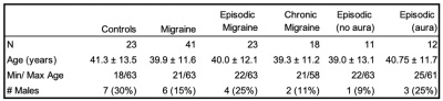 |
White matter microstructural alterations in chronic, episodic, and aura migraine
Benjamin Ades-Aron, Sait Ashina, Bettina Conti, Yvonne Lui, Mia Minen, Dmitry Novikov, Timothy Shepherd, Els Fieremans
Migraine is associated with increased risk for stroke and white matter abnormalities, though the exact nervous tissue pathology remains poorly understood. This study used diffusion kurtosis imaging, a clinically feasible extension of diffusion tensor imaging, to compare white matter microstructural changes in migraineurs to healthy controls, and between their different subtypes: chronic, episodic, with and without aura. Using a voxel-wise statistical approach, we found that axial and radial kurtosis were significantly altered depending on migraine subtype. While radial kurtosis is significantly reduced in all migraine patients compared to controls, axial kurtosis is increased in episodic migraines with and without aura, suggesting different underlying pathology.
|
17:24
|
0294.
 |
Evolution of cerebral hemodynamics and metabolism across the early lifespan in patients with sickle cell disease
Spencer Waddle, Lori Jordan, Meher Juttukonda, Chelsea Lee, Niral Patel, Manus Donahue
Sickle cell disease (SCD) is an inherited hemolytic anemia with altered hemodynamics and increased stroke risk. However, lifetime trends in SCD cerebral hemodynamics and metabolism are poorly understood. We used non-invasive functional measures of cerebral blood flow (CBF) from arterial spin labeling, and oxygen extraction fraction (OEF) from T2-Relaxation-Under-Spin-Tagging MRI, to quantify hemo-metabolic patterns in controls (n=64) and SCD patients (n=125) across the early lifespan (6-40 years). CBF decreases with age in healthy controls (-3.2 mL/100g/min per decade) but increases in patients (5.2 mL/100g/min per decade). OEF was elevated in SCD, showing a similar slope with age as controls.
|
17:36
|
0295.
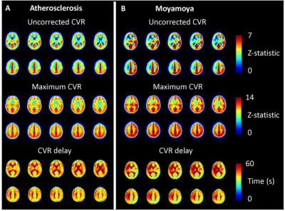 |
Differences in temporal cerebrovascular reactivity responses between patients with atherosclerotic and non-atherosclerotic intracranial disease: implications for optimal hypercapnic reactivity experiments
Meher Juttukonda, Larry Davis, Spencer Waddle, Sarah Lants, Matthew Fusco, Manus Donahue
Intracranial stenosis may be due to atherosclerotic or idiopathic non-atherosclerotic mechanisms, and each condition may incur different consequences on cerebral hemodynamics. The purpose of this study was to use a time regression analysis approach applied to hypercapnic BOLD fMRI data to evaluate how cerebrovascular reactivity (CVR) timing and maximum CVR may differ between patient groups and with vasculopathy extent. Time-to-maximum CVR may be lengthened in territories supplied by stenotic vessels in both patient groups; however, maximum CVR may be reduced on average only in patients with non-atherosclerotic disease, potentially indicating that arteriolar smooth muscle and/or endothelial function may differ substantially between conditions.
|
17:48
|
0296.
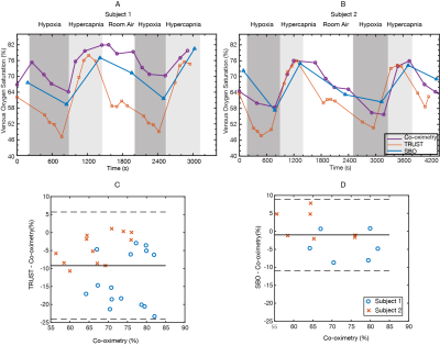 |
In-vivo validation of T2- and susceptibility-based SvO2 measurements with jugular vein catheterization under hypoxia and hypercapnia
Xin Miao, Krishna Nayak, John Wood
This study aimed to validate T2- and susceptibility-based cerebral venous oxygen saturation (SvO2) measurements with the clinical standard, jugular vein catheterization. T2-relaxation-under-tagging (TRUST) and susceptibility-based oximetry (SBO) were performed on two healthy subjects with jugular catheterization and eleven subjects without catheterization under baseline, hypoxia and hypercapnia. TRUST tightly agreed with the jugular reference under hypercapnia but significantly underestimated SvO2 under baseline and hypoxia. Bias between SBO and the reference was independent on the physiological state. A proportional bias was observed comparing TRUST and SBO. The results suggested caution for inter-subject comparison of absolute SvO2 measurements using either TRUST or SBO.
|
|
