What's New in the Spinal Cord?
Oral
Neuro
Monday, 13 May 2019
| Room 518A-C |
16:00 - 18:00 |
Moderators: Jiwon Oh |
16:00
|
0297.
 |
Investigating tissue microstructure using NODDI and histology: A chronic injury study on ex vivo macaque spinal cords

Andrew Bauman, Andrew Yung, Jie Liu, Junhao Liu, Qingan Zhu, Piotr Kozlowski, Wolfram Tetzlaff
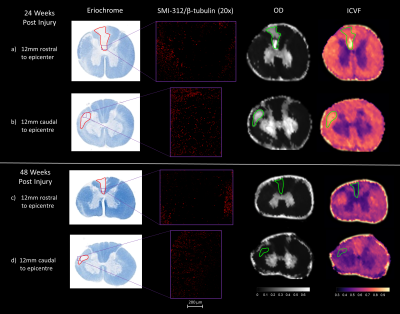
In this study we sought to understand whether Neurite Orientation Dispersion and Density Imaging (NODDI) could accurately delineate the extent of microstructure damage in an ex-vivo macaque spinal cord. We used histology as a gold standard for delineating the margin between damaged and undamaged tissue. Qualitative analysis exposed disagreements between NODDI maps and histology in areas of damage. This suggests that NODDI may not be appropriate for this model of chronic spinal cord injury.
|
16:12
 |
0298.
 |
Finger-Tapping Task fMRI in the Human Cervical Spinal Cord at 7T
Alan Seifert, Yazhuo Kong, Karla Miller, Irene Tracey, S Johanna Vannesjo
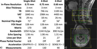
Functional MRI of the spinal cord is challenging due to its small size and deep anatomical location. Increasing field strength enhances BOLD signal and improves SNR, but B0 distortions produced by the lungs and vertebral column are amplified, presenting additional challenges in protocol optimization. Barry et al. have successfully performed resting-state fMRI at 7T; here, we present observations of robust, well-localized motor task activation in the human cervical spinal cord at 7T. We assessed single-shot and multi-shot EPI at two different resolutions. Multi-shot EPI achieved finer resolution and less spatial distortion in this preliminary 7T spinal cord task fMRI study.
|
16:24
|
0299.
 |
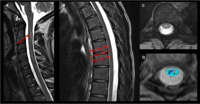
Advanced Magnetic Resonance techniques in Post-Mortem Human Spinal Cord Injury: Correlations with Histopathology
Sarah Morris, Valentin Prevost, Piotr Kozlowski, Andrew Yung, Andrew Bauman, Zahra B., Caron Fournier, Allan Aludino, Lisa Parker, Kevin Dong, Femke Streijger, G. R. Moore, Brian Kwon, Cornelia Laule
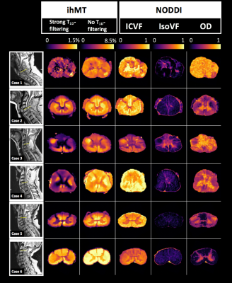
Spinal cord injury prognosis assessments rely on subjective clinical evaluations and often poorly predict outcome; quantitative imaging biomarkers for spinal cord injury evaluation would aid clinical decision making. Our study applied two advanced MRI techniques to the imaging of post-mortem human spinal cord injury samples. We compared in-homogeneous magnetisation transfer and NODDI metric maps with six histological stains to relate the MR image contrast to biological correlates. We found a correlation trend between ihMT signal with strong T1D-filtering and Luxol Fast Blue optical density (myelin phospholipid stain) in white and grey matter.
|
16:36
|
0300.
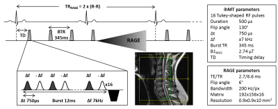 |
Cervical Spine inhomogeneous Magnetization Transfer (ihMT) Imaging Using ECG-Triggered 3D Rapid Acquisition Gradient-Echo (ihMT-RAGE)
Thomas Troalen, Virginie Callot, Gopal Varma, Arash Forodighasemabadi, Maxime Guye, David Alsop, Guillaume Duhamel, Olivier Girard
Inhomogeneous magnetization transfer (ihMT) is a promising MRI modality that provides high sensitivity and specificity to myelinated tissue. Demyelinating pathologies in the central nervous system could then be addressed by this technique. The goal of this work is to demonstrate that the combination of ihMT preparation with Rapid Acquisition Gradient Echo (ihMT-RAGE), as recently proposed for the brain, can be adapted to an ECG-triggered 3D exploration of the cervical spinal cord within a clinically-compatible scan time. The in-vivo ihMT results from healthy cervical spinal cord demonstrate the great potential of the ihMT-RAGE technique for future investigations of degenerative spinal cord pathologies.
|
16:48
|
0301.
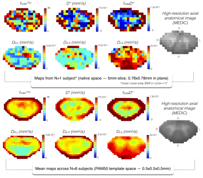 |
Intra-Voxel Incoherent Motion at 7T to quantify human spinal cord microperfusion: pitfalls and promises
Simon Levy, Stanislas Rapacchi, Aurélien Massire, Thorsten Feiweier, Maxime Guye, Thomas Troalen, Virginie Callot
Spinal cord microperfusion assessment in human is challenging but would greatly help characterize tissue integrity and surgery decision-making. Intra-Voxel Incoherent Motion (IVIM) microperfusion measurement is promising but remains highly Signal-to-Noise ratio (SNR) demanding. Monte-Carlo simulations show that IVIM two-step segmented fitting approach is less accurate than directly fitting the bi-exponential representation to all b-values. Simulations also help quantify required SNR and estimation errors to measure IVIM parameters in the context of low perfusion. Exploiting 7T SNR gain, large number of repetitions and group average, IVIM was able to unveil the gray matter higher microperfusion-related pattern, compared to white matter, in agreement with brain studies.
|
17:00
|
0302.
 |
Cervical spinal cord diffusion MRI and intraspinal space restriction at the occipito-cervical junction in mucopolysacharidoses patients
Igor Nestrasil, Rene Labounek, Carol Nguyen, Ivan Krasovec, Jan Valosek, Alena Svatkova, Julien Cohen-Adad, Christophe Lenglet, Chester Whitley
The overall goal of this project is to establish novel MRI parameters for reliable detection of cervical spinal cord (CSC) microstructural abnormalities in patients with Mucopolysaccharidosis (MPS) that develop prior to the clinical manifestation of spinal cord damage. Quantitative analysis of diffusion MRI (dMRI) may characterize microstructural alterations of CSC with high sensitivity. In this study measures of CSC microstructure were determined by dMRI using a protocol based on the RESOLVE (REadout Segmentation Of Long Variable Echo trains) sequence. Derived diffusion metrics were then related to the anatomical measures of the cervical spine in patients with MPS.
|
17:12
|
0303.
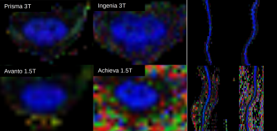 |
A Multi-site/multi-scanner DTI study of the adult spinal cord
Devon Middleton, Joshua Fisher, Adam Flanders, Feroze Mohamed, John Woo, Mark Elliot, Scott Faro, Laura Krisa
This study presents DTI data collected for the complete cervical and thoracic spinal cord in healthy adult subjects as part of a multi-site/multi-scanner study. Thirty adult subjects were imaged with four different scanners including 1.5T and 3T field strengths and variability in DTI metrics was examined.
|
17:24
|
0304.
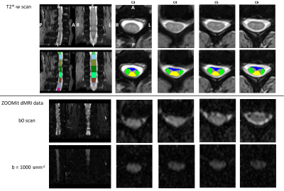 |
Column-specific microstructural changes in patients with non-myelopathic degenerative compression of the cervical spinal cord revealed by diffusion MRI
Jan Valošek, René Labounek, Tomáš Horák, Alena Svátková, Petr Kudlicka, Pavel Hok, Jan Kocica, Christophe Lenglet, Petr Hluštík, Josef Bednarík, Petr Bednarík
While delineation of microstructural changes in white matter (WM) columns of cervical spinal cord (CSC) in patients with non-myelopathic degenerative CSC compression (NMDCSCC) remains a challenge for most current MRI techniques, High Angular Resolution Diffusion Imaging (HARDI) protocols promise to overcome this issue. Thus, our group utilized novel HARDI-ZOOMit protocol to extract metrics from diffusion tensor and ball-and-stick models in three major CSC columns. HARDI-ZOOMit protocol was able to detect column-specific significant differences between healthy controls and patients with NMDCSCC with more complex abnormalities in ventral CSC columns in C3-C6 levels.
|
17:36
|
0305.
 |
Establishing a Relationship Between Pain and Spinal Cord Demyelination using Magnetization Transfer Imaging and Thermal Sensory Testing
Nadia Barakat, Steven Staffa, Leslie Benson, Mark Gorman, David Zurakowski, David Borsook
Myelitis is a demyelinating disorder of the spinal cord . It can occur as an isolated syndrome or in the context of an autoimmune condition such as MS. Pain is a significant problem in myelitis and has a major impact on treatment response and rehabilitation efforts. Magnetization Transfer Imaging has the ability to provide a marker for myelin content. Defining a relationship between pain and demyelination could lead to improved disease outcome. Our results showed significant differences in spinal cord MTR (C1 to T12) and pain (heat/cold stimuli) between patients and controls, and strong correlation between MTR and heat detection thresholds.
|
17:48
|
0306.
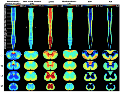 |
Construction of a quantifiable rat spinal cord atlas and tract delineation using agglomerative clustering
Harris Nami, Ariane Saliani, Aldo Zaimi, Tanguy Duval, Christian Perone, Nikola Stikov, Julien Cohen-Adad
With qMRI becoming the norm in obtaining quantitative values for most MRI studies, there is still a need for its validation as new techniques are constantly being implemented. With this in mind, we propose a matter microstructure atlas of the rat spinal cord based on high-resolution histology. This atlas contains morphometric data such as axon density, axon diameter and g-ratio. Furthermore, a clustering algorithm was implemented to generate white matter tracts delineation based on axon morphometry, possibly leading to an improvement on outdated atlases. The proposed atlas is open access and can be used for quantitative comparisons with qMRI studies.
|
|
