Novel Neuroimaging Techniques
Oral
Neuro
Thursday, 16 May 2019
| Room 510A-D |
08:15 - 10:15 |
Moderators: Armen Kocharian, Yunhong Shu |
08:15
 |
1040.
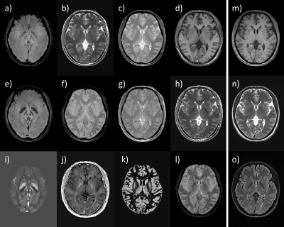 |
STrategically Acquired Gradient Echo (STAGE) Imaging for Standardized Multi-Contrast and Quantitative Brain Data Acquisition: Validating T1 mapping and Preliminary Clinical Results

Yongsheng Chen, M. Marcella Lagana, Shuang Xia, E. Mark Haacke
One major thrust in radiology today is imaging standardization with rapidly acquired quantitative multi-contrast information. This is critical for multi-center trials, for the collection of big data and the use of artificial intelligence in evaluating the data. STAGE is one such method that can provide more than ten qualitative and quantitative pieces of information in roughly 5 minutes or less at 3T. In this work, we introduce several new image contrasts derived from STAGE and validate STAGE T1 mapping using the ISMRM/NIST phantom as well as in vivo data. We also present some preliminary pathological results.
|
08:27
|
1041.
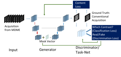 |
OneforAll: Improving the Quality of Multi-contrast Synthesized MRI Using a Multi-Task Generative Model
Guanhua Wang, Enhao Gong, Suchandrima Banerjee, Huijun Chen, John Pauly, Greg Zaharchuk
To improve the quality and efficiency of multi-contrast MR neuroimaging, a new Multi-Task Generative Adversarial Network (GAN) is proposed to synthesize multiple contrasts using a uniformed network. The cohort of 104 subjects consisting of both healthy and pathological cases is used for training and evaluation. For both the subjective and non-subjective evaluation, the proposed method achieved improved diagnostic quality compared with state-of-the-art synthetic MRI image reconstruction methods based on model-fitting and also previously shown deep learning methods.
|
08:39
|
1042.
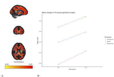 |
White matter microstructure changes as a function of time-of-day: a biomarker of accumulating sleep pressure?
Irene Voldsbekk, Daniel Roelfs, Atle Bjørnerud, Inge Groote, Ivan Maximov
There is an increasing awareness of time-of-day (TOD) effects in MRI. The underlying causes of these fluctuations may be related to diurnal physiological variations, reflecting the processes that govern transition between sleep and wake. We probed TOD effects in a prospective manner, scanning 47 healthy individuals in the morning and the same evening using a five-shell diffusion-weighted imaging protocol at 3T, where a number of important Zeitgeber signals were rigorously controlled. We found significant changes in an number of derived DWI parameters (FA, MD, AWF, MK), and speculate that these changes are pointing towards underlying mechanisms of sleep-wake homeostasis.
|
08:51
|
1043.
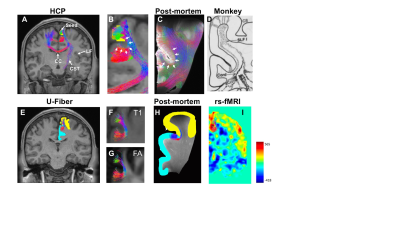 |
Whole Brain Mapping of Subcortical U-fibers in the Human Connectome Project Data
Jing Li, Zifei Liang, Ying-Chia Lin, Choong Heon Lee, Weihong Zhang, Steven Baete, Yulin Ge, Jiangyang Zhang
Although reconstruction of subcortical U-fibers from diffusion MRI data has been demonstrated before, systematic examination of U-fibers inthe brain remain challenging. In this study, we used a detailed cortical parcellation map with 78 distinct cortical regions to map more than 200 U- fibers in 60 subjects from the Human Connectome Project dataset. The results were compared to post-mortem results and chemical tracer results in monkey brains for selected fibers. We also found moderate correlations between fiber streamline numbers and resting state connectivity. By mapping all subject data to the MNI space, maps of group average U-fiber were generated.
|
09:03
|
1044.
 |
T2 Weighted Whole-Brain Intracranial Vessel Wall Imaging at 3 Tesla with Cerebrospinal Fluid suppression
Lei Zhang, Yanjie Zhu, Yiu-cho Chung, Lijie Ren, Na Zhang, Xin Liu
T2 weighting intracranial vessel wall MR imaging provides good contrast to discriminate various important plaque components and is a good tool to differentiate intracranial vasculopathy. However, the strong CSF signal in T2-weighted images interfere the depiction of the vessel wall. In this study, we proposed to use T2IR preparation module combined with T2-weighted SPACE for whole brain intracranial vessel wall imaging, the T2IR pulses were used to suppress CSF while minimizing its effect on the signal reduction from T2w SPACE. This new technique was first evaluated in healthy volunteers and then tested on some stroke patients.
|
09:15
|
1045.
 |
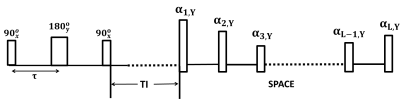
Stirred CSF Measured by Low b-value DTI
Yoshitaka Bito, Kuniaki Harada, Hisaaki Ochi, Kohsuke Kudo
Cerebrospinal fluid (CSF) plays an important role in clearance systems of the brain. There have been many studies on bulk flow of CSF; however, there have been few studies on complex flow like pseudo-random movement of CSF. In this paper, we investigate locally “stirred” CSF by using low b-value diffusion tensor imaging (DTI). Measured DTI shows inhomogeneous physiology of CSF, including extremely high and anisotropic diffusion tensors around the middle-cerebral artery. It demonstrates that the low b-value DTI will be useful in estimating local drainage of CSF.
|
09:27
|
1046.
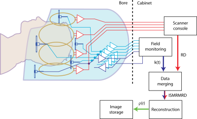 |
Integration of field monitoring for neuroscientific applications - SNR, acceleration and image integrity
David Brunner, Simon Gross, Thomas Schmid, Alexander Boegel, Benjamin Dietrich, Yoojin Lee, Bertram Wilm, Christian Mirkes, Christoph Barmet, Klaas Pruessmann
Full integration of field monitoring capability in an MRI system’s hard- and software by co-designing the two subsystems for neuroscientific applications is demonstrated along with its benefits in cutting edge fast imaging. The system entails a custom brain receiver array with integrated field-probes monitoring k-space during readout as well as the subsequent data processing pipeline for image reconstruction. All data is aggregated in an ISMRMRD data container for open access.
|
09:39
|
1047.
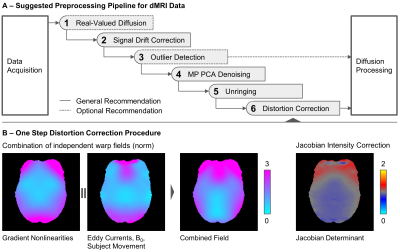 |
A Joint Recommendation for Optimized Preprocessing of Connectom Diffusion MRI Data
Cornelius Eichner, Qiuyun Fan, Susie Huang, Derek Jones, Evgeniya Kirilina, Michael Paquette, Umesh Rudrapatna, Chantal Tax, Qiyuan Tian, Nikolaus Weiskopf, Alfred Anwander
The possibility of acquiring diffusion MRI data with extremely high resolution and strong diffusion weighting imposes new challenges on the preprocessing. In a joint effort, all research institutions with the availability of a human MRI system with 300 mT/m maximum gradient strength compared their respective processing pipelines and converged towards a joint recommendation for high-quality dMRI preprocessing. The joint pipeline represents the current state-of-the-art of dMRI image processing and will be made available to other researchers. The result of this joint effort promises better reproducibility of diffusion MRI results.
|
09:51
|
1048
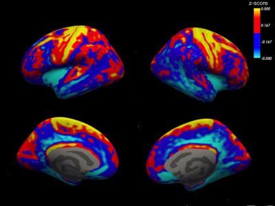 |
Myeloarchitectonic mapping of cortical gray matter with 3D inhomogeneous magnetization transfer (ihMT)
Fanny Munsch, Gopal Varma, Manuel Taso, Olivier Girard, Arnaud Guidon, Guillaume Duhamel, David Alsop
Advances in inhomogeneous magnetization transfer (ihMT) imaging have enabled improved volumetric imaging of gray matter. We performed a cortical surface-based analysis of the ihMT ratio (ihMTR) and the inverse ihMT ratio (ihMTRinv) to investigate the distribution of myelin in cortical gray matter. IhMT MRI, and especially ihMTRinv, displayed regional differences in cortical myelination, in agreement with postmortem studies. These findings support the myelin sensitivity and specificity of ihMTRinv and its use for gray matter characterization.
|
10:03
|
1049.
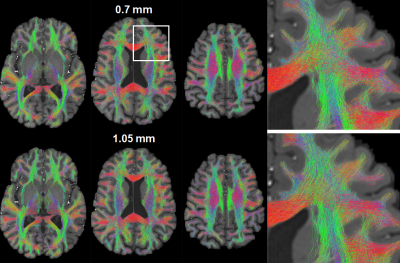 |
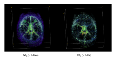
Submillimeter whole brain diffusion MRI at 7 Tesla using simultaneous multislice imaging
Xiaoping Wu, Eddie Auerbach, Kamil Ugurbil
Acquisition of whole-brain diffusion MRI at submillimeter resolutions has proven to be highly beneficial for probing the brain’s structure and connectivity. Here, we investigate how simultaneous multislice (SMS) imaging, a single-shot acquisition that has great success in large-scale cohort studies such as the Human Connectome Projects (HCP), can be used to achieve submillimeter whole-brain diffusion at 7 Tesla. We acquired SMS diffusion at 0.7-mm isotropic resolution using a standard body gradient. Our results show that these submillimeter data can be used to generate a whole-brain tractogram and a connectome comparable to those of the 1.05-mm HCP-style acquisition using denoising approaches.
|
|
