Thursday, 16 May 2019
| Room 518A-C |
16:00 - 18:00 |
Moderators: Kader Oguz, Eugene Yu |
16:00
|
1239.
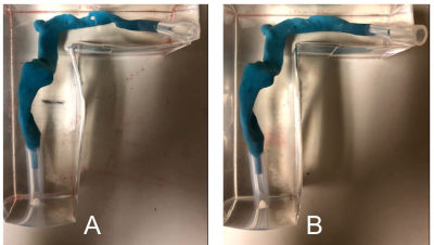 |
Investigation of blood flow causing pulsatile tinnitus in a patient-specific benchtop flow model

Keerthi Valluru, Henrik Haraldsson, Evan Kao, Joseph Leach, Alexandra Wright, Megan Ballweber, Karl Meisel, David Saloner, Matthew Amans
We created 3D printed patient-specific cerebral venous flow models of a patient with pulsatile tinnitus (PT) caused by idiopathic intracranial hypertension whose PT was resolved by CSF removal via lumbar puncture (LP). Prior to and almost immediately after the LP, she underwent MR imaging consisting of anatomic and flow evaluation sequences. Benchtop flow models were generated from both the pre and post LP data sets. We performed sound measurements and flow analysis of these models to identify flow features that correlate with sound production. Very similar features were seen both in vitro and in vivo in the pre-LP (PT) state that were absent in the post-LP (no PT) state. Patient-specific flow models may be an appropriate in vitro surrogate for venous causes of PT that could enable concrete determination of the links between geometry, blood flow, and PT.
|
16:12
 |
1240.
 |
Brain and head-and-neck MRI in immobilization masks: a novel and practical setup for radiotherapy
Stefano Mandija, Federico D'Agata, Robin Navest, Alessandro Sbrizzi, Cornelis Raaijmakers, Rob Tijssen, Marielle Philippens, Enrica Seravalli, Joost Verhoeff, Jan Lagendijk, Cornelis van den Berg
For radiotherapy treatment planning, it is essential to perform the MRI/CT exams in treatment position. For this purpose, thermoplastic immobilization masks are used for brain and head-and-neck radiotherapy. However, since standard immobilization masks do not fit in the diagnostic MR head/neck coils, suboptimal surface-loop coils leading to poor image quality are used in clinical practice. Here, we present a new immobilization setup. This setup has several advantages compared to state-of-the-art setups: it fits in the diagnostic head/neck MR coils, it allows diagnostic image quality in treatment position, high SNR, homogenous signal, restricted motion (about 1 mm) and accurate inter-fraction repositioning.
|
16:24
|
1241.
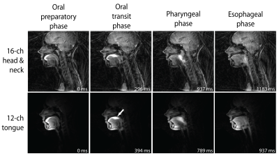 |
A 12-channel flexible receive coil for accelerated tongue imaging
Luuk Voskuilen, Michel Italiaander, Paul de Heer, Alfons Balm, Ferdinand van der Heijden, Gustav Strijkers, Ludi Smeele, Aart Nederveen
Acceleration techniques necessary for real-time MRI of swallowing and diffusion imaging of the tongue require multiple coil elements and decrease SNR. Therefore, we designed a 12-channel flexible tongue coil with a higher density of elements compared to the conventional head and neck coil. The SNR in the tongue coil is better, while the G-factor is lower compared to the conventional coil. Real-time MRI displayed better image quality with fewer radial streaking artefacts using the tongue coil. A multiband-SENSE factor of 2 was feasible for diffusion-weighted imaging with the tongue coil.
|
16:36
|
1242.
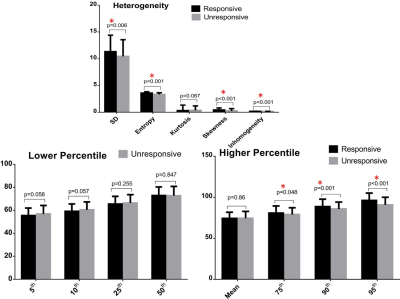 |
Baseline Multiparametric Volumetric T2RT Histogram Analysis: Can It Be Used to Predict Therapy Response in Patients with Thyroid Associated Ophthalmopathy (TAO) Undergoing Intravenous Methylprednisolone
ping liu, jing zhang
Prompt recognition the characteristics of extraocular muscles (EOMs) representing responding to intravenous methylprednisolone pulses (ivMP) is crucial for identifying optimal, patient-tailored treatment options in the era of personalized, precision medicine for thyroid-associated ophthalmopathy (TAO). This study attempts to predict the therapy response using the baseline histogram features of EOMs from volumetric T2 relaxion time (T2RT) texture analysis. Multivariate logistic regression analysis suggested that 90th and 95th percentile T2RT, skewness, entropy and inhomogeneity were significant predictors of ivMP response. Volumetric T2RT histogram analysis is a feasible and promising tool for predicting the response of ivMP therapy in patients with TAO.
|
16:48
|
1243.
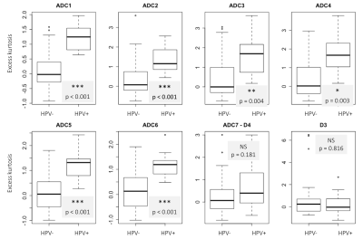 |
Diffusion Weighted Imaging to differentiate human papillomavirus positive from human papilloma virus negative head and neck squamous cell carcinoma: how do b-values influence results?
Bénédicte Delattre, Vincent Lenoir, Yacine M'Rad, Minerva Becker
This study investigates whether and how the choice of b-values influences ADC ability to differentiate between human papillomavirus (HPV) positive (HPV+) from HPV negative (HPV-) head and neck squamous cell carcinoma (HNSCC) using quantitative histogram parameters of tumor ADC maps obtained with different combinations of b-values and with monoexponential and bi-exponential models (IVIM). Results show that ADC calculated with 2 b-values, b0 and b1000, are sufficient and that ADC calculated only with high b-values (combination excluding b0) are not able to differentiate both tumor types.
|
17:00
|
1244.
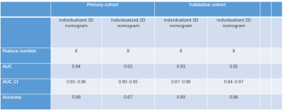 |
MRI-based radiomics nomogram in differentiating sinonasal mucosal melanoma from sinonasal lymphoma
Did Not Present
Linying Guo, Zuohua Tang, Yang Song, Guang Yang, Jing Zhang, Yucheng Pan, zebin xiao, Zhongshuai Zhang
Differentiating sinonasal mucosal melanoma (SNMM) from sinonasal lymphoma before surgery is important. However, it is a challenge to perform preoperative diagnosis through either histological biopsy or conventional MRI. In this study, an MRI-based individualized 3D nomogram through radiomics was constructed, and its value was explored in the differentiation between the two entities. The nomogram shows satisfactory efficiency in the diagnosis, and its performance was better than that of human radiologists. This study demonstrates that noninvasive MRI-based radiomics could be helpful in the preoperative differentiation of SNMM from sinonasal lymphoma.
|
17:12
|
1245.
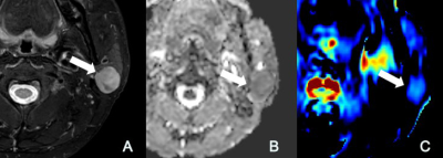 |
Differentiation of Malignant and Benign Parotid Tumors with Amide Proton Transfer-Weighted MR Imaging
Yu Chen, Tong Su, Zhengyu Jin, Zhentan Xu, Zhizheng Zhuo, Zhuhua Zhang, Huadan Xue
This retrospective study evaluated the diagnostic efficacy of diffusion-weighted imaging (DWI) and amide proton transfer-weighted (APTw), for differentiating between malignant, pleomophic adenomas and Warthin’s tumours. APTw imaging can differentiate Warthin’s tumors and malignant tumors of parotid. ADC can differentiate Warthin’s tumors and pleomorphic adenomas. APT combined with ADC had the better performance to differentiate the three common kinds of parotid tumours.
|
17:24
|
1246.
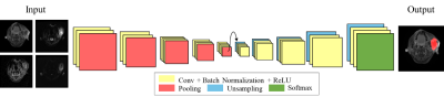 |
Detection of malignant parotid gland tumors using multi-modality MRI and deep learning: diffusion versus T1 contrast-enhanced imaging
Yi-Ju Chang, Chun-Jung Juan, Yi-Jui Liu, Teng-Yi Huang
The study presents an automatic recognition method for parotid gland tumor. We used a convolution neural network to conduct the segmentation of parotid gland tumor and classifications of tumor types. We also designed eight combinations of various MRI contrasts to compare the results of recognition for parotid gland tumor. We compared results obtained using various combinations of MR images as the input of the convolutional neural network and found that diffusion-related parameters and contrast-enhanced T1 images played the primary role of the prediction accuracy.
|
17:36
|
1247.
 |
Probabilistic structural atlas of human lateral parabrachial nucleus, medial parabrachial nucleus and vestibular nuclei complex using in vivo 7 Tesla MRI.
Kavita Singh, Iole Indovina, Jeffrey Staab, Marta Bianciardi
Parabrachial and vestibular nuclei are anatomically and functionally connected brainstem gray-matter structures involved in autonomic and vestibular functions. Their assessment in research and clinical investigations is difficult due to limited image-resolution/contrast of clinical scanners and the absence of probabilistic atlas of these structures. We delineated these nuclei in single-subject multi-contrast 1.1mm-resolution 7Tesla MRI of healthy humans and generated a validated in-vivo probabilistic atlas of these nuclei in stereotaxic space. Upon coregistration to clinical-MRI, this atlas might improve the evaluation of lesions and assessment of connectivity-pathways underlying autonomic and vestibular mechanisms in a broad-set of clinical conditions relating to these nuclei.
|
17:48
|
1248
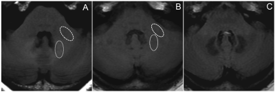 |
Nasopharyngeal Irradiation can Increase Signal Intensity on T1-Weighted MRI of the Dentate Nucleus in Patients with Nasopharyngeal Malignancies
Video Permission Withheld
Rongbiao Tang, Mark Haacke, Qingrou Wang, Naying He, Ke-min Chen, Fuhua Yan
Patients with nasopharyngeal malignancies (NPM) are generally treated by nasopharyngeal irradiation. In order to evaluate curative effect, gadolinium-based contrast agents (GBCAs)-based MRI examinations are always performed repeatedly. Whether the nasopharyngeal irradiation affects the T1 signal intensity (SI) in DN remains unclear. 68 NPM patients and 68 suitable control patients were enrolled. We found that the uptake rate of gadolinium from the NPM was significantly higher than that from control patients. We speculated that the nasopharyngeal irradiation increases the T1 SI by the damage to the blood–brain barrier (BBB).
|
|
