Pediatrics: Image Acquisition
Oral
Neuro
Tuesday, 14 May 2019
| Room 510A-D |
08:15 - 10:15 |
Moderators: Julie Guerin |
08:15
|
0380.
 |
The Role of Diffusion Tensor Imaging in the Characterisation of Paediatric Brain Tumours - a Multi-Centre Study

Heather Rose, Christopher Bennett, Jan Novak, Lesley MacPherson, Shivaram Avula, Theodoros Arvanitis, Chris Clark, Simon Bailey, Dipayan Mitra, Dorothee Auer, Richard Grundy, Andrew Peet
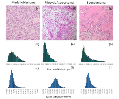
CNS tumours are the most common solid tumour in paediatrics and the most common cause of childhood cancer deaths. The diagnostic role of Diffusion Tensor Imaging (DTI) in patients with either medulloblastoma (MB), pilocytic astrocytoma (PA) or ependymoma (EP) was investigated. Fractional anisotropy (FA) and mean diffusivity (MD) means were found to be significantly different between tumour groups, as determined by one-way ANOVA (p=0.0002 and <0.0001). MD distributions enabled classification of tumour type, using linear discriminant analysis (LDA), with an average accuracy of 80%. DTI metrics were shown to provide an insight into the structure of paediatrics CNS tumours with LDA classification using MD demonstrating improved accuracy over FA.
|
08:27
|
0381.
 |
New insights into the development of white matter microstructure across childhood and adolescence from ultra-strong gradients
Sila Genc, Chantal Tax, Erika Raven, Maxime Chamberland, Greg Parker, Derek Jones
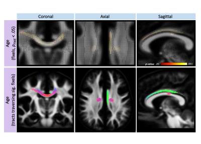
Recent advancements in diffusion-weighted imaging analysis techniques, such as fixel-based analysis (FBA), have improved our understanding of fibre-specific patterns of white matter development over childhood. Here, we investigate differential patterns of fibre properties with age and sex, using multiple b-value sampling schemes. The body of the corpus callosum and cingulum undergo significant development over the ages of 8-18, and FBA was robust to these patterns across sampling schemes. However, apparent fibre density (AFD) may be overestimated in clinical sampling schemes for older children, suggesting that higher b-values may improve AFD estimation in emerging complex fibre configurations developing with age.
|
08:39
|
0382.
 |
White matter fibrography of the extremely preterm brain: longitudinal connectome changes from childhood to adolescence.
Ryan McNaughton, Hernan Jara, Mina Botros, Baiyu Zhou, Stephan Anderson, Osamu Sakai, Edward Sung, Robert Joseph, Karl Kuban, Michael O'Shea
Purpose: To study comparatively and longitudinally the connectome changes from childhood (age 10 years) to adolescence (age 15 years) using white matter fibrography (WMF). Methods: WMF was used to generate the connectomes of 9 extremely preterm born individuals using MRIs obtained at ages 10 and 15 years. Results: The most noticeable connectome change was a marked increase in the fiber density accompanied by fiber thinning. Conclusion: As anticipated, WMF connectomics of the extremely preterm brain demonstrate clearly observable WM architectural changes from 10 to 15 years of age from sparse fiber-thick to dense fiber-thin.
|
08:51
|
0383.
 |
Evaluation of multi-shell diffusion MRI acquisition strategy on quantitative analysis of multi-compartment models
Xiaodong Zhang, Sudeep Patel, Chun-Xia Li
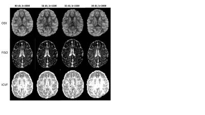
Multi-shell diffusion MRI (dMRI) allows for analyzing the water diffusion signal using multi-compartment diffusion models, providing more specific characterization of tissue microstructure in grey matter and white matter than standard diffusion tensor imaging (DTI). However, the traditional multi-shell dMRI data acquisition usually demands high gradient strength and long scanning duration and its application is hindered for subjects like fetuses and infants in which fast imaging and reduced gradient strength is required. In the present study, the quantification analysis of NODDI and DBSI was evaluated using different hybrid diffusion imaging (HYDI) acquisition strategy for fast imaging on a clinical 3T setting. The results demonstrated that the data acquisition time for multi-shell dMRI can be reduced dramatically using HYDI gradient encoding strategy, while the quality of derived NODDI, DBSI, and DTI indices is generally maintained, suggesting quantitative analysis of multi-compartment models are applicable for developmental study of whole brain fetuses and infants by using multi-shell dMRI with HYDI encoding scheme.
|
09:03
 |
0384.
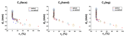 |
Electrocorticography-combined diffusion tract quantification model to improve benefit-risk assessment in pediatric epilepsy surgery
Min-Hee Lee, Nolan O'Hara, Csaba Juhasz, Eishi Asano, Jeong-Won Jeong
The present study proposes a novel diffusion weighted imaging (DWI) tract classification methodology which integrates DWI-maximum a posteriori probability (DWI-MAP) analysis with Kalman filter in order to predict an optimal margin of cortical resection balancing postoperative benefit (seizure freedom) and risk (motor deficit in face, hand and leg) in pediatric epilepsy surgery. The predicted margins provided high Fisher’s exact test probability, 0.92 (0.94) of successful avoidance of motor deficits with (or without) seizure freedom. This finding demonstrates the translational value of a DWI tract classification approach in quantitative benefit-risk assessment to achieve ultimate goal of pediatric epilepsy surgery.
|
09:15
|
0385.
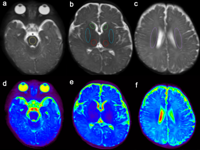 |
Early identification of neonatal mild hypoxic-ischemic encephalopathy by Amide Proton Transfer magnetic resonance imaging: A Pilot Study
Did Not Present
Sijin Chen, Yingjie Mei, Yikai Xu
Neonatal hypoxic-ischemic encephalopathy (HIE) is a major complication of perinatal asphyxia, with high morbidity and morbidity. APT imaging is a potential technique for detecting in vivo characterization of the internal environment during hypoxic-ischemic brain injury. We investigated the feasibility of APT in differentiating neonatal mild HIE from normal age-matched infants, and to explore the changes in the internal environment in neonatal mild HIE. The results indicate that APT imaging for neonatal mild HIE is a useful and feasible technique with diagnostic capability.
|
09:27
|
0386.
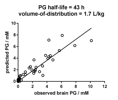 |
Long half-life of propylene glycol in neonatal brain: an MRS study
Petra Pouwels, Monique van de Lagemaat, Laura van de Pol, Bregje Witjes, Inge Zonnenberg
Neonatal convulsions are preferably treated with intravenous phenobarbital that contains propylene glycol (PG) as solvent. Very high concentrations of brain PG have been observed with quantitative MRS, especially when low-concentrated phenobarbital medication was used. PG can have serious adverse effects, and the half-life is longer in neonates than in adults. Based on given medication and the interval until MRS examination we estimated a PG half-life in neonatal brain that is at least 30 hours and maybe up to 43 hours. This shows that extremely high and potentially toxic PG concentrations will persist longer than expected in the neonatal brain.
|
09:39
|
0387.
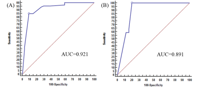 |
A MR-based nomogram for predicting cerebral palsy in periventricular leukomalacia children before 2 years old
Tingting Huang, Zhe Liu, Chao Jin, Haoxiang Jiang, Heng Liu, Wei Xing, Yunhu Wu, Xiaocheng Wei, Gang Zhang, Jian Yang
Early identification of cerebral palsy (CP) in children with periventricular leukomalacia (PVL) is crucial for prescribing the indispensable treatment and rehabilitation. In this study, we visually assessed PVL-associated MR signs, i.e. T2/T2 Flair hyperintense in the centrum ovale, posterior limb of internal capsule, pedunculus cerebri and thalamus, and found they were independent predictors of CP. Based on these signs, a MR-based nomogram for predicting CP in PVL children aged less than 2 yr was developed. Results indicated that the area under receiver operating characteristic curve, sensitivity and specificity for this nomogram were 0.921, 91.2% and 83.3%, respectively. These suggested the potential role of our MR nomogram in predicting the CP outcome of PVL children before 2 years old.
|
09:51
|
0388
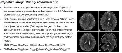 |
Silent MR Can Improve the Success Rate of Infant Examination: The Application Value of an MRI Acoustic Reduction Technique in Infants
Video Permission Withheld
Wei Xia, Xi Zhu, Weiqiang Dou
Although the data for the objective image quality of silent MR do not correspond with the subjective image quality scores, these data can greatly improve the success rate of the examination, reduce acoustic noise, improve patient comfort, and allow for higher overall diagnostic usefulness when compared with those of conventional MR. In summary, silent MR is more suitable for the infant brain than conventional MR.
|
10:03
|
0389.
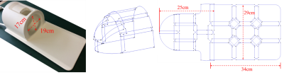 |
A 24-channel head and spine array for 3T pediatric MRI under 3-year-old
Ye Li, Guangzu Xu, Dengbin Wang, Hui Zheng, Nan Li, Xiangming Hou, Yang Xin, Guobin Li, Qiang He, Xiaoliang Zhang, Xin Liu, Hairong Zheng
Pediatric MRI is an important tool for clinical diagnosis and research. It demands high spatial resolution and short scan time to increase success rates. To obtain high resolution images within resonable scan time, a dedicated 24-channel head and spine coil array was designed and fabricated for patient under 3-year-old in this work. Compared to a commercial 24-channel head and neck adult coil, the proposed coil offers higher resolution, image quality and shorter scan time in patient studies.
|
|
