Blood Brain Barrier, Glymphatics & CSF Flow
Oral
Neuro
Wednesday, 15 May 2019
| Room 518A-C |
08:15 - 10:15 |
Moderators: Nivedita Agarwal, Karl-Olof Lovblad |
08:15
 |
0746.
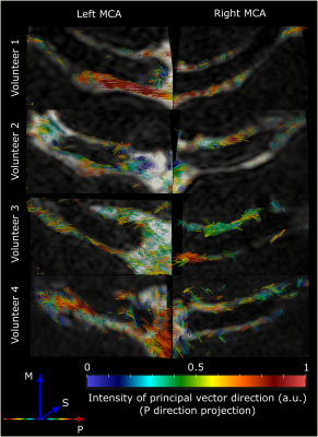 |
High resolution T2-prepared MRI enables non-invasive assessment of CSF flow in perivascular spaces of the human brain

Lydiane Hirschler, Roxana Aldea, Léonie Petitclerc, Itamar Ronen, Patrick de Koning, Mark van Buchem, Matthias van Osch
Recently, flow of cerebrospinal fluid (CSF) has been shown to play an important role in the transport of brain metabolites, ushering in the concepts of glymphatics and intramural periarterial drainage. Failure of waste-drainage has been linked to incurable neurodegenerative diseases such as Alzheimer’s disease and cerebral amyloid angiopathy. The lack of a non-invasive imaging technique for the investigation of perivascular drainage mechanisms in the human brain strongly hinders the assessment of brain clearance in human subjects. In this study, we present the first non-invasive technique to visualize the CSF-flow along the middle cerebral artery (MCA), as well as along perivascular spaces of smaller arteries in the human brain.
|
08:27
|
0747.
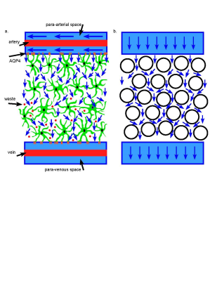 |
A Novel MRI Phantom to Study Interstitial Fluid Transport in the Glymphatic System
Michal Komlosh, Dan Benjamini, Nathan Williamson, Ferenc Horkay, Elizabeth Hutchinson, Peter Basser
The glymphatic system transports cerebral spinal fluid throughout the brain to clear metabolic and cellular waste during sleep. While there is growing recognition of the critical role this system plays in maintaining normal brain health and in explaining pathology, there are no known noninvasive imaging methods to measure and characterize the efficacy of glymphatic transport in vivo. In this study, we designed, constructed, and tested a glymphatic transport magnetic resonance imaging (MRI) flow phantom. Using it, we determined it may be possible to detect interstitial glymphatic flows via diffusion MRI acquisition methods.
|
08:39
|
0748.
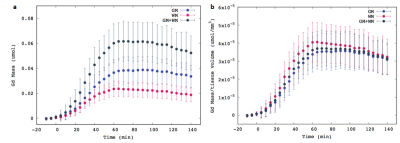 |
Gd-DOTA mass distribution from the Glymphatic Pathway into rat brain measured by 3D DCE-MRat 9.4T
Xiaodan Liu, Sunil Koundal, Simon Sanggaard, Helene Benveniste, Hedok Lee
The recently discovered the "glymphatic pathway" for brain waste clearance, comprises a brain-wide peri-vascular transit passageway for facilitating CSF-ISF ‘cross-talk’, in an AQP4 water channel driven manner. In this study, we report a method to calculate time-resolved 3D Gd-DOTA mass distribution maps to depict the glymphatic transport. We detected higher mass retention in GM compared to WM, due to the tissue volume differences. High Gd uptake in the cerebellum coincides with the high expression of AQP4 water channels in this area. These findings emphasize the importance of the anatomical glymphatic network with high Gd uptake capacity may be related with the distribution and functionality of AQP4.
|
08:51
 |
0749.
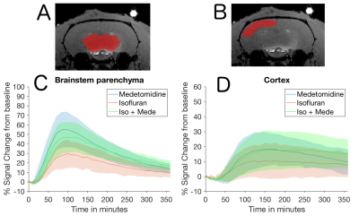 |
Brain state and CSF production define glymphatic washout in the rat brain
Martin Segeroth, Lydia Wachsmuth, Franziska Albers, Cornelius Faber
The Glymphatic System, which is essential for brain-wide waste clearance, is enhanced during sleep. Here, we investigate the effect of brain state and CSF formation using constrast enhanced MRI in a rat model. Our data support the hypothesis that sleep enhances glymphatic clearance due to better CSF intrusion into the brain parenchyma caused by higher CSF formation and smaller arteries. Higher diffusion in cortex associated with altered brain state during sleep influence CSF distribution within parenchyma.
|
09:03
|
0750.
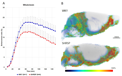 |
Impaired glymphatic transport and loss of peri-arterial AQP4 expression in spontaneously hypertensive stroke prone rats compared to normotensive WKY controls
sunil koundal, Simon Sanggaard, Yuechuan Xue, Xiaodan Liu, Joanna Wardlaw, Maiken Nedergaard, Hedok Lee , Helene Benveniste
Cerebral small vessel disease (CSVD) is one of the important vascular factor contributing to the cognitive impairment and dementia. Clinically, CSVD hallmarks includes MR white matter hyperintensities and dilated perivascular spaces. Brain-wide perivascular transit passageways for CSF, also known as Glymphatic system has recently been described as cerebral metabolic waste clearance pathway. We evaluated Glymphatic transport by DCE-MRI in middle aged spontaneously hypertensive stroke prone (SHRSP) rats and normal Wistar Kyoto (WKY) rats, which demonstrated significant impairment of Glymphatic transport in brain parenchyma in 7-month old SHRSP rats in comparison to controls.
|
09:15
|
0751.
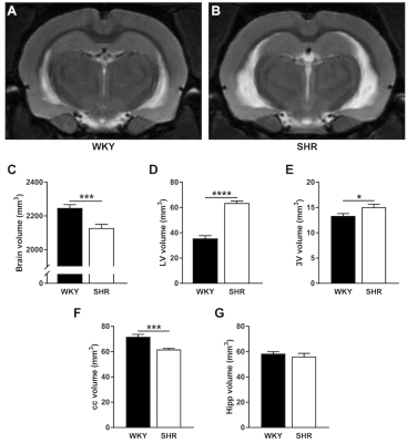 |
Elucidating brain water management in hypertension: a preclinical MRI study
Daphne Naessens, Bram Coolen, Judith de Vos, Ed van Bavel, Gustav Strijkers, Erik Bakker
Hypertension has been associated with alterations in vascular function and an imbalanced water management of brain tissue, yet the underlying mechanisms for these pathologies remain unclear. To further elucidate possible mechanisms of the fluid homeostasis in the brain, we performed non-invasive MR imaging on normotensive and spontaneous hypertensive rats. We assessed total brain and regional brain volumes, as well as diffusional and water exchange properties of the brain tissue.
|
09:27
|
0752.
 |
Sleep-related changes in diffusivity overnight: a window into glymphatic activity in humans?
Ruth O'Gorman Tuura, Carina Volk, Valeria Jaramillo, Reto Huber
This study examined overnight changes in diffusivity as a proxy marker for clearance in the glymphatic system, a recently proposed pathway for waste clearance in the brain. In 18 healthy adults, the mean diffusivity increased overnight in multiple brain regions, consistent with the hypothesised expansion of the extracellular space during sleep. In contrast, the diffusivity within CSF decreased overnight, possibly due to temperature effects. The overnight reduction in CSF diffusivity did not correlate with sleep parameters, but the overnight increase in diffusivity in the brain was positively correlated with the percentage of time spent in REM sleep, assessed with EEG.
|
09:39
|
0753.
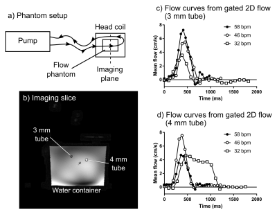 |
Accuracy of real-time quantitative flow imaging in small diameters using compressed sensing at 7T: a phantom study
Johannes Töger, Mads Andersen, Karin Markenroth Bloch
Recent studies have shown that cerebrospinal fluid (CSF) flow is strongly affected by respiration, which may potentially be used for future diagnosis and treatment follow-up in diseases such as normal pressure hydrocephalus and congenital malformations. However, quantitative measurements of respiratory effects on CSF flow in small diameters are not currently available. Therefore, this abstract shows a phantom validation of flow measurement using a radial golden-angle real-time flow sequence, reconstructed using compressed sensing. Results show that mean velocities can be quantified with a small underestimation, suggesting that the protocol is promising for future study of respiratory effects on CSF flow.
|
09:51
|
0754.
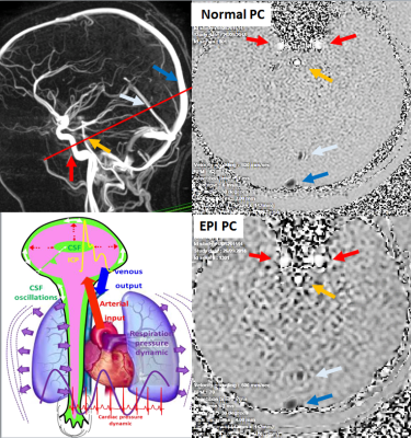 |
Real-time phase contrast magnetic resonance imaging for assessment of cerebral haemodynamics during breathing.
Olivier Balédent, Pan Liu, Armelle Lokossou, Fall Sidy, Serge Metanbou, Malek Makki
To investigate cerebral blood flows by EPI Phase Contrast (EPI-PC) and Normal Phase contrast sequences, in volunteers. Cerebral arterial and venous blood flows were calculated by homemade software. Arterial cerebral blood flows measured by EPI-PC were significantly higher than those measured by normal PC. Whereas venous cerebral blood flows were not different between the two technics. Post processing of continuous EPI-PC flow signal provides blood flows curves during inspiration and expiration. Blood flows during inspiration were respectively higher than during expiration. EPI-PC flow quantify arterial and venous intracranial blood flows in few seconds without any cardiac or respiratory gating.
|
10:03
|
0755.
 |
Macroscopic Hyperdynamic CSF Flow and CSF Volume Analysis in Children and Adolescents with Congenital Heart Disease
Vincent Lee, William Reynolds, Julia Wallace, Nancy Beluk, Ashok Panigrahy
In this study we investigate whether there are differences in CSF volumes between CHDs and healthy controls, as well as examining any correlation between CSF volume and CSF flow, and whether this relationship is altered in CHDs. We showed that certain CSF volumes are elevated in CHD. We also observed that increased flow metrics are correlated with both increased volume in some CSF compartments and decreased volume in other CSF compartments. Lastly, there maybe interplay between CSF flow and volume within CHDs.
|
|
