Advances in Flow Imaging
Oral
Cardiovascular
Monday, 13 May 2019
| Room 516C-E |
08:15 - 10:15 |
Moderators: Michael Hope, Ruth Lim |
08:15
 |
0086.
 |
Acute effects of nicotine-free e-cigarette aerosol inhalation on vascular function detected by multi-parametric quantitative MRI

Alessandra Caporale, Michael Langham, Shampa Chatterjee, Alyssa Johncola, Felix Wehrli
Electronic cigarette (e-cig) vaping has been causally associated with arterial stiffening, oxidative stress, and impaired vasodilatory capacity. Here we investigated acute effects of nicotine-free e-cig vaping in terms of aortic pulse-wave velocity, a marker of arterial stiffness, and measures of peripheral and neurovascular reactivity to induced hypoxia, by means of a quantitative MRI protocol. Flow mediated dilation, a marker of endothelial function, was significantly impaired after vaping; moreover, the peripheral vascular response to cuff-induced ischemia and neurovascular reactivity were altered. Considering the increasing use of e-cig among youth, these results underscore the urgency of further investigation.
|
08:27
 |
0087.
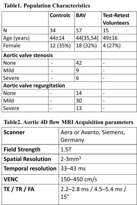 |
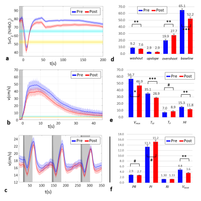
Stochastic Flow Co-expression Signatures: A novel concept for volumetric 4D flow assessment with application to aortic valve disease
Mohammed S.M. Elbaz, Michael Scott, Alex Barker, Patrick McCarthy, Chris Malaisrie, Jeremy Collins, Robert Bonow, James Carr, Michael Markl
Studies have shown an impact of aortic valve disease, as bicuspid aortic valve (BAV), on altered aortic blood flow. Nevertheless, aortic flow changes can be complex making objective visual assessment a challenging task. Existing quantitative flow metrics are useful, but each reflects only partial components of the overall complex flow changes. Here we propose a novel concept that uniquely captures the signature of normal and altered volumetric aortic flow changes derived from 4D Flow MRI. We demonstrated the high reproducibility and the feasibility of the derived flow signature in capturing distinctly altered flow signatures in the aorta of BAV patients.
|
08:39
|
0088.
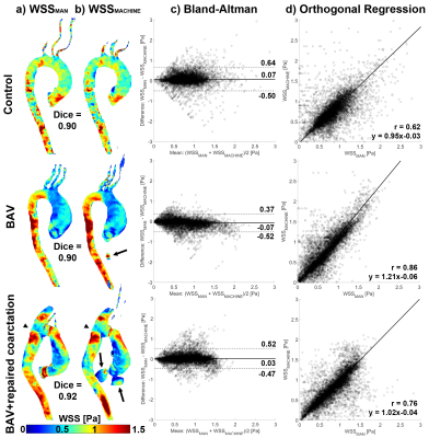 |
Machine learning for automatic three-dimensional segmentation of the aorta in 4D flow MRI
Martijn Froeling, Emile Farag, R. Nils Planken, Tim Leiner, Pim van Ooij
In this study we present a machine learning convolutional neural network (CNN) for automatic segmentation of the aorta used for peak systolic wall shear stress (WSS) assessment from 4D flow MRI data. The automated three-dimensional WSS profiles (WSSMACHINE) were compared with WSS calculated using manually (WSSMAN) created segmentations. Bland-Altman and orthogonal regression analysis revealed good agreement between WSSMAN and WSSMACHINE in terms of small mean differences and slopes and intercepts close to unity and zero respectively. The CNN has the ability to drastically accelerate aortic segmentation from 4D flow MRI data, which will greatly improve the clinical applicability of WSS.
|
08:51
|
0089.
 |
5D Flow Tensor MRI for Mapping Reynolds Stresses in the Aorta
Jonas Walheim, Hannes Dillinger, Sebastian Kozerke
We present a 5D Flow MRI approach for mapping the Reynolds stress tensor in the in-vivo aorta within 6 minutes. First and second statistical moments of fluctuating velocities are encoded using six different velocity encoding gradient directions embedded in a Cartesian Golden angle undersampling scheme with data-driven motion detection and locally low-rank imaging reconstruction. It is demonstrated that this approach permits a time-efficient assessment of velocity vector fields, turbulent kinetic energy and Reynolds shear stresses of the aorta in-vivo.
|
09:03
|
0090.
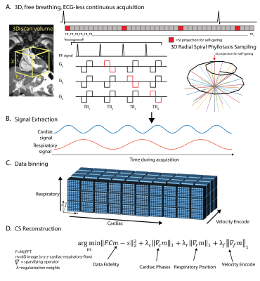 |
5D flow MRI: A free-running, fully self-gated, radial imaging framework for cardiac and respiratory motion-resolved assessment of 3D blood flow dynamics
Liliana Ma, Jérôme Yerly, Christopher Roy, Davide Piccini, Lorenzo Di Sopra, James Carr, Matthias Stuber, Michael Markl
Recent advances have enabled fully self-gated high resolution imaging using a Free-running framework, where data is continuously collected, retrospectively binned into cardiac and respiratory phases, and reconstructed using multi-dimensional compressed sensing (CS) for efficient functional and anatomical imaging of the heart. Here, we propose a novel expansion of this framework to cardiac and respiratory motion-resolved 3D flow imaging— or 5D flow MRI. The findings of this study show that 5D flow MRI is feasible in-vitro and in-vivo and can depict cardiac and respiratory-resolved 3D hemodynamics.
|
09:15
|
0091.
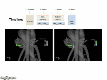 |
Chronological 4D Flow MRI Assessment of Pulmonary Artery Stenosis Stent Treatment
Ryan Pewowaruk, Klarka Mendrisova, Luke Lamers, Chris Francois, Alejandro Roldán-Alzate
Branch pulmonary artery stenosis, a common complication after surgical repair of congenital heart disease, is treated with intravascular stenting. As the acute and chronic effects of stenting are unknown this study uses 4D Flow MRI to serially monitor hemodynamics in a porcine model of pulmonary artery stenosis. Intervention increases flow through the stenosed artery, but not to a normal value. Chronologically, stenosis flow is found to increase immediately after stenting and then remain constant.
|
09:27
|
0092.
 |
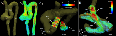
4D Flow MRI Quantification of Congenital Shunts: Comparison to Invasive Catheterization
Daniel Kupsky, Howaida El-Said, Laith Alshawabkeh, Seth Kligerman, Albert Hsiao
When used for quantification of intracardiac and extracardiac shunts, 4D Flow MRI has been shown to have high reproducibility between measurement location and observers, and high consistency with MRI measurements of stroke volume. However, there has been little data showing its relationship to invasive measurements obtained during catheter angiography, which serves as the clinical reference standard at many institutions. We retrospectively evaluated patients who underwent 4D Flow MRI and invasive right heart catheterization during clinical work-up of congenital heart disease. 4D Flow measurements correlated extremely well with invasive measurements of shunt fraction by oximetry and cardiac output by Fick calculation in patients without a shunt.
|
09:39
|
0093.
 |
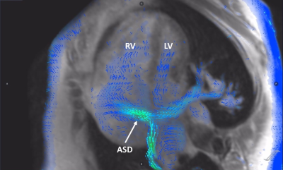
Comprehensive hemodynamic assessment in non-sedated neonates with CHD using motion-robust 3D radial phase-contrast MR
Eric Schrauben, Jessie Lim, Datta Goolaub, Davide Marini, Michael Seed, Christopher Macgowan
The complex hemodynamics in congenital heart disease (CHD) are difficult to visualize and quantify in neonates and young infants. Here we present a novel motion robust and respiratory-resolved acquisition and reconstruction pipeline that addresses the need for rapid, high spatial resolution imaging in these patients. 3D cardiac flow is visualized and quantification comparison with 2D PC measurements is exhibited. This technique opens the door for more comprehensive investigations into the wealth of hemodynamic information not normally considered in surgical planning and follow-up evaluations of CHD.
|
09:51
|
0094.
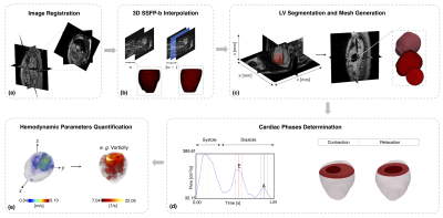 |
Sensitivity Study of 4D flow Left Ventricular Hemodynamics Parameters in Healthy Volunteers and Dilated Cardiomyopathy Patients
Pamela Franco, Julio Sotelo, Bram Ruijsink, David Nordsletten, Eric Kerfoot, Joaquín Mura, Cristian Tejos, Daniel Hurtado, Sergio Uribe
Segmentation of the Left Ventricle (LV) from MRI and 4D flow datasets is an essential step for quantifying clinical indices and hemodynamic parameters. Automatic methods for heart segmentation are generally validated using cardiac volumetric and global ejection fraction. However, little is known about the changes of hemodynamic parameters when subjected to different segmentations. In this study, we present a sensitivity assessment of left intraventricular hemodynamic parameters in healthy volunteers and Dilated Cardiomyopathy (DCM) patients using a finite element quantification approach using 4D flow MRI data, when subjected to changes of LV segmentations.
|
10:03
|
0095.
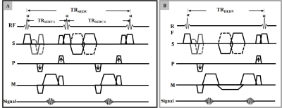 |
Simultaneous Measurement of Myocardial and Blood Flow Velocities using a Dual Echo Dual Velocity Encoding (DEDV) Phase-Contrast MRI (PC-MRI) Approach for Evaluating Left Ventricular (LV) Diastolic Function
Afis AJALA, Jiming Zhang, Erick Buko, Luning Wang, Debra Dees, Janie Swaab, Benjamin Cheong, Pei-Herng Hor, Raja Muthupillai
PC-MRI based Left ventricular (LV) diastolic indices such as E/Em ratio are conventionally estimated from two separate PC acquisitions: One sensitive to myocardium and the other to blood velocities. This ensures optimal velocity to noise ratio and zero velocity aliasing. A rapid single acquisition (dual echo dual VENC PC-MRI method) sensitive to both low and high velocities was developed. Preliminary result on 7 healthy subjects demonstrated a high agreement of LV diastolic indices obtained from the proposed and conventional method.
|
|
