MR Angiography & Vessel-Wall Imaging
Oral
Cardiovascular
Wednesday, 15 May 2019
| Room 516C-E |
15:45 - 17:45 |
Moderators: Kevin Johnson, M. Eline Kooi |
15:45
|
0976.
 |
Sub-millimeter, Non-contrast 3D Coronary MRA for Assessment of Arterial Stenosis: Initial Clinical Experience and Comparison with Coronary CT Angiography

Imran Rashid, Aurelien Bustin, Teresa Correia, Giulia Ginami, Radhouene Neji , Karl Kunze, Tevfik Ismail, Ronak Rajani, Claudia Prieto, Rene Botnar
Coronary CT angiography (CCTA) is an established diagnostic imaging modality for the assessment of patients with suspected coronary disease, where high resolution (sub-millimeter) imaging is required for the accurate detection and quantification of stenosis. To date, achieving such resolutions using coronary MRA (CMRA) has not been possible due to prohibitively long scan times. However, accelerated imaging using image-based navigators and non-rigid motion compensated patch-based undersampled reconstruction techniques have enabled sub-millimeter coronary MR imaging in acceptable acquisition times. In this feasibility study, we demonstrate that accelerated sub-millimeter CMRA holds promise for the detection and exclusion of significant coronary artery stenosis.
|
15:57
|
0977.
 |
Clinical Evaluation of Cine Fast Interrupted Steady-State (FISS) Arterial Spin Labeling for Dynamic MR Angiography of the Heart and Great Vessels
Robert Edelman, Emily Aherne, Amit Pursnani, Jianing Pang, Ioannis Koktzoglou
Evaluation of hemodynamic patterns is often an essential component of the cardiac MRI exam. We hypothesized that cine fast interrupted steady-state (FISS) ASL could prove advantageous for demonstrating flow patterns in the heart and great vessels and tested this technique in 19 patients undergoing cardiac MRI for standard indications. We found that cine FISS ASL is a robust, efficient imaging technique that is easily incorporated into cardiac MRI protocols and shows promise for depicting abnormal blood flow patterns in patients with a variety of cardiovascular disorders, including aortic stenosis, hypertrophic cardiomyopathy, and congenital shunts.
|
16:09
 |
0978.
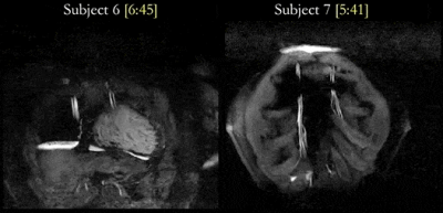 |
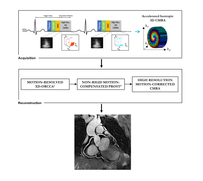
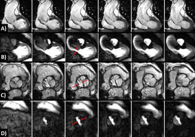
Highly Accelerated 3D Whole-Heart Isotropic Sub-Millimeter CMRA with Non-Rigid Motion Correction
Aurelien Bustin, Teresa Correia, Imran Rashid, Gastao Cruz, Radhouene Neji, René Botnar, Claudia Prieto
Whole-heart sub-millimeter isotropic coronary magnetic resonance angiography (CMRA) provides detailed information of the coronary arteries and surrounding vessels. Recently, a patch-based reconstruction technique (3D PROST) has been proposed to achieve sub-millimeter isotropic resolution CMRA in a predictable scan time. However, this approach only corrects for 2D translational respiratory motion of the heart and image quality can be affected by residual non-rigid motion. Here we propose to integrate 3D PROST into a highly accelerated non-rigid motion correction framework to achieve high quality whole-heart free-breathing isotropic sub-millimeter Cartesian CMRA in a clinically feasible scan time. The feasibility of the proposed method was tested in seven healthy subjects and two patients with suspected coronary artery disease.
|
16:21
 |
0979.
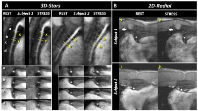 |
“3D-Stars” Cine MRI for the Coronary Arteries: Feasibility of Volumetric Endothelial Function Assessment
Gabriele Bonanno, Robert G Weiss, Davide Piccini, Jérôme Yerly, Sahar Soleimanifard, Li Pan, Xiaoming Bi, Allison G Hays, Matthias Stuber, Michael Schär
2D coronary cine MRI has been shown to noninvasively assess coronary endothelial dysfunction, which is an early manifestation of atherosclerosis and a predictor of future acute events. However, atherosclerosis is a diffuse process and a 2D approach can only provide local measures. Recently, we introduced “3D-Stars”, a free-breathing golden-angle 3D stack-of-stars cine sequence with isotropic spatial resolution to image the proximal and mid segments of the right coronary artery. Here, we show excellent image quality at rest and during isometric-handgrip stress acquisitions and feasibility for assessing normal coronary endothelial function along the vessel in a small cohort of healthy subjects.
|
16:33
|
0980.
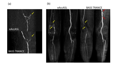 |
A comparison of Enhanced Acceleration-Selective Arterial Spin Labeling (eAccASL) and Background Suppressed Single shot TFE-TRANCE (BASS TRANCE) for the peripheral arteries
Natsuo Konta, Shuhei Shibukawa, Makoto Obara, Yuta Akamine, Tomohiko Horie, Takashi Okazaki, Yui Nagafuji, Tetsu Niwa, Yutaka Imai
We evaluated the peripheral MR angiography (MRA) of 8 healthy volunteers using conventional and enhanced acceleration-selective arterial spin labeling (cAccASL, eAccASL), and Background suppressed single shot TFE-TRANCE (BASS TRANCE). In eAccASL, additional 180°refocusing pulses and motion sensitized gradients (MSGs) will compensate for B1 inhomogeneity and eddy current effects at 3.0 Tesla. Contrast ratios for BASS TRANCE was the highest values, compared to cAccASL and eAccASL. However, qualitative assessment showed eAccASL could provide more robust visualization than cAccASL and similar visualization to BASS TRANCE. Therefore, eAccASL can provide the more robust peripheral MRA without any gating.
|
16:45
|
0981.
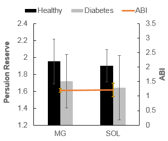 |
Contrast Free Methods for Vascular Assessment in Lower Extremities of Diabetes
Jie Zheng, Yongsheng Chen, Sara Gharabaghi, Mark Haacke, Ran Li, Masoud Edalati, Mary Hastings, Mohamed Zayed
The purpose of this study is to develop a new contrast-free MR angiography technique without using any ECG triggering for the vascular assessment of lower extremities in patients with diabetes. Healthy volunteers and diabetes underwent both MR angiography and skeletal muscle perfusion imaging at rest and during an isometric contraction exercise. The angiography, perfusion, and even possible calcification of vessels can be assessed in one imaging session in patients with diabetes.
|
16:57
|
0982.
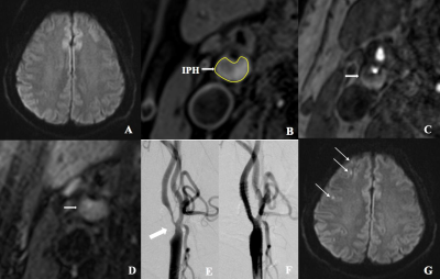 |
Associations between carotid intraplaque hemorrhage and new ipsilateral ischemic lesions after carotid artery stenting: A quantitative study with multi-contrast MRI
Did Not Present
Jiang Lin, Aihua Ji, Peng Lv
The risk of cerebral embolism after carotid artery stenting (CAS) in patients with carotid intraplaque hemorrhage (IPH) is still controversial. This study further investigated the relationship between IPH and new ipsilateral ischemic lesions (NIIL) after CAS, and performed a volumetric MRI analysis of IPH for predicting the risk of NIIL following CAS. We confirm that carotid IPH is associated with the incidence of NIIL following CAS. Quantification of IPH volume with MRI can be useful for predicting the risk of NIIL after CAS.
|
17:09
|
0983.
 |
Automatic AHA Classification of Carotid Atherosclerotic Lesions in Multicontrast MR Images using Deep Learning
Jifan Li, Shuo Chen, Xihai Zhao, Yuan Chun, Rui Li
In this study, we aimed to develop a convolutional neural network (CNN) to classify carotid atherosclerotic lesions in high-resolution multicontrast MR images automatically using the modified American Heart Association (AHA) classification scheme as criteria. The network was trained on a large number of plaque images combined with lesion type labeled by experienced radiologists. Transfer learning was utilized to take the advantage of state-of-the-art CNN pre-trained on ImageNet dataset. The accuracy of lesion type classification achieved 85.1% with preprocessing and fine-tuning of the network.
|
17:21
|
0984
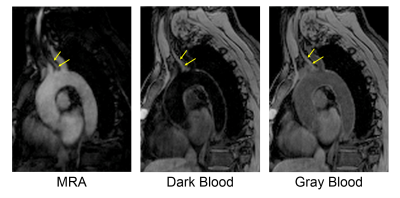 |
ECG- and Navigator-Free 3D Multi-Contrast Aortic Vessel Imaging with MR Multitasking
Video Permission Withheld
Jaime Shaw, Anthony Christodoulou, Zhehao Hu, Shlee Song, Marcel Maya, Bin Sun, Xiaoming Bi, Fei Han, Debiao Li, Zhaoyang Fan
MR imaging has the potential to provide a comprehensive assessment of atherosclerotic disease of the aortic arch with both luminal and vessel wall imaging approaches, but typically have long scan times. MR Multitasking is useful for expediting acquisitions that have otherwise been complicated by navigator gating or ECG triggering. In this work, we present an MR multitasking technique that simultaneously produces MRA and multi-contrast vessel wall images with a single ECG- and navigator-free 3D acquisition. Four healthy control subjects and four patients with neurovascular disorders and suspected aortic atherosclerosis were scanned. The 3D, free-breathing, non-ECG sequence provided images at time points for MRA, dark blood, and gray blood contrasts.
|
17:33
|
0985.
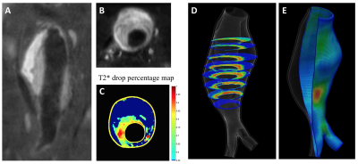 |
MRI-based Analysis of Morphologic, Inflammatory, and Mechanical Stress Factors that Govern Abdominal Aortic Aneurysm Progression Rates
Chengcheng Zhu, Joseph Leach, David Saloner, Michael Hope
Clinical management of abdominal aortic aneurysm (AAA) disease is based on the maximal aneurysm diameter. Novel markers including vascular inflammation, intraluminal thrombus (ILT) composition and mechanical vessel wall stress have been correlated to AAA risk. Prior studies have typically focused on a single marker. We have developed a comprehensive AAA assessment based on high-resolution black-blood MRI and followed 41 patients for 1.9±0.6 years. Our results showed that both active ILT change and inflammation (identified by USPIO uptake) were strongly associated with AAA growth and/or intervention, and wall stress varied in a sub-group of patients. Such comprehensive assessment may improve AAA patient risk stratification.
|
|
