Myocardial Perfusion
Oral
Cardiovascular
Thursday, 16 May 2019
| Room 516C-E |
16:00 - 18:00 |
Moderators: Merlin Fair, Liang Zhong |
16:00
 |
1229.
 |
Initial Clinical Evaluation of Quantitative Ultra-high Resolution First-pass Spiral Perfusion Imaging with Whole Heart Coverage in Patients with Ischemic Heart Disease at 3T

Yang Yang, Austin Robinson, Roshin Mathew, Christopher Kramer, Michael Salerno
First-pass contrast-enhanced myocardial perfusion imaging is a useful noninvasive tool to evaluate patients with coronary artery disease, but current techniques are still limited in spatial-temporal resolution, ventricular coverage and absolute quantification which reduces the sensitivity to detect perfusion differences between the endocardium and epicardium and quantify ischemic burden. In this study, we designed a dual-density dual-contrast spiral pulse sequence to achieve quantitative ultra-high resolution first-pass spiral perfusion imaging with whole heart coverage at 3T and further tested in 9 patients with suspected CAD undergoing cardiac catheterization.
|
16:12
|
1230.
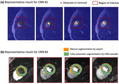 |
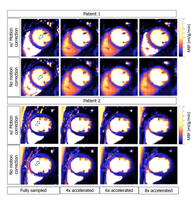
Rapid Automatic Quantification of Myocardial Blood Flow in Free-breathing Myocardial Perfusion MRI without the Need for Motion Correction: A Novel Spatio-temporal Deep Learning Approach
Zulma Sandoval, John Van Dyke, Prateek Malhotra, Rohan Dharmakumar, Behzad Sharif
It can be argued that the most significant technical impediment for wider clinical adoption of fully-quantitative cardiac perfusion MRI is the lack of a fully-automatic post-processing workflow across all scanner platforms. In this work, we present an initial proof-of-concept based on a deep-learning approach for quantification of myocardial blood flow that eliminates the need for motion correction, hence enabling a rapid and platform-independent post-processing framework. This is achieved by optimizing/training a cascade of deep convolutional neural networks to learn the common spatio-temporal features in a dynamic perfusion image series and use it to jointly detect the myocardial contours across all dynamic frames in the dataset.
|
16:24
|
1231.
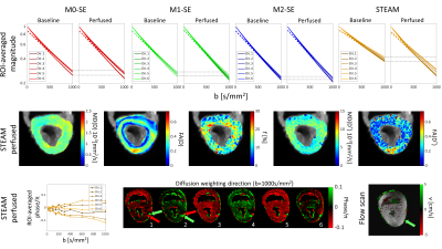 |
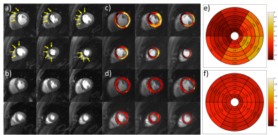
On Probing Intravoxel Incoherent Motion in the Heart using Spin Echo versus Stimulated Echo Diffusion Weighted Imaging
Georg Spinner, Christian Stoeck, Linda Mathez, Constantin von Deuster, Christian Federau, Sebastian Kozerke
Intravoxel Incoherent Motion (IVIM) imaging in the heart remains challenging resulting in large variation of the perfusion surrogates in practice. In the present work, IVIM sensitivity of standard and motion-compensated Spin Echo (SE) and Stimulated Echo Acquisition Mode (STEAM) diffusion-weighted imaging approaches are analyzed using Monte Carlo simulations and perfused porcine heart experiments. An extended IVIM model is proposed to account for the microstructural properties of the myocardium. It is demonstrated that motion-compensated SE sequences provide insufficient perfusion sensitivity. In contrast, STEAM allows delineating hypoperfused myocardium using experimental imaging data.
|
16:36
|
1232.
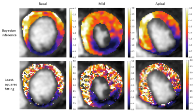 |
Hierarchical Bayesian myocardial perfusion quantification
Cian Scannell, Amedeo Chiribiri, Adriana Villa, Marcel Breeuwer, Jack Lee
Myocardial perfusion can be quantified from dynamic contrast-enhanced MRI. This facilitates a non-invasive, automated, fast and user-independent evaluation of myocardial blood flow. However, due to the relatively low SNR, low temporal resolution and short scanning time the model fitting can yield unreliable parameter estimates. To counter-act this, simplified models and segmental averaging are used. In this work, Bayesian inference is employed. The inclusion of both spatial prior knowledge and prior knowledge of the kinetic parameters improves the reliability of the parameter estimation. This allows the generation of accurate high-resolution voxel-wise quantitative perfusion maps that clearly delineate areas of ischaemia.
|
16:48
|
1233.
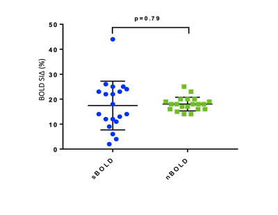 |
Improving the accuracy of Blood Oxygen Level Dependent Imaging using FLASH normalised T2 prepared SSFP acquisition
Betty Raman, Kenneth Chan , Rina Ariga , Moritz Hundertmark, Masliza Mahmod , Stefan Neubauer , Aaron Hess, Elizabeth Tunnicliffe
T2-prepared steady state free precession (SSFP) Blood Oxygen Level Dependent (BOLD) imaging is a promising technique for ischaemia detection but has wide normal ranges, in part because the signal intensity is heart-rate dependent. Fast low angle shot (FLASH) readouts can be interleaved with the SSFP readouts to normalise SSFP images, rendering it heart-rate insensitive. In this study, we examined the variability and reproducibility of standard and FLASH-normalised T2-prepared SSFP BOLD in healthy subjects. FLASH-normalised T2-prepared SSFP BOLD produced a highly reproducible normal BOLD range with an effect size similar to that of the standard method and with considerably reduced variability.
|
17:00
 |
1234.
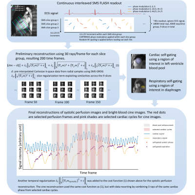 |
Whole heart ungated myocardial perfusion imaging with steady-state radial SMS readout and without magnetization preparation
Ye Tian, Jason Mendes, Brent Wilson, Edward DiBella, Ganesh Adluru
Here we propose a framework for dynamic contrast enhanced myocardial perfusion MRI using an interleaved slice-group radial simultaneous multi-slice acquisition without magnetization preparation or ECG gating. The unique acquisition and reconstruction framework can provide 9 image slices with high spatial resolution cover the whole heart, and have comparable normal-ischemic tissue contrast with saturation recovery prepared radial readout, but is more efficient and robust. Also bright-blood cine images at the same slice positions can be reconstructed that can improve the cardiac MRI efficiency.
|
17:12
|
1235.
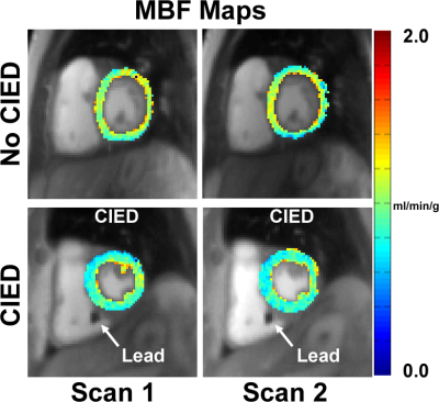 |
Compressed Sensing Accelerated Wideband Myocardial Perfusion Pulse Sequence for Patients with a Cardiac Implantable Electronic Device
KyungPyo Hong, Lexiaozi Fan, Jeremy Collins, Daniel Lee, Daniel Kim
We have recently developed a 2-fold accelerated wideband myocardial perfusion MRI pulse sequence using TGRPPA for imaging patients with a cardiac implantable electronic device (CIED). In patients with rapid heart rates and/or during stress, it may be necessary to further accelerate the perfusion scans to adequately sample the heart. This study describes a 5-fold accelerated wideband myocardial perfusion pulse sequence using compressed sensing and how repeatable it is for quantification of resting myocardial blood flow (MBF) measurement in patients with and without a CIED.
|
17:24
|
1236.
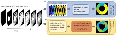 |
High-resolution quantification of myocardial perfusion using spatial correlations
Judith Lehnert, Christoph Kolbitsch, Gerd Wübbeler, Amedeo Chiribiri, Cian Scannell, Tobias Schafffter, Clemens Elster
Pixel-wise quantification of myocardial perfusion by dynamic contrast-enhanced magnetic resonance imaging allows for a non-invasive, observer independent and reproducible evaluation of the perfusion with high spatial resolution. The method suggested here, exploits the spatial smoothness of the perfusion using a spatial Tikhonov regularization to cope with the low signal-to-noise ratio of the data. This allows us to obtain quantitative perfusion values with high spatial resolution to detect small ischemic regions. The parameter regulating the strength of regularization is determined from the L-curve criterion and does not require any manual adjustments. The feasibility of the method is demonstrated in three patients.
|
17:36
|
1237.
 |
Simultaneous 13N-Ammonia and gadolinium first-pass myocardial perfusion with hybrid PET-MR imaging: a clinical feasibility study.
Muhummad Sohaib Nazir, Sarah-May Gould, Xenios Milidonis, Eliana Reyes, Tevfik Ismail, Radhouene Neji, Sébastien Roujol, Jim O'Doherty, Hui Xue, Sally Barrington, Reza Razavi, Paul Marsden, Peter Kellman, Sven Plein, Amedeo Chiribiri
Hybrid PET-MR imaging allows simultaneous measurement of myocardial blood flow (MBF) under identical physiological conditions. We sought to determine feasibility of simultaneous 13N-Ammonia PET and dynamic contrast-enhanced cardiovascular magnetic resonance (CMR) in healthy volunteers. 13N-Ammonia and gadolinium contrast were administered simultaneously during adenosine stress. Mean global stress MBF values for PET and CMR were 2.58 ± 0.11ml/g/min and 2.60 ± 0.47ml/g/min respectively. On a per territory basis, there was a moderate correlation (r = 0.63, p=0.03) and CMR underestimated PET MBF by 0.34ml/g/min (95% limits of agreement -0.49 to +1.18). Future studies in patients with CAD is warranted.
|
17:48
|
1238.
 |
High-Resolution motion-corrected 2D Myocardial Perfusion MRI using Locally Low Rank and Wavelet Sparsity Constraints
Joao Tourais, Torben Schneider, Xenios Milidonis, David Higgins, Javier Sanchez-Gonzalez, Gastao Cruz, Claudia Prieto, Chris Saunderson, Louise Brown, Sven Plein, Amedeo Chiribiri, Jouke Smink, Teresa Correia
First-pass perfusion cardiac MR (FPP-CMR) allows the assessment of ischemic heart disease. However, conventional FPP-CMR has limited spatial resolution and requires breath-holding. Moreover, diagnostic accuracy may be compromised due to dark-rim artifacts. Here, we propose a free-breathing quantitative high-resolution FPP-CMR framework that combines dynamically variable undersampling with a motion-corrected reconstruction approach that uses spatial and temporal constraints. The proposed motion-corrected strategy improves image sharpness and quantification of myocardial blood flow from free-breathing acquisitions. The highly undersampled acquisitions allow short acquisition windows and facilitate higher spatial resolution images, which are less sensitive to dark-rim artifacts, thus improving the diagnostic accuracy of FPP-CMR.
|
|
