Preclinical Exploratory Body Methods
Oral
Body: Breast, Chest, Abdomen, Pelvis
Thursday, 16 May 2019
| Room 513D-F |
16:00 - 18:00 |
Moderators: Lindsey Crowe, Sabrina Ronen |
16:00
|
1219.
 |
Quantitative 129Xe MRI detects early impairment of gas-exchange in a rat model of pulmonary arterial hypertension

Rohan Virgincar, John Nouls, Simone Degan, Xinyu Xiong, Ziyi Wang, Yi Qi, Sudarshan Rajagopal, Bastiaan Driehuys
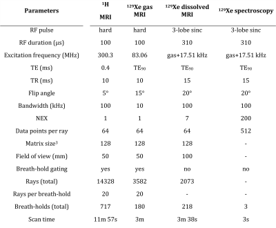
Hyperpolarized 129Xe MRI is capable of regional mapping of gas-exchange and has found application in a wide range of lung disorders. Here, we apply 129Xe gas exchange MRI and dynamic spectroscopy at 7 Tesla to study a rat model of pulmonary arterial hypertension. 129Xe spectroscopy and gas-exchange imaging showed reduced uptake by RBCs early in the progression of the disease, and at later time points was accompanied with increased uptake by barrier tissues, structural abnormalities, edema, and ventilation defects. Imaging results were compared to H&E histology, which showed evidence of vascular remodeling.
|
16:12
|
1220.
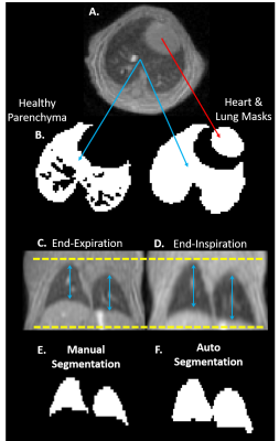 |
Retrospectively Gated UTE MRI with Deep-Learning Segmentation to Quantify Preclinical Lung Fibrosis Severity
Ian Stecker, Sneha Sitaraman, Matt Freeman, Jinbang Guo, Emily Martin, Chase Hall, Tim Weaver, Zackary Cleveland
Fibrosis contributes to morbidity and mortality in an array of pulmonary diseases, and no therapies exist to halt or reverse its progression. This paucity of treatment strategies results from poorly understood etiology, making it essential to develop animal models that fully mimic human disease and identify noninvasive, quantitative markers for fibrosis severity for use in animal models and patients. Here we report an image reconstruction and analysis pipeline, combining retrospectively gated ultrashort echo time (UTE) MRI and deep-learning segmentation to quantify lung fibrosis progression in Surfactant Protein C (SP-C) knock-in models based on mutations that predispose humans to lung fibrosis.
|
16:24
 |
1221.
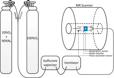 |
Dynamic BOLD Responses of Renal Tissues to interleaved Hypoxia and Hyperoxia in Healthy Rats as Measured by MRI at 7T
Kaixuan Zhao, Yingjie Mei, Guixiang Yang, Zhengce Wang, Lili Zhou, Ed X. Wu, Yanqiu Feng
Renal oxygen status is tightly correlated with renal (patho)physiology. How the renal oxygen is regulated in response to interleaved hypoxia and hyperoxia has not been well investigated. In this work we utilized dynamic BOLD MRI with temporal resolution of 9 seconds to track the renal tissues oxygen responses in healthy rats, thereafter, characterizing the renal oxygen changes by a mathematic model. In the experiment, significant T2* overshot was observed in outer medulla at the initial stage of re-oxygenation. Significant difference of model parameters compared among renal cortex, outer medulla and inner medulla was observed.
|
16:36
|
1222.
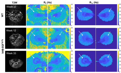 |
Spin-lock Relaxation Rate Dispersion Reveals Spatiotemporal Changes Associated with Tubulointerstitial Fibrosis in Murine Kidney
Feng Wang, Daniel Colvin, Suwan Wang, Hua Li, Raymond Harris, Ming-Zhi Zhang, John Gore
Currently there are no reliable non-invasive means for assessing the severity and progression of fibrosis in kidneys. Here we evaluate spin-lock MR imaging with different locking fields for detecting progressive renal fibrosis in an hHB-EGFTg/Tg mouse model. We fit the dispersion of spin-lock relaxation rates R1ρ at different locking fields (frequencies) to a model of exchanging water pools and assessed the value of the derived quantities for detecting tubulointerstitial fibrosis in kidney, including the transverse relaxation rate (R2), the asymptotic value of R1ρ at high locking frequency (R1ρinf), an exchange rate-weighted parameter Sρ, and the inflection frequencies ωinfl in the dispersion.
|
16:48
 |
1223.
 |
Magnetization Transfer Imaging is Insensitive to Decrease in Renal Perfusion: A Graded Ischemia Study on Swine
Kai Jiang, Christopher Ferguson, John Woollard, James Krier, Xiangyang Zhu, Lilach Lerman
The impact of renal perfusion on magnetization transfer imaging (MTI) was evaluated on seven pigs that underwent consecutive MRI at baseline, 50%, 75%, and 100% acute renal artery stenosis as well as reperfusion. Renal perfusion, R2*, apparent diffusion coefficient (ADC), stiffness, and MT ratio (MTR) were measured. During graded ischemia, decline in renal perfusion was accompanied with elevated R2*, decreased ADC and stiffness. In contrast, no change was observed in the MTR. In conclusion, MTI is insensitive to decrease in renal perfusion and may offer a relatively reliable technique for measurement of renal structural changes.
|
17:00
|
1224.
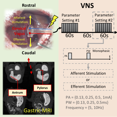 |
Contrast-enhanced MRI reveals differential effects of afferent and efferent vagus nerve stimulation on gastric motility
Kun-Han Lu, Jiayue Cao, Robert Phillips, Terry Powley, Zhongming Liu
We used in vivo contrast-enhanced MRI to evaluate how vagus nerve stimulation (VNS) could modulate and coordinate the motility of the antrum and pylorus. We tested cervical VNS with different settings (afferent or efferent) and parameters (amplitude, duration, and frequency) and found differential effects on the motility. Our results suggest that selective stimulation of vagal afferent using a monophasic pulse train is a more effective strategy to facilitate and coordinate the antro-pyloric motility than selective stimulation of the vagal efferent. These results could shed lights on the selection of VNS settings for modulating gastric functions.
|
17:12
|
1225.
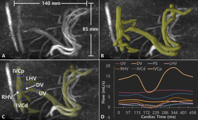 |
Fetal Hepatic Flow in Sheep Assessed using 4D flow MRI
Eric Schrauben, Brahmdeep Saini, Jack Darby, Stacey Holman, Jia Yin Soo, Mitchell Lock, Sunthara Perumal, Michael Seed, Janna Morrison, Christopher Macgowan
Assessment of fetal hepatic flow poses difficult technical problems due to the small nature of vessels, slow blood flow within the liver, and sources of motion present in utero. Here, 4D flow MRI is coupled with specialized animal preparation to capture 3D fetal hepatic hemodynamics in a late gestation sheep model of human pregnancy. Full vasculature segmentation, fetal liver shunt visualization, and comprehensive measurement of hepatic circulation is demonstrated.
|
17:24
|
1226.
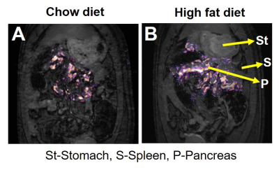 |
Metabolic Imaging of Pancreas in a High Fat Diet Fed Obese Rodent Model
Jadegoud Yaligar, Sanjay Kumar Verma, Rengaraj Anantharaj, Venkatesh Gopalan, Giang Le Thi Thu, Kavita Kaur, S Sendhil Velan
Onset of hyperglycemia and lipotoxicity are associated with reduced β-cell mass and function compromising insulin secretion leading to insulin resistance. It is technically challenging to image pancreas in rodents by conventional imaging approaches. In this study we have utilized GdDOTA-diBPEN to image the pancreas in control and high fat diet (HFD) fed rat model. HFD fed rodent model shows increase in pancreatic fat and functional βcell volume during compensatory phase which is due to the adaptive response to HFD. Longitudinal assessment of pancreatic fat accumulation and βcell mass and function will provide better insights associated with β-cell dysfunction.
|
17:36
|
1227.
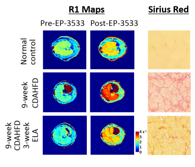 |
Multi-Parametric MRI of Liver Fibrosis and Treatment Response in a Rat Model of Nonalcoholic Steatohepatitis (NASH)
Iris Zhou, Veronica Clavijo Jordan, Nicholas Rotile, Smitha Krishnan, Hema Krishnan, Gunisha Arora, Hannah Slattery, Noah Warner, Christian Farrar, Bryan Fuchs, Peter Caravan
Noninvasive MR quantification of matrix using a collagen-binding molecular MR probe and tissue stiffness by MR elastography were used to assess liver fibrosis and the anti-fibrotic effects of elafibranor or diet change in a rat model of nonalcoholic steatohepatitis (NASH). Reduction of fibrosis after diet change or elafibranor treatment were detected by EP-3533 collagen imaging and stiffness measurements and correlated with morphometricassessment of fibrosis from histology and with hydroxyproline content as a biochemical surrogate for total liver collagen. Multi-parametric MRI can be used to characterize disease progression and serve as sensitive tool for evaluating treatment response.
|
17:48
|
1228.
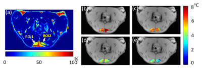 |
Magnetic resonance temperature imaging in activated brown adipose tissue of rat
Chuanli Cheng, Chao Zou, Qian Wan, Yangzi Qiao, Changjun Tie, Min Pan, Xin Liu, Hairong Zheng
Brown adipose tissue (BAT) is a special adipose tissue which burns fat and dissipates energy in heat. As a result, it is considered to be the next potential therapeutic target for treating obesity and other metabolic diseases. The ability of heat dissipation of BAT is thought to be highly related to the BAT activity. In this study, we intended to use MR thermometry to map the temperature change distribution in BAT after activated by Norepinephrine. The preliminary results show the PRFS based thermometry can observe the temperature change in BAT after activation, implying that MR thermometry might be a useful tool to characterize the BAT activity.
|
|
