Liver
Oral
Body: Breast, Chest, Abdomen, Pelvis
Thursday, 16 May 2019
| Room 518A-C |
13:45 - 15:45 |
Moderators: Victoria Chernyak, Jeong-Hee Yoon |
13:45
 |
1155.
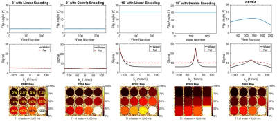 |
Motion-Robust, High-SNR Fat Quantification using a Variable Flip Angle Approach

Ruiyang Zhao, Yuxin Zhang, Xiaoke Wang, Kevin Johnson, Scott Reeder, Diego Hernando
Current chemical shift encoded (CSE)-MRI techniques enable quantification of liver fat using 3D breath-hold methods. However, many patients are unable to sustain breath-holds, leading to motion artifacts that cause variability and bias in fat quantification. A recently developed 2D sequential CSE-MRI technique is motion-robust with short temporal footprint but suffers from low SNR due to the small flip angles used to avoid T1 bias. To overcome this limitation, we propose a novel centric encoding variable flip angle (CEVFA) technique that minimizes T1 bias in fat quantification while achieving higher SNR performance and validate this technique in phantoms and in-vivo scans.
|
13:57
|
1156.
 |
Multimodal MRI Combined with Liver Volume for Quantitative Assessment of Liver Function in Patients With Cirrhosis
Chenxia Li, Haitian Liu, Xiang Li, Rong Wang, Jian Yang, Yuelang Zhang
This study aim to combine multimodal MRI with liver volume for quantitative assessment of liver function in patients with cirrhosis. Eighty-five subjects underwent GD-EOB-DTPA enhanced MR and IVIM-DWI.Through statistical analysis, RE, D* and Vliver/Vspleen from modeling group were selected as optimal indicators. Using multiple regression to establish a multi-parametric model for liver function assessing: F(x). The ROC analysis of the validation group showed that it had an AUC of 0.973 in distinguishing Child-Pugh A group and Child-Pugh B and C group. The results showed it could evaluate the overall liver function and expected to be a method for preoperative local liver function assessment.
|
14:09
 |
1157.
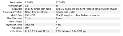 |
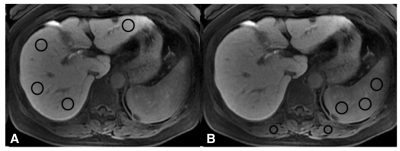
Improved robustness of R2* mapping using spin-density projection for liver iron quantification at 3 Tesla
Xiaozhou Liu, MD, Ivan Pedrosa, MD PhD, Diego Hernando, PhD, Scott Reeder, MD PhD, Takeshi Yokoo, MD PhD
In patients with iron overload, liver iron concentration can be measured by R2- or R2*-MRI. While gradient-recalled-echo (GRE) based R2*-MRI holds practical advantages over spin-echo based R2-MRI, the R2* technique is more challenging in patients with severe iron overload with rapid signal decay. We introduce a technique known as spin density projection (SDP), which uses liver-to-subcutaneous fat spin density ratios as an internal reference to improve robustness of R2* fitting. We performed a computer simulation and retrospective analysis of GRE imaging data in 45 patients with suspected iron overload and compared R2* fitting without and with SPD. We found that SPD increased robustness by reducing standard deviation of R2* estimation.
|
14:21
|
1158.
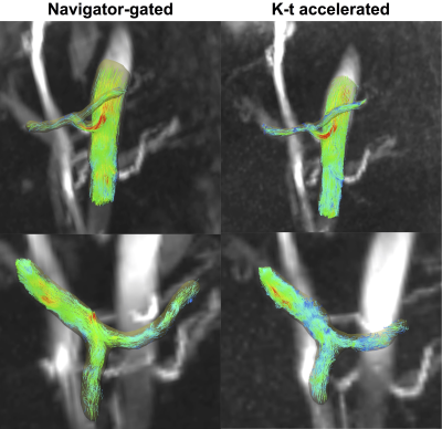 |
Flow quantification in portal hypertension with 4D flow: comparison of a free breathing k-t accelerated sequence to a respiratory-navigated sequence
Octavia Bane, Daniel Stocker, Paul Kennedy, Stefanie Hectors, Scott Friedman, Thomas Schiano, Maria Isabel Fiel, Swan Thung, Emilie Bollache, Susanne Schnell, Aaron Fischman, Michael Markl, Bachir Taouli
The purpose of our prospective study was to compare a free-breathing k-t GRAPPA accelerated 4D flow sequence with a standard navigator-gated sequence, and to correlate hemodynamic parameters to the hepatic venous pressure gradient (HVPG) in patients with chronic liver disease. The k-t accelerated sequence had significant (three-fold) reduction in acquisition time, while obtaining equivalent image quality and quantitative parameters in the large vessels as the standard sequence. However, there was systematic under-estimation of velocities in small arteries. Time-average velocity in the superior mesenteric vein measured with the k-t sequence was positively correlated with HVPG.
|
14:33
|
1159.
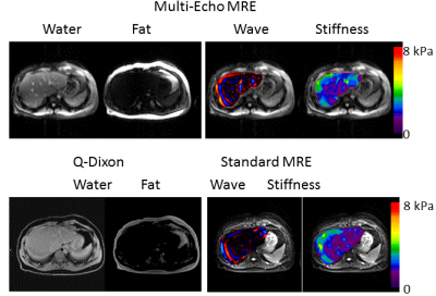 |
Simultaneous Fat, T1 and Stiffness Quantification Using Multi-Echo, Variable Flip Angle, Spoiled-Gradient-Echo, Magnetic Resonance Elastography
Yuan Le, Joshua Trzasko, Kevin Glaser, Yuxiang Zhou, Joseph Hoxworth, Bradley Bolster, Jr., Joel Felmlee, Richard Ehman, Jun Chen
Hepatic fat fraction measurements using Dixon MRI and stiffness measurements using MRE provide important liver fat and fibrosis biomarkers for evaluating liver diseases. We have developed Dixon MR Elastography to measure fat fraction and stiffness in a single acquisition. In addition, by also using variable flip angles this new technique can simultaneously measure stiffness, T1, and fat fraction, thus providing even more clinically useful information in one scan. Promising results were obtained in a fat/water/gel phantom and six healthy volunteers.
|
14:45
|
1160.
 |
Early analysis of hepatic PDFF and liver stiffness results in the Strong Heart Study, investigating possible factors affecting nonalcoholic fatty liver disease in an American Indian population
Walter Henderson, Ying Zhang, Justin Dvorak, Tim Mower, Donovan Beswick, Sanjay Narotam, Alvin Silva, Tanya Wolfson, Danielle Batakis, Ashley Louie, Yesenia Covarrubias, Cynthia West, Tauqeer Ali, Richard Devereux, Jonathan Weinsaft, Jason Umans, Rohit Loomba, Shelley Cole, Claude Sirlin, Michael Middleton
In this analysis of early data from the Strong Heart Study, we show that advanced MRI estimates of proton-density fat fraction (PDFF) and liver stiffness can be acquired successfully in a population of American Indians. Diagnosis of diabetes, and whether participants self-report binge-drinking are collected. Too few cases have been enrolled to permit significance testing, but PDFF values are higher for diagnosed diabetes, and stiffness values are higher for self-reported binge drinking. This study offers a unique opportunity to study the possible roles of diabetes and binge drinking in nonalcoholic fatty liver disease for an at-risk population of American Indians.
|
14:57
|
1161.
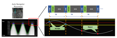 |
Highly Accelerated Prospectively Gated Free Breathing Dynamic Abdominal Imaging with Cartesian Sampling
Janio Szklaruk, Priya Bhosale, Kang Wang, Ty Cashen, Jingfei Ma, Ersin Bayram
Dynamic abdominal imaging has the requirements of high spatial-temporal resolution, large spatial coverage, and acquisition timing with the contrast injection. Golden angle radial sampling in combination with compressed sensing and parallel imaging has been reported for free breathing dynamic volumetric imaging. However, radial sampling suffers from streaking artifacts, inflexible FOV prescription, and long reconstruction time. In this study, we report a Cartesian sampling technique that combines compressed sensing, parallel imaging, temporal view sharing, and navigator for prospective gating. The potential of the technique for free breathing dynamic abdominal imaging is demonstrated in both volunteers and patients.
|
15:09
|
1162.
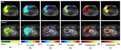 |
In Vivo Multicomponent 3D-T1? Relaxation Mapping of Human Liver under Free-breathing
Azadeh Sharafi, Rahman Baboli, Krishna Shanbhogue, Sonja Olsen, Tobias Block, Hersh Chandarana, Ravinder Regatte
Chronic hepatic disease damages the liver and the subsequent wound-healing process results in liver fibrosis which can ultimately progress to cirrhosis. In this study, we evaluated bi-exponential 3D-T1ρ mapping for in-vivo liver applications under free breathing on a standard clinical 3T scanner, employing combinations of golden-angle radial sampling and parallel imaging. The proposed free-breathing multi-component 3D-T1ρ mapping technique has potential for assessment of liver inflammation and fibrosis, and it could serve as future imaging biomarker for disease-modifying liver-fibrosis therapies.
|
15:21
|
1163.
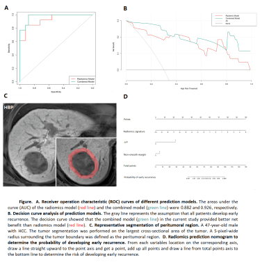 |
Radiomics analysis of peritumoral tissue on gadoxetic acid-enhanced MR imaging for prediction of early recurrence in hepatocellular carcinoma
Zhen Zhang, Song Bin
Postoperative recurrence has become the main obstacle in prognosis of HCC patients, even after successful curative therapy. Our study analyzed the radiomics features derived from peritumoral tissue on gadoxetic acid-enhanced MR images, combined with clinical characteristics and subjective imaging findings, to evaluate their ability to preoperatively predict early recurrence of HCC after surgical resection. An AUC of 0.882 for the radiomics signature and an improved AUC of 0.926 for integrated radiomics nomogram were obtained. These results suggest that radiomics features can accurately and objectively predict early recurrence of HCCs after curative resection from preoperative MR images.
|
15:33
|
1164.
 |
Gadoxetate acid disodium-enhanced MRI: Multiple arterial phases using differential sub-sampling with cartesian ordering (DISCO) may achieve more optimal late arterial phases than the single arterial phase imaging
Did Not Present
Yi Wei, Hehan Tang, Xiaocheng Wei, Bin Song
Differential sub-sampling with cartesian ordering (DISCO) is a high spatial-temporal resolution MR technique that combines multiple features: a dual echo SPGR echo acquisition for DIXON water-fat separation, a pseudo-random variable density k-spacesegmentation, parallel imaging and a view sharing reconstruction. Gadoxetate acid disodium is a specific hepatobiliary contrast agent, however, the high frequency happening of transient severer motion can impair arterial phase (AP) images. In this study, we prospectively determine multiple AP imaging using DISCO can improve the capturing rate of well-timed late AP with less motion artifact in in comparison to single AP imaging using LAVA-Flex.
|
|
