Pancreas: Quantative Tissue Properties & MRCP
Oral
Body: Breast, Chest, Abdomen, Pelvis
Tuesday, 14 May 2019
| Room 516C-E |
15:45 - 17:45 |
Moderators: Verena Obmann, Manuel Taso |
15:45
 |
0638.
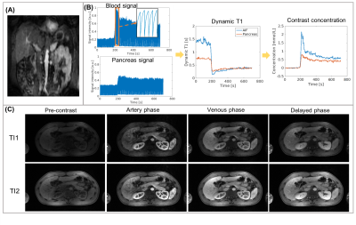 |
Whole-abdomen Free-breathing Quantitative Dynamic Contrast Enhanced (DCE) MR Imaging of Pancreatic Ductal Adenocarcinoma (PDAC) with Fast T1 Mapping Using Multitasking: A Pilot Study

Nan Wang, Anthony Christodoulou, Srinivas Gaddam, Lixia Wang, Yibin Xie, Zixin Deng, Zhengwei Zhou, Wensha Yang, Zhaoyang Fan, Richard Tuli, Simon Lo, Andrew Hendifar, Stephen Pandol, Debiao Li
PDAC is the 3rd leading cause of cancer-related death in the US with poor prognoses. Although conventional DCE-MRI techniques have demonstrated high sensitivity and specificity in tumor delineation, the diagnosis and prognosis of PDAC continues to be challenging with currently available imaging tools. In this work, we proposed a novel Multitasking DCE technique enabling free-breathing acquisition, 3D whole-abdomen coverage, high temporal resolution (500 ms), and dynamic T1 mapping to allow for accurate quantification of tissue perfusion and vascular properties of PDAC. The in vivo feasibility of the proposed technique is demonstrated in healthy subjects and patients with PDAC.
|
15:57
|
0639.
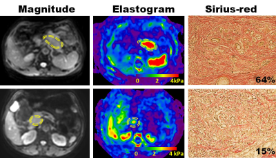 |
The Stiffness as Obtained by MR Elastography Correlates with the Stroma Proportion and Prognosis of Resectable Pancreatic Adenocarcinoma
Did Not Present
yu shi, xianyi zhang, xiaoli cai, fei yang
One of the defining characteristics of pancreatic adenocarcinoma (PDAC) is abundant desmoplastic stroma. Our study showed that pancreatic stiffness determined by magnetic resonance elastography (MRE) is a promising technique to predict the stroma proportion and shows potentials to predict prognosis of resectable PDAC.
|
16:09
|
0640.
 |
Precise stiffness-based detection of pancreatic carcinoma by tomoelastography
Stephan Marticorena Garcia, Rosa Schmuck, Bahra Marcus, Christian Burkhardt, Jing Guo, Bernd Hamm, Jürgen Braun, Ingolf Sack
Pancreatic stiffness was investigated using multifrequency MR elastography (MRE) and tomoelastography data processing in healthy controls (CTR) and patients with pancreatic ductal adenocarcinoma (PDAC). In healthy volunteers, tomoelastography was highly reproducible and showed no significant influences of region and age on pancreatic stiffness. Furthermore, we show that PDAC can be detected as stiff masses with full separation of MRE-values between CTR and PDAC. MRE-based tumor volume correlated excellently with CT-volumetry. Tomoelastography is well suited for boundary detection of pancreatic tumors within standardized, quantitative and contrast-agent free imaging examinations.
|
16:21
|
0641.
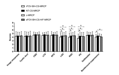 |
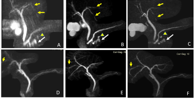
Evaluation of the clinical usefulness of 3-dimensional Magnetic Resonance Cholangiopancreatography with compressed sensing in patients with pancreatobiliary disease
Xiangtian Zhao, Mengyue Huang, Jinxia Zhu, Bernd Kühn, Weijie Wang, Jingliang Cheng
This study aimed to compare the acquisition time, image quality, and diagnostic performance of three three-dimensional (3D) magnetic resonance cholangiopancreatography (MRCP) with compressed-sensing (CS) prototype protocols with those of conventional MRCP in a large cohort of patients with suspected pancreaticobiliary disorders, and to explore the effect of the in-plane phase-encoding direction on the image quality of small-field-of-view (FOV) breath-hold (BH) CS-MRCP (sFOV-BH-CS-MRCP). We found that CS-MRCP provides comparable image quality and diagnostic performance with significantly shortened scan time, and sFOV-BH-CS-MRCP should be acquired in the head-feet phase-encoding direction instead of the left-right direction.
|
16:33
 |
0642.
 |
Can two breath-hold 3D-MRCP replace the conventional 3D-MRCP?
Ming He, Huadan Xue, Jiazheng Wang, Zhengyu Jin
This study was to evaluate and compare the image quality and diagnostic performance of three MRCP protocols, including BH-3D-CS-MRCP, Gradient Spin Echo (BH-GraSE-3D-MRCP) and navigator-triggered (NT) MRCP.
|
16:45
|
0643.
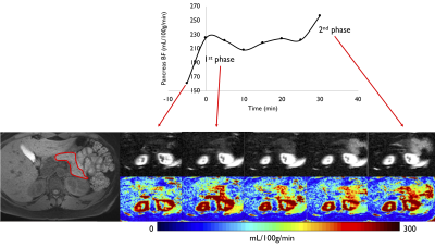 |
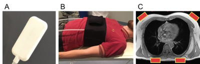
Pancreatic perfusion modulation following glucose stimuli revealed by non-invasive Arterial Spin Labeling (ASL) MRI
Manuel Taso, Koenraad Mortele, Fotini Papadopoulou, Martin Smith, David Alsop
Being able to non-invasively monitor pancreatic perfusion changes is essential for its clinical translation, but it can also prove useful in the evaluation of endocrine disorders such as diabetes. While some perfusion modification following a glucose challenge was observed with PET, ASL has not yet succeeded. We propose here an investigation of pancreatic perfusion modulation following an oral glucose challenge with background-suppressed pCASL at 3T, highlighting perfusion modulation that could be linked to pancreatic endocrine function. Such paradigm could either serve as a tool for studying endocrine disorders or provide a glucose-enhanced scan that increases SNR for diagnostic purposes.
|
16:57
|
0644.
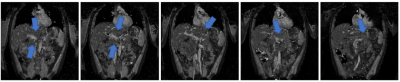 |
Magnetic Resonance Fingerprinting of the Normal Pancreas
Joshua Kaggie, Eva Mendes Serrao, Dimitri Kessler, Mary McLean, Bruno Carmo, Guido Buonincontri, Rolf Schulte, Evis Sala, Kevin Brindle, Amy Frary, Martin Graves, Ferdia Gallagher
MR imaging of the pancreas is challenging due to its retroperitoneal deep-sited location in the abdomen. In addition to its position, the pancreas is subject to breathing motion artifact, which limits the clinical value of pancreatic MRI. Patients with pancreatic cancer are usually very frail, which limits their tolerance to long examinations or breath-hold MRI measurements. MR Fingerprinting (MRF) is an innovative measurement technique that provides qualitative data and quantitative parameter maps from a single acquisition with the potential to reduce exam times. MRF is technically challenging due to limitations in processing capabilities, which we assess in this work.
|
17:09
|
0645.
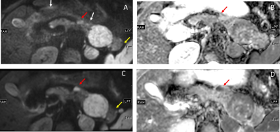 |
Comparison of reduced field-of-view (rFOV) and full FOV (fFOV) diffusion-weighted imaging (DWI) in the assessment of insulioma: image quality and WHO grading
Ming He, Huadan Xue, Zhengyu Jin, Jiazheng Wang, Jin Xu
Reduced-FOV DWI (rFOV-DWI) of the pancreas has been applied in small cohorts and demonstrated improved image quality, but it has not been studied in the detection and characterization of insulioma. In this study, we compared the imaging quality (IQ) of rFOV-DWI and full FOV DWI sequence in insulioma detection. We also explored the correlation between the ADC value and WHO classification.
|
17:21
|
0646.
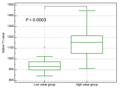 |
Pancreatic T1 Mapping and Extracellular Volume Fraction in Patients with Impaired Glucose Tolerance
Yoshifumi Noda, Satoshi Goshima, Yusuke Tsuji, Kimihiro Kajta, Yuta Akamine, Tomoyuki Okuaki, Masatoshi Honda, Hiroshi Kadohara, Nobuyuki Kawai, Hiroshi Kawada, Yukichi Tanahashi, Masayuki Matsuo
The presence of pancreatic fibrosis is a representative feature of the pancreas in patients with impaired glucose tolerance (IGT). The extracellular volume fraction (ECV) is reported to be associated with cardiac and hepatic fibrosis. In this study, we evaluated the feasibility of the ECV of the pancreas based on T1 mapping for the assessment of HbA1c values. Our results showed that increased ECV of the pancreas was significantly correlated with HbA1c values, so the ECV of the pancreas could serve as a potential imaging biomarker for the assessment of patients with IGT.
|
|
