Quantitative Kidney Imaging
Oral
Body: Breast, Chest, Abdomen, Pelvis
Thursday, 16 May 2019
| Room 510A-D |
16:00 - 18:00 |
Moderators: Jingfei Ma |
16:00
 |
1199.
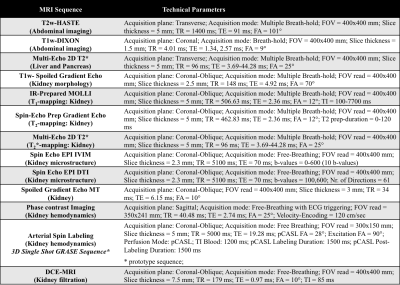 |
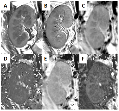
The multi-centre iBEAt study: A comprehensive multi-parametric MR imaging biomarker panel for Diabetic Kidney Disease

Kanishka Sharma, Fotios Tagkalakis, Irvin Teh, Christopher Kelly, David Shelley, Virva Saunavaara, Dmitry Kuznetsov, Anil Karihaloo, Michael Mansfield, Mark Gilchrist, Roberto De Blasi, Mark Ibberson, Maria-Alexandra Olaru, Bernd Ku¨hn, Nicolas Grenier, Steven Sourbron
There is a major clinical need for better biomarkers to identify Diabetic Kidney Disease (DKD) patients at risk of progression. iBEAt is a prospective multi-centre cohort study in 500 patients aiming to determine if MRI biomarkers can provide prediction of progression. We have developed a dedicated MRI biomarker panel for iBEAt interrogating body composition, renal morphology and tissue structure, hemodynamics and filtration. Here we present the details as well as example data and preliminary results on the ISMRM/NIST phantom, and on healthy volunteers.
|
16:12
|
1200.
 |
Intravoxel-Incoherent-Motion Diffusion Weighted MR Imaging for Early Assessment of Graft Function in Kidney Transplant
Yi-Hsin Tsai, Yung-Chieh Chang, Mu-Chih Chung, Hao-Chung Ho, Clayton Chi-Chang Chen, Jyh-Wen Chai
There are currently many different criteria for defining delayed graft function in patients with renal transplant, some of which require subjective decision-making, others require observation over time. We propose the intravoxel incoherent motion MR sequence as a method to acquire objective, sensitive biomarkers early after transplantation. The proposed method is shown to provide information of rapid-moving fluid in addition to conventional slow diffusion from the renal parenchema, and have the potential to assess the renal graft condition from both diffusion-based and perfusion-based view.
|
16:24
|
1201.
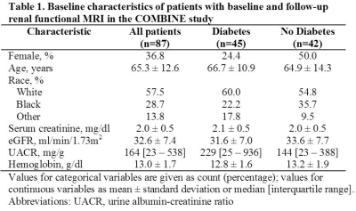 |
Lower ADC Values are Associated with Annual Loss of Renal Function in Individuals with Advanced CKD
Anand Srivatsava, Wei Li, Tamara Isakova, Stuart Sprague, COMBINE Investigators, Pottumarthi Prasad
We present data from a multi-center trial involving patients with advanced CKD (stage 3B & 4) with multiple etiologies. BOLD and Diffusion MRI data was acquired at baseline and repeated 1 year later. Renal function was monitored on a quarterly basis to evaluate progression. Our data show for the first time a strong association of ADC with annual loss of renal function. Further ADC values showed differences when individuals were stratified by diabetes and progression status. BOLD MRI did not demonstrate similar association.
|
16:36
|
1202.
 |
Unbiased MRI Assessment of Renal Tubular Volume Fraction with Data-Driven IVIM
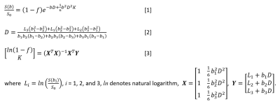
Joao Periquito, Min-Chi Ku, Kathleen Cantow, Erdmann Seeliger, Bert Flemming, Thomas Gladytz, Dirk Grosenick, Thoralf Niendorf, Andreas Pohlmann
T2* mapping does not fully represent renal tissue oxygenation. Tubular volume fraction changes should be considered to correct T2*. Diffusion weighted imaging provides information about in-vivo evaluation of water mobility which can be linked to three sources: tissue water diffusion, blood flow, and tubular flow. In this work we explore the feasibility of assessing tubular volume fraction changes using the non-negative least squares (NNLS) approach that is data-driven and requires no a priori knowledge
|
16:48
|
1203.
 |
Fast hybrid IVIM-DK imaging in the kidney
Did Not Present
Kao-Lang Liu, Kuo-How Huang, Chin-Chen Chang, Wen-Chau Wu
The application of intravoxel incoherent motion (IVIM) and diffusion kurtosis (DK) MRI has been hindered largely by the long scan time required to sample over multiple b-values and/or diffusion-encoding directions. In this study, a novel method is described to expedite hybrid IVIM-DK imaging in the kidneys. Scan time is reduced by acquiring minimally-required non-zero b-values, while index calculation is made more time-efficient by using closed-form solution to replace nonlinear fitting. Experimental data demonstrated feasibility with b = 0/400/800/1600 s/mm2. Measurement variability was found greater among diffusion-encoding directions than between repeats, suggesting non-trivial structural anisotropy in the kidneys.
|
17:00
|
1204.
 |
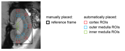
Renal multi-parametric MRI: Ready to launch? A reproducibility study
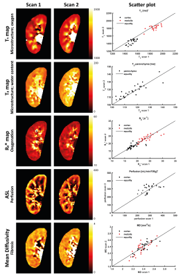
Anneloes de Boer, Anita Harteveld, Peter Blankestijn, Clemens Bos, Suzanne Franklin, Martijn Froeling, Jaap Joles, Marianne Verhaar, Hans Hoogduin, Tim Leiner
Functional MRI of the kidneys is a promising tool for diagnosis, prognosis and treatment monitoring in kidney disease, but there is urgent need for technical validation of renal MRI parameters, including reproducibility. We performed repeated measurements of a multi-parametric renal MRI protocol, including oxygenation, diffusion, perfusion and relaxometry, in healthy volunteers at 3T. This allows for mutual comparison of parameters in terms of reproducibility. As a first step to large scale studies, we have determined reproducibility and shown feasibility of renal quantitative, multi-parametric MRI within a clinically acceptable scan time.
|
17:12
|
1205
 |
Recombinant expression and synthesis of a targeted human contrast agent for quantitative renal MRI
Video Permission Withheld
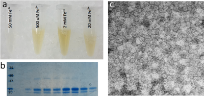
Kasey Emoto, Edwin Baldelomar, Maria Veronica Clavijo-Jordan, Jennifer Charlton, Courtnie Yokono, Kevin Bennett
This study demonstrates the synthesis and use of a human recombinant cationic ferritin nanoparticle, synthesized in ecoli, as contrast agent for targeted renal imaging. Injected nanoparticles accumulated in the glomerular basement membrane in a mouse, allowing measurement of nephron endowment using gradient echo imaging and automated segmentation. Use of human recombinant contrast agents may allow improved biocompatibility for clinical translation.
|
17:24
 |
1206.
 |
Radiomic analysis of MRI in Clear Cell Renal Carcinoma: Non-invasive prediction of High Grade Histology
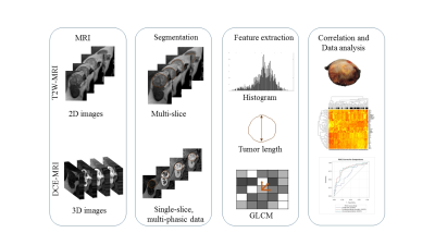
Durgesh Dwivedi, Yin Xi, Ananth Madhuranthakam, Michael Fulkerson, Alberto Diaz de Leon, Yee Ng, Matthew Lewis, Jeffrey Cadeddu, Aditya Bagrodia, Vitali Margulis, Payal Kapur, Ivan Pedrosa
Clear cell RCC (ccRCC), the most common and aggressive subtype of kidney cancer is a very heterogenous disease. Percutaneous biopsies are limited to determine tumor grade, particularly in larger, heterogeneous tumors. MRI can diagnose ccRCC histology with reasonable accuracy. However, efforts to distinguish indolent from aggressive ccRCCs using MRI have produced only modest results. Here we apply quantitative radiomic analysis based on first-order (histogram) and second-order (texture feature) statistics to MRI data for the prediction of high grade ccRCC. Our findings suggest that radiomic analysis of MRI data can help in the prediction of tumor grade in ccRCC.
|
17:36
|
1207.
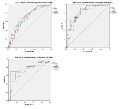 |
Value of Whole-lesion MR T2WI Texture Analysis in Diagnosis of Renal Oncocytoma from Localized RCC Subtypes: Comparison with Characteristic Imaging Signs
Yichen Wang, Fei Xu, Jin Zhang, Yan Chen
Accurate imaging diagnosis of renal oncocytomas remains a challenge for radiologists. Our study retrospectively collected MR imaging of 37 renal oncocytomas and 131 Stage I renal cell carcinoma, compared the diagnostic efficacy between whole-lesion MRI texture analysis and traditional imaging features. Tumor central scar or segmental enhancement inversion showed low sensitivity with high specificity. While whole-lesion MRI texture analysis showed better performance in diagnosis of renal oncocytomas, especially in differentiating oncocytomas with chrommphobe RCC. Whole-lesion MR T2WI texture analysis can be an applicable method in diagnosis of renal oncocytomas.
|
17:48
|
1208.
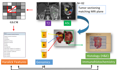 |
Non-Invasive Assessment of Intratumor Heterogeneity in Clear Cell Renal Cell Carcinoma using Quantitative MRI-Derived Texture Features
Durga Udayakumar, Durgesh Dwivedi, Ze Zhang, Yin Xi, Tao Wang, Ananth Madhuranthakam, Payal Kapur, Asghar Hajibeigi, Allison Joyce, Qurratulain Yousuf, Michael Fulkerson, Alberto Diaz de Leon, Matthew Lewis, Jeffrey Cadeddu, Aditya Bagrodia, Vitali Margulis, James Brugarolas, Ivan Pedrosa
Intra and inter-tumor heterogeneity pose a challenging task for predicting tumor behavior due to the limited understanding of the molecular mechanism of clear cell renal cell carcinoma (ccRCC) development. The purpose of the present study was to correlate non-invasive quantitative measures of heterogeneity on MR imaging with histopathologic signatures of aggressiveness and gene expression heterogeneity in ccRCC. MRI derived Haralick texture features offer objective, quantitative measures of ccRCC aggressiveness, which can help compensate for the limitations of percutaneous biopsies in the tissue characterization of larger, heterogeneous tumors and assist in the implementation of active surveillance and neoadjuvant therapy protocols.
|
|
