Traumatic, Neoplastic & Degenerative Musculoskeletal Diseases
Oral
Musculoskeletal
Wednesday, 15 May 2019
| Room 513D-F |
13:30 - 15:30 |
Moderators: Bernard Dardzinski, Ashley Williams |
13:30
|
0861.
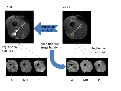 |
Diffusion tensor imaging and T2 measurements are sensitive to changes in muscle tear due to healing

John Biglands, Steven Tanner, Ai Lyn Tan, John Ridgway, Paul Emery, Thorsten Feiweier, Philip Robinson, Andrew Grainger, Philip O'Connor
The aim of this study was to investigate the ability of diffusion tensor imaging (DTI) and T2 measurements to detect changes due to healing in athletes with muscle tears. 13 professional athletes were imaged within 2 days of injury and at the point that they returned to training. Regions of interest depicting the tear were drawn at visit 1 and were propagated to parameter maps from both visits taking into account motion between images. The study showed significant differences in DTI parameters and T2 values between visits, implying that these measures may be useful quantitative markers in assessing muscle healing.
|
13:42
|
0862.
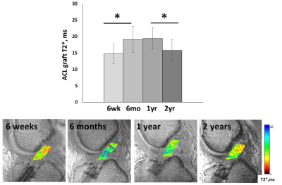 |
T2* and UTE-T2* Evaluations of Anterior Cruciate Ligament Graft Maturation
Ashley Williams, Gordhan Mahtani, Constance Chu
This study tests the hypothesis that quantitative T2* and UTE-T2* are sensitive to ACL graft changes reflective of histological stages of human ACL graft incorporation following ACL reconstruction. T2* variability at 6 months suggests transition from early stage incorporation to remodeling, while stable values from 6 months to 1 year are consistent with remodeling. ACL graft T2* and UTE-T2*, respectively, from two different cohorts decreased between 1 and 2 years suggesting continued graft maturation during the second year. T2* and UTE-T2* of ACL graft incorporation may help to inform decisions concerning return to sports and work activities following ACL reconstruction.
|
13:54
|
0863.
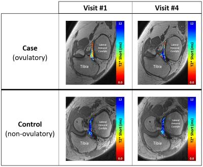 |
Changes in ACL T2* over the course of a menstrual cycle: a new biomarker for ACL injury risk?
Erin Argentieri, Tatum Braun, Ryan Breighner, Alissa Burge, Joseph Nguyen, Matthew Koff, Ellen Casey, Hollis Potter
This study assessed changes in ACL T2* metrics throughout the menstrual cycle. In the pre-ovulatory phase, ovulatory case subjects exhibited significant shortening of T2*S and PS in comparison to visit #4 (post-ovulatory phase). Non-ovulatory control subjects displayed no significant changes over time. Results of the current study suggest that there is a shift in bound water (T2*S) within the ACL from pre- to post-ovulatory phases. Shifts in tissue water content have been associated with altered mechanical properties and changes in ligament stiffness may alter proprioceptive sense and contribute to increases in laxity and risk of ACL-injury within the pre-ovulatory phase.
|
14:06
 |
0864
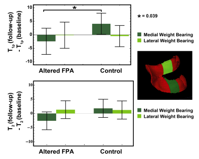 |
Gait retraining as a conservative treatment for medial knee OA: preliminary findings
Video Permission Withheld
Valentina Mazzoli, Scott Uhlrich, Elka Rubin, Feliks Kogan, Brian Heargraves, Scott Delp, Gary Beaupre, Garry Gold
Osteoarthritis is a major societal burden and is associated with pain and disability. To cope with the “osteoarthritis epidemic” and its high associated costs, there is a need for new conservative treatments. This study investigates the potential of gait retraining with altered foot progression angle as one inexpensive conservative treatment for medial knee osteoarthritis. Our results show that this treatment may be effective in reducing pain and improving the MRI outcomes in osteoarthritis patients. This suggests the potential of personalized gait retraining with altered foot progression angle as an inexpensive and effective conservative method for the management of osteoarthritis patients.
|
14:18
 |
0865
 |
Assessment of an In Vitro Model of Rotator Cuff Tendinopathy Using 3D-UTE MRI Biomarkers with Biochemical and Histological Correlation
Video Permission Withheld
Tan Guo, Rachel High, Qingbo Tang, Jonathan Wong, Yajun Ma, Adam Searleman, Sarah To, Lidi Wan, Saeed Jerban, Jiang Du, Eric Chang
Changes in extracellular matrix are seen in cuff tendinopathy, and in particular, alterations in collagen proportion and property are characteristic. Tendon contains an abundance of short T2 components that rapidly decay to background levels. Thus, UTE sequences are better suited for quantitative evaluation of tendon compared with standard clinical MR sequences. In this study, we assess the sensitivity of multiple UTE biomarkers for the evaluation of rotator cuff degeneration in a controlled laboratory experiment.
|
14:30
|
0866.
|
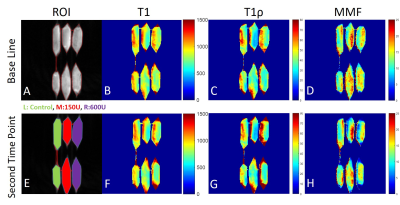
Usefulness of enhancement pattern analysis of non-contrast perfusion using arterial spin labeling (ASL) for the evaluation of painful shoulder diseases
Katsumi Nakamura, Fumihide Arakami, Tomoko Nishi, Kennichi Inoue, Satoshi Masuda, Akiyoshi Yamamoto, Asaka Aso, Mitsue Miyazaki
We investigated the usefulness of non-contrast perfusion obtained using arterial spin labeling (ASL) for the evaluation of various shoulder diseases. In 137 consecutive patients, the subacromial space was divided into four areas, in which ASL enhancement patterns were evaluated. Most of the full thickness tear of rotator cuff demonstrated diffuse enhancement. Most of the adhesive capsulitis showed the anterior area enhancement to be consistent with rotator interval. Acromioclavicular (AC) arthritis showed dominant enhancement in the AC joint. The specific enhancement pattern of ASL is well correlated with the pathology of shoulder diseases, which is highly useful for the differential diagnosis.
|
14:42
|
0867.
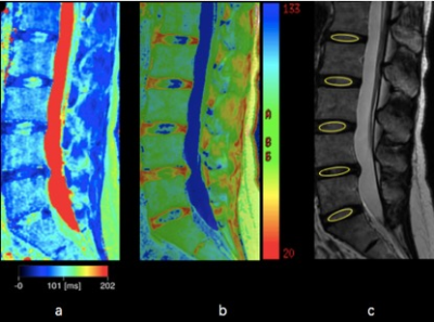 |
The use of relaxometry measurements in synthetic MR in assessing the lumbar intervertebral disk degeneration in patients with chronic low back pain
Yuwei Jiang, Lu Yu, Xiaojie Luo, Chunmei Li, Jianxun Qu, Min Chen
Our purpose was to verify the use of synthetic MR in quantitative analysis of lumbar intervertebral disk degeneration in patients with chronic low back pain. Twenty-four patients underwent MR examination including conventional T2map and synthetic MR. Our results showed strong correlations between the two techniques. These absolute parameters obtained can reflect the intervertebral disk degeneration grades quantitatively. Synthetic MR is efficient and reproducible in the assessment of disk degeneration.
|
14:54
|
0868.
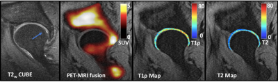 |
[18F]-NaF PET-MR Imaging Reveals Bone-Cartilage Interactions in Hip Osteoarthritis
Radhika Tibrewala, Emma Bahroos, Hatef Mehrebian, Sarah Foreman, Thomas Link, Valentina Pedoia, Sharmila Majumdar
In Osteoarthritis (OA), degeneration of articular cartilage can be accompanied by changes in bone structure. In this study, we analyzed the distribution of [18F]-NaF uptake and blood flow in the femur and acetabulum in 10 hip OA patients and studied associations between bone remodeling and cartilage composition in the presence of morphological abnormalities using PET-MR, quantitative MR and femur shape modeling. Our results showed associations of Standardized Uptake Values (SUV), blood flow to bone (Kpat) with patient reported pain, when different bone shapes like shaft thickness and coxa valga were taken into account.
|
15:06
|
0869.
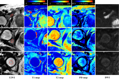 |
Differential Diagnosis Between Normal Marrow and Metastasis of Pelvic Bones Using Relaxation Maps from Synthetic MRI
Yadong Cui, Yue Lin, Chunmei Li, Jianxun Qu, Bing Wu, Min Chen
Synthetic MRI enables quantification of T1, T2 and proton density (PD) value. The purpose of our study was to assess the feasibility for distinguishing bone metastasis from normal marrow with synthetic MRI. Our study found that T1 value of metastasis was significantly higher than those of red and yellow marrow. The T2 value of metastasis was significantly lower than those of red and yellow marrow. We concluded that synthetic MRI could be used for differentiating bone metastasis from normal marrow.
|
15:18
|
0870
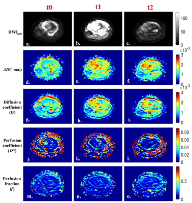 |
IVIM Analysis in Response Evaluation of Osteosarcoma Treated with Neoadjuvant Chemotherapy: Correlation with Histopathological Necrosis
Video Permission Withheld
Esha Baidya Kayal, Devasenathipathy Kandasamy, Kedar Khare, Raju Sharma, Sameer Bakhshi, Amit Mehndiratta
Histological necrosis is the current gold standard for response evaluation in osteosarcoma treated with neoadjuvant chemotherapy (NACT). However it is applicable only after tumor resection on completion of NACT. Thus, a non-invasive early marker of NACT response is desirable. We performed NACT response prediction and evaluation using Intra-voxel Incoherent Motion (IVIM) Diffusion weighted MRI and histogram analysis. IVIM parameters and its histogram analysis revealed clinically useful information in characterizing and predicting chemotherapy response to Osteosarcoma.
|
|
