MRI of Normal Musculoskeletal Physiology, Exercise & Biomechanics
Oral
Musculoskeletal
Tuesday, 14 May 2019
| Room 518A-C |
08:15 - 10:15 |
Moderators: Hermien Kan, Valentina Mazzoli |
08:15
 |
0410.
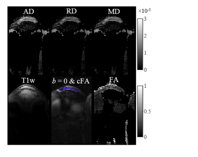 |
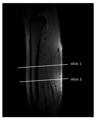
In Vivo Diffusion MRI Evaluation of Human Patellar Tendon Microstructure and Microcirculation

Kenneth Wengler, Takeshi Fukuda, Dharmesh Tank, David Komatsu, Megan Paulus, Elaine Gould, Mingqian Huang, Mark Schweitzer, Xiang He
The patellar tendon (PT) is essential for knee extension and patellar tendinopathy causes persistent knee pain in ~14% of recreational athletes. Microstructure and microcirculation play a critical role in the progression of tendinopathy and can be non-invasively assessed by DTI and IVIM MRI, respectively. In this study, the novel ste-RS-EPI diffusion MRI protocol originally developed for Achilles tendon imaging was applied to image healthy PT. For the first time, DTI and IVIM images were acquired of PT for microstructure and microcirculation evaluation.
|
08:27
|
0411.
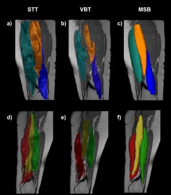 |
Advantages of tractography for the evaluation of intramuscular variances in human thigh muscles
Johannes Forsting, Robert Rehmann, Marlena Rohm, Martijn Froeling, Lara Schlaffke
Muscle diffusion tensor imaging can indirectly provide information about muscular microstructure and architecture, which plays an increasing role in the evaluation of neuromuscular disease progression and treatment monitoring. The separation of different muscles is essential to evaluate intermuscular differences and variances. Here we have compared three methods to assess diffusion metrics of thigh muscles and showed, that tractography shows less variance in diffusion metrics than parameter maps. For the observed parameters FA, MD, RD and λ1 we found significant main effects of muscle, when using tractography, which was not found with manual annotation using ROI assessment of the parameter maps.
|
08:39
|
0412.
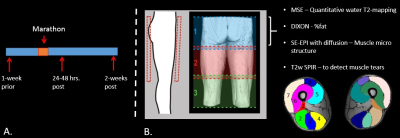 |
Quantitative multi-parametric MRI reveals micro-structural changes in upper-leg muscles after running a marathon
Melissa Hooijmans, Jithsa Monte, Martijn Froeling, Sandra van den Berg-Faay, Adrianus Bakermans, Vincent Aengevaeren, Mario Maas, Thijs Eijsvogels, Aart Nederveen, Gustav Strijkers
Quantitative MR techniques have shown promise for detection of muscle micro trauma. Muscle injury and recovery involve many pathophysiological processes including, inflammation, regeneration and fibrosis, therefore multi-parametric approaches are critically needed. This study used a multi-parametric quantitative approach to assess micro structural changes in the upper leg muscles after running a marathon on an individual muscle basis as well as on a localized level. Our results indicate that diffusion indices are highly sensitive to detect micro-structural changes on a localized and whole volume basis and that this approach could prove valuable for improved outcome prediction and risk-assessment of sports-related-injuries.
|
08:51
|
0413.
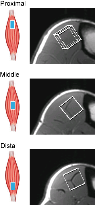 |
Phosphocreatine recovery in the human tibialis anterior after dynamic exercise shows a proximo-distal gradient, which is not explained by acetylcarnitine differences.
Linda Heskamp, Mark van Uden, Tom Scheenen, Arend Heerschap
The phosphocreatine (PCr) recovery rate of the tibialis anterior muscle after isometric exercise shows a strong gradient along the length of the muscle. In this study we demonstrate that this also holds for dynamic exercise of the TA. To further examine if mitochondrial metabolism is involved we determined acetylcarnitine (AC) levels along the length of the TA, but no differences were detected. Together with previous correlations found for blood oxygenation by NIRS experiments and perfusion by IVIM we conclude that muscle perfusion and oxygenation are main components determining the PCr recovery gradient in the TA.
|
09:03
|
0414.
 |
Simultaneous Measurement of Perfusion and T2* in Calf Muscle at 7T with Dynamic Exercise using Radial Acquisition.
Sultan Zaman Mahmud, Bruce Gladden, Andreas Kavazis, Robert Motl, Thomas Denney, Adil Bashir
Impairments in oxygen delivery and consumption can lead to reduced muscle endurance and physical disability. Perfusion, a measure of microvascular blood flow, provides information on nutrient delivery. T2* provides information about relative tissue oxygenation. Changes in these parameters following stress, such as exercise, can yield important information about imbalance between delivery and consumption. In this study we implemented golden angle radial MRI acquisition technique to simultaneously quantify muscle perfusion and T2* at 7T, and demonstrate assessment of spatial and temporal changes in these parameters within calf muscles during recovery from plantar flexion exercise.
|
09:15
|
0415.
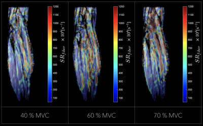 |
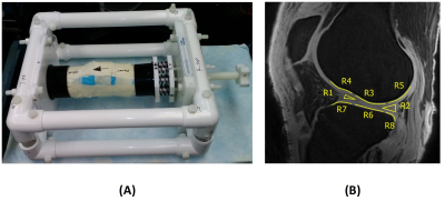
Mapping of Muscle Strain Rate at Varying Force Levels of Isometric Contraction, with Compressed-Sensing Velocity-Encoded Phase-Contrast MR Imaging.
Shantanu Sinha, Vadim Malis, Usha Sinha
Strain rate (SR) tensor mapping can be conveniently computed from velocity encoded phase contrast (VE-PC) imaging. The study of the variation of strain rate indices with force output (% Maximum Voluntary Contraction (MVC)) can provide additional information similar to stress-strain relationships measured at the whole muscle level. However, such studies have been limited by the long VE-PC sequence time precluding its use at high MVCs. We have developed a compressed sensing VE-PCI technique to enable acquisitions across a range of MVCs. Successful SR mapping for 30-70%MVC on six subjects is reported here.
|
09:27
|
0416.
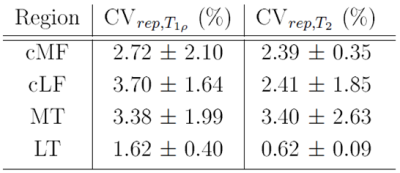 |
Quantitative MR Relaxation Imaging of Cartilage Compositional Response to Exercise
Dimitri Kessler, Joshua Kaggie, James MacKay, Alexandra Morgan-Roberts, Robert Janiczek, Martin Graves, Fiona Gilbert
We introduce a method for imaging changes in healthy knee cartilage composition after joint loading with exercise that could be extended for use in subjects with joint disease. Six healthy participants were imaged before and at multiple time-points after a mild joint loading exercise. Our study demonstrates that quantitative MR mapping of T1ρ and T2 relaxation times of femorotibial cartilage is repeatable and able to demonstrate changes in cartilage composition in response to short-term joint loading exercise. Imaging cartilage compositional recovery after joint loading may present a new way of detecting early alterations in cartilage homeostasis associated with osteoarthritis.
|
09:39
|
0417.
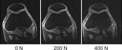 |
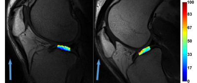
Quantification of patellofemoral cartilage deformation and contact area changes in response to static loading via high-resolution MRI with prospective motion correction
Thomas Lange, Hans Meine, Elham Taghizadeh, Benjamin Knowles, Norbert Südkamp, Maxim Zaitsev, Kaywan Izadpanah
Patellofemoral cartilage deformation and contact area changes in response to in situ loading were measured with high-resolution MRI. In situ loading was realized with a pneumatic loading device and motion artifacts were corrected with prospective motion correction based on optical tracking of the knee cap. Semi-automatic cartilage segmentation based on deep learning proved essential for robust quantification of the load-induced changes. Cartilage thickness and contact area showed significant and weight-dependent changes in response to loading. The patellofemoral deformation and contact mechanism under loading might be used for investigation of the knee biomechanics and as a biomarker of early-stage cartilage degeneration.
|
09:51
|
0418.
 |
Detecting articular cartilage and meniscus deformations under mechanical load using magnetization transfer ultrashort echo time (MT-UTE) modeling: ex vivo study
Saeed Jerban, Yajun Ma, Akhil Kasibhatla, Jonathan Lee, Yanjun Chen, Tan Guo, Lidi Wan, Eric Chang, Jiang Du
Ultrashort echo time (UTE) MRI can be used for quantitative assessment of compressive tissues in knee joint such as articular cartilage (AC) and meniscus. In osteoarthritis, the mechanical properties of AC and menisci are altered prior to gross morphological changes. In this study, magnetization transfer UTE imaging (UTE-MT) with two-pool modelling was used to detect deformation levels of AC and meniscus under load. Compression resulted in significant increases of macromolecular fraction (MMF) in AC and menisci of young, but not elderly donors. Our results indicate that biomechanical changes vary with age and can be detected with UTE-MT MRI.
|
10:03
|
0419.
 |
Magic-Angle Effect on In Vivo T2 Mapping of Cartilage
Victor Casula, Olli Pekka Aro, Petri Paakkari, Stefan Zbyn, Mika Nevalainen, Mikko Nissi, Miika Nieminen
Magic-angle effect in T2 relaxation time of knee cartilage was determined in vivo at clinical magnetic field strength. T2 was measured in healthy volunteer’s articular cartilage on a 3 T clinical scanner at two different orientations of the normal direction of the articular surface with respect to the main magnetic field. Magic-angle effect produced significant variations in the T2 values of healthy cartilage (up to 102% difference in deep cartilage), clearly demonstrating the importance of taking the orientation into account in clinical studies when interpreting cartilage degeneration using T2 mapping.
|
|
