Cutting-Edge Advances in Cancer Imaging
Oral
General Cancer Imaging
Monday, 13 May 2019
| Room 520A-F |
13:45 - 15:45 |
Moderators: Mukund Seshadri |
13:45
 |
0192.
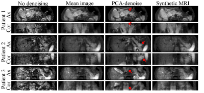 |
Model-free denoising of multi b-value diffusion-weighted MR images using principal component analysis: simulations and in vivo results

Oliver Gurney-Champion, David Collins, Andreas Wetscherek, Mihaela Rata, Remy Klaassen, Hanneke van Laarhoven, Kevin Harrington, Uwe Oelfke, Matthew Orton
We present a principal component analysis (PCA) toolkit for mode-free denoising of multi b-value diffusion-weighted images for clinical use. In simulations, PCA-denoising suppressed the random noise equally well (up to 55%) as synthetic MRI. Contrary to synthetic MRI (systematic error up to 29% of total signal intensity), PCA-denoising did not introduce any systematic errors (<2%). In volunteer and patient image data, PCA-denoising resulted in sharper and less noisy images than synthetic MRI, which resulted in sharper and clearer tumour boundaries. In conclusion, our PCA-denoising toolkit is promising for denoising b-value images for clinical use.
|
13:57
|
0193.
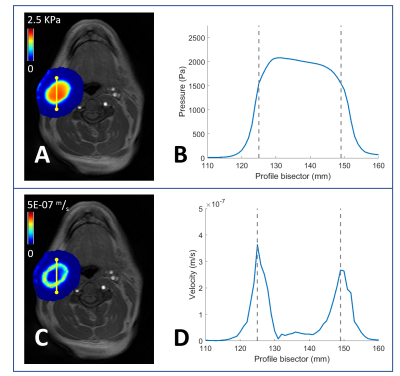 |
DCE MRI-Based Computational Modeling of Interstitial Fluid Pressure and Velocity in Head and Neck Cancer: Initial Analysis
Eve LoCastro, Yonggang Lu, Ramesh Paudyal, Yousef Mazaheri, Vaios Hatzoglou, Amaresha Shridhar, Alan Ho, Nancy Lee, Joseph Deasy, Amita Shukla-Dave
We applied computational fluid modeling to head-and-neck cancer patients' DCE-MRI data using permeability maps from extended Tofts' model and tumor geometry from imaging. Interstitial fluid pressure (IFP) maps generated from computational fluid modeling depict heterogeneous distribution of elevated IFP and velocity in tumor tissue. We found significant correlation between tumor volume and IFP.
|
14:09
 |
0194.
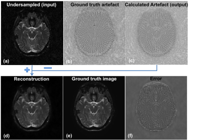 |
Deep residual learning of radial under sampling artefacts for real-time MR image guidance during radiotherapy
Bjorn Stemkens, Cheryl Sital, Max Blokker, Tom Bruijnen, Jan Lagendijk, Rob Tijssen, Cornelis van den Berg
MRI-guided radiotherapy using hybrid MR-Linac systems, requires high spatiotemporal resolution MR images to guide the radiation beam in real time. Here, we investigate the concept of deep residual learning of radial undersampling artifacts to decrease acquisition time and minimize extra reconstruction time by using the fast forward evaluation of the network. Within 8-10 milliseconds most streaking artifacts were removed for undersampling rates between R=4 and R=32 in the abdomen and brain, facilitating real-time tracking for MR-guided radiotherapy.
|
14:21
|
0195
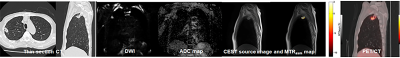 |
Amid Proton Transfer-Weighted Imaging vs. Diffusion-Weighted Imaging vs. FDG-PET/CT: Capability for Management of Solitary Pulmonary Nodules
Video Permission Withheld
Yoshiharu Ohno, Masao Yui, Takeshi Yoshikawa, Shinichiro Seki, Katsusuke Kyotani, Takamichi Murakami
We hypothesized that APTw imaging had equal or better potential for diagnosis of SPNs and prediction of postoperative recurrence prediction in postoperative lung cancer patients, when compared with DWI and FDG-PET/CT. In addition, multiparametric approach among all three methods had better potential than single-parametric approach on each method in this setting. The purpose of this study was to compare the diagnosis and prediction capabilities of pulmonary nodules among single- and multi-parametric approaches by APTw imaging, DWI, and FDG-PET/CT.
|
14:33
|
0196.
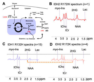 |
A Noninvasive Comparison Study between Human Gliomas with IDH1 and IDH2 Mutations by MR Spectroscopy
Xin Shen, Natalie Voets, Sarah Larkin, Nick de Pennington, Puneet Plaha, Richard Stacey, James Mccullagh, Christopher Schofield, Stuart Clare, Peter Jezzard, Tom Cadoux-Hudson, Olaf Ansorge, Uzay Emir
The oncogenes that are expressed in gliomas reprogram particular pathways of glucose, amino acid, and fatty acid metabolism. Mutations in the isocitrate dehydrogenase genes (IDH1/2) in diffuse gliomas are associated with abnormally high 2-hydroxyglutarate (2-HG) levels. Non-invasive measurement of 2-HG via in vivo 1H magnetic resonance spectroscopy (MRS) can be used to differentiate mutant cytosolic IDH1 from mitochondrial IDH2 in gliomas.
|
14:45
|
0197.
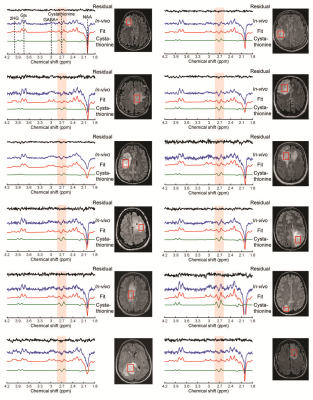 |
Cystathionine as a marker for 1p/19q codeleted gliomas by in vivo magnetic resonance spectroscopy
Francesca Branzoli, Clément Pontoizeau, Anna Luisa Di Stefano, Dinesh Deelchand, Romain Valabregue, Stéphane Lehéricy, Marc Sanson, Chris Ottolenghi, Malgorzata Marjanska
Molecular markers such as mutation in isocitrate dehydrogenase (IDH) and codeletion of chromosome arms 1p and 19q (1p/19q codeletion) have highly benefited diagnosis and prognosis in brain gliomas. However, the biological effects of 1p/19q codeletion are still not clear. We report selective accumulation of cystathionine in IDH-mutated, 1p/19q codeleted gliomas observed by edited 1H magnetic resonance spectroscopy. Noninvasive detection of cystathione enables identification of glioma subtypes in vivo and opens up the possibility of investigating nonivasively cancer-specific metabolic pathways.
|
14:57
|
0198.
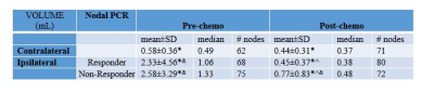 |
The effects of neoadjuvant chemotherapy on axillary lymph node volume and compacity in breast cancer patients: an MRI study
Renee Cattell, Krupal Patel, Thomas Ren, James Kang, Pauline Huang, Jason Ha, Ashima Muttreja, Jules Cohen, Haifang Li, Lea Baer, Cliff Bernstein, Sean Clouston, Roxanne Palermo, Timothy Duong
Axillary lymph node involvement in breast cancer is associated with higher risk of distant metastasis and recurrence. This study evaluated whether MRI can be used to longitudinally monitor effects of neoadjuvant chemotherapy on axillary lymph nodes in situ. Nodal volume and compacity were analyzed with respect to treatment responders and non-responders. Comparisons with unaffected nodes were made. Neoadjuvant chemotherapy significantly reduced nodal volume of the affected nodes in both responders and non-responders. Nodal volumes of the responders normalized whereas those of the partial-responders did not normalize completely. This approach may prove useful for monitoring cancer treatment effects on nodal morphology.
|
15:09
|
0199.
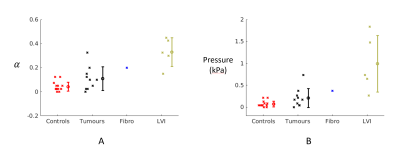 |
Lymphovascular invasion correlates with elevated tumor pressure as quantified by MR Elastography: initial results from a breast cancer trial
Daniel Fovargue, Sweta Sethi, Jack Lee, Marco Fiorito, Adela Capilnasiu, Stefan Hoelzl, Jurgen Runge, Jose de Arcos, Keshthra Satchithananda, Arnie Purushotham, David Nordsletten, Ralph Sinkus
Gauging metastatic propensity is crucial as it impacts decision making in oncology (e.g. whether a patient should receive surgery immediately or neoadjuvant chemotherapy). Interstitial fluid pressure (IFP) is known to correlate with microvascular invasion, a proxy for metastatic potential. However, no current imaging biomarkers correlate with this. We present a method to noninvasively calculate a total tumor pressure (which IFP contributes to). The method reconstructs pressure via nonlinear biomechanics and MR Elastography and is validated in simulations and phantoms. Elevated pressure values from a cohort of 16 breast cancer patients correlate with lymphovascular invasion possibly providing a much sought-after biomarker.
|
15:21
|
0200.
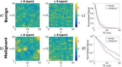 |
Noninvasive MRI mapping of malignant infiltration into lymph nodes
Inês Santiago, João Santinha, Andrada Ianus, Antonio Galzerano, Maria Barata, Nickolas Papanikolaou, Antonio Beltran, Celso Matos, Noam Shemesh
Mapping malignant infiltration into lymph nodes can have a critical impact on patient decision making. We predicted that multigradient echo (MGE) experiments could reflect cellularity and cell-size changes due to underlying susceptibility distributions. Lymph nodes extracted from rectal cancer patients exhibited the predicted non-monotonic and non-mono-exponential MGE signal decay, which provided insights into the underlying microstructure. A simple model distinguished benign from malignant nodal tissue and the differences were at least partially explained by differences in cellularity and cell size. These results can impact lymph node staging accuracy, as already corroborated by our pilot results in-vivo, upon rectal cancer staging, at 1.5T.
|
15:33
|
0201.
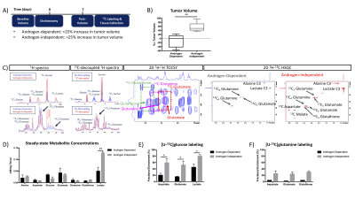 |
Androgen Independence Leads to Altered Metabolism in Prostate Cancer Cell and Murine Models
Jinny Sun, Justin Delos Santos, Robert Bok, Romelyn Delos Santos, Mark Van Criekinge, Daniel Vigneron, Renuka Sriram, John Kurhanewicz
This study demonstrated significant increases in flux through glycolysis, oxidative metabolism, and glutaminolysis associated with androgen independence using patient-derived cell lines and a treatment-driven murine model. This data supports using a combination of hyperpolarized [1-13C]pyruvate, [2-13C]pyruvate and [5-13C]glutamine to noninvasively discriminate between androgen-dependent and androgen-independent prostate cancer in future patient studies using hyperpolarized 13C MRI.
|
|
