Interrogating the Tumor Microenvironment
Oral
General Cancer Imaging
Tuesday, 14 May 2019
| Room 511BCEF |
13:30 - 15:30 |
Moderators: Yoon Seong Choi, Gene Kim |
13:30
|
0500.
 |
Dextran-based CEST MRI for detecting extradomain-B fibronectin in pancreatic cancer

Jiaqi Lu, Zheng Han, Jia Zhang, Yuguo Li, Jing Liu, Kenji Fujiwara, Peter Zijl, Lei Zhang, Guanshu Liu
A dextran-peptide conjugate was developed for MR molecular imaging of pancreatic ductal adenocarcinoma (PDAC) through its overexpressed microenvironment biomarker, extradomain-B fibronectin (EDB-FN). Dextrans can be directly detected by chemical exchange saturation transfer (CEST) MRI without the need for radionuclide- or metallic labeling. In addition, large molecular weight dextran, dextran 10 (MW~ 10 kD), provides an approximately fifty times higher sensitivity per molecule than a single glucose unit. The potential of this highly biocompatible diamagnetic probe is demonstrated in a murine syngeneic allograft PDAC tumor model.
|
13:42
|
0501.
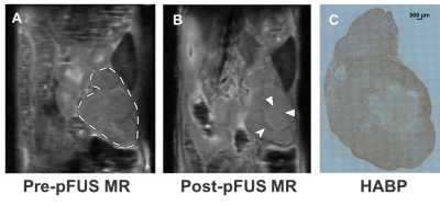 |
Multi-Parametric MRI assessment of pulsed focused ultrasound treated pancreas tumor
Ezekiel Maloney, Yak-Nam Wang, Ravneet Vohra, Tatiana Khohklova, Stella Whang, Helena Son, Joshua Park, Kayla Gravelle, Yasser Hussaini, Stephanie Totten, Joo Ha Hwang, Donghoon Lee
Pancreatic ductal adenocarcinoma (PDA) is characterized by excessive levels of hyaluronan and collagen, resulting in a dense fibroinflammatory stroma that inhibits penetration of chemotherapeutic drugs into the tumor. Pulsed focused ultrasound (pFUS) treatment has shown promising results in disrupting the dense stroma and reducing the interstitial fluid pressure of PDA. The purpose of this study is to noninvasively assess response to pFUS treatments using quantitative MRI and to correlate the MRI results with histopathology data.
|
13:54
 |
0502.
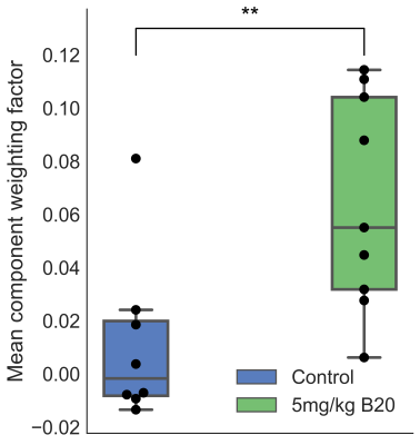 |
Dynamic Oxygen-Enhanced MRI (dOE-MRI) with group ICA detects increased oxygenation in murine tumours treated with VEGF-ablation therapy
Firas Moosvi, Jennifer Baker, Andrew Yung, Piotr Kozlowski, Andrew Minchinton, Stefan Reinsberg
Tumours treated with VEGF ablation sustain changes to their vasculature, which can result in tissue oxygenation changes. This work uses dynamic oxygen enhanced MRI (dOE-MRI) to assess oxygenation of murine SCCVII tumours treated with B20-4.1.1 (murine anti-VEGF antibody) relative to controls. T1-weighted parameter maps and a modified ICA quantitative analysis technique, groupICA, describe an increase in B20-treated tumour oxygenation.
|
14:06
|
0503.
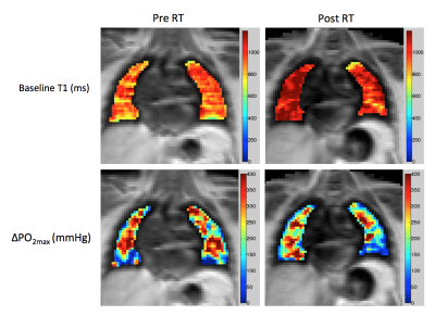 |
Dynamic OE-MRI Mapping of Lung Parenchymal Radiotherapy Effects in Non-Small Cell Lung Cancer Patients
Michael Dubec, Ahmed Salem, Yvonne Watson, Ross Little, Corinne Faivre-Finn, Julian Matthews, Marcel van Herk, James O'Connor, Geoff Parker
Radiation induced lung disease (RILD) can occur following thoracic irradiation, chemotherapy and immunotherapy in lung cancer patients, manifesting as inflammation, radiation pneumonitis or fibrosis. Oxygen Enhanced (OE)-MRI provides spatial information on oxygen delivery to lung tissue. Five non-small cell lung cancer patients underwent OE-MRI pre and post radiotherapy. Statistically significant changes were measured between pre and post radiotherapy scans in both the contralateral and ipsilateral lungs. These findings indicate that OE-MRI can assess changes in lung function following radiotherapy, with potential application for personalised therapy and reduction of toxicity.
|
14:18
|
0504.
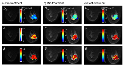 |
Prediction of Breast Cancer Response to Neoadjuvant Chemotherapy with High b-Value Non-Gaussian Diffusion MRI
Muge Karaman, Shunan Che, Guangyu Dan, Zheng Zhong, Xinming Zhao, Han Ouyang, Xiaohong Zhou
Neoadjuvant chemotherapy has been used to extend surgical options by downstaging tumor in both locally advanced and operable breast cancer. An early imaging assessment of tumor response to neoadjuvant chemotherapy is critical for timely tailoring personalized treatment strategies. In this study, we investigate whether the changes in the parameters derived from a non-Gaussian diffusion model – continuous-time random-walk (CTRW) model – are predictive of pathologic response in women undergoing neoadjuvant chemotherapy. Our results demonstrate the high predictive performance of the combined changes in CTRW parameters from pre-treatment levels as early as the second cycle of chemotherapy.
|
14:30
|
0505.
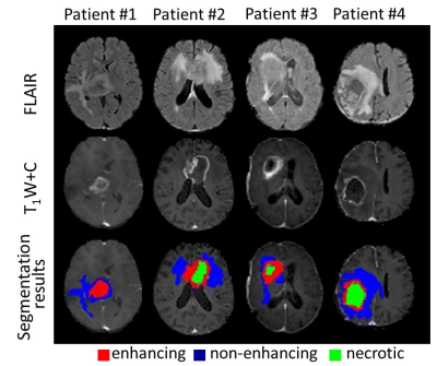 |
Utilizing deep learning for automatic longitudinal assessment of brain tumor response based on RANO criteria
Idan Bressler, Dafna Ben Bashat, Orna Aizensein, Felix Bokestein, Deborah Blumenthal, Moran Artzi
The aim of this study was to implement a deep-learning approach for automatic therapy response assessment in patients with high-grade-glioma (HGG), based on the response-assessment in neuro-oncology (RANO) criteria. A total of 135 conventional MRI scans from 67 patients were included. A neural network with a U-net architecture was trained for identification and subsegmentation of lesion components. The similarity coefficient score between segmentation results and ground truth was 0.88±0.06. Consistency in therapy response assessment was obtained in the majority of cases. These results demonstrate the potential applicability of the proposed method for automatic therapy response assesment in patients with HGG.
|
14:42
|
0506.
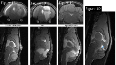 |
Multiparametric Advanced Fast Imaging (MAFI) to Characterize Tumor Habitat in Orthotopic Mouse Models of Pediatric Brain Tumors
Jenna Steiner, Angela Pierce, Andrea Griesinger, Bethany Veo, Aaron Knox, Nathan Dahl, Adam Green, Nicholas Foreman, Rajeev Vibhakar, Natalie Serkova
Brain tumors are the second most common malignancy in childhood (exceeded only by leukemia). Clinically, multiparametric MRI is now considered to be the neuroimaging standard for detecting brain tumors. Pediatric brain tumors have a diverse array of clinical manifestations, cellular and molecular phenotypes, and tumor habitats. There is an unmet need to develop human-faithful pediatric mouse models and fast high-resolution physiological MRI for their detection and characterization. Here, we report on a non-gadolinium, Multiparametric Advanced Fast Imaging (MAFI) approach followed by radiomics analysis to detect, characterize and differentiate three distinct brain tumor subtypes in mouse patient-derived xenograft (PDX) models.
|
14:54
|
0507.
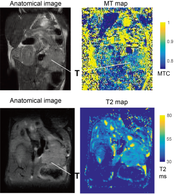 |
Identification of early stage murine pancreatic tumors by a combinatorial approach employing SPEN DWI, FLAIR T1 and hyperpolarized 13C MRI
Ricardo Martinho, Qingjia Bao, Stefan Markovic, Dina Preise, Keren Sasson, Avigdor Scherz, Lucio Frydman
Pancreatic ductal adenocarcinoma has a poor prognosis. This study explored the use of a multimodal screening approach on a preclinical mouse PDAC model that included T1 and T2 mapping, SPatiotemporal ENcoding (SPEN) and EPI-based DWI, and MT methods, and hyperpolarized 13C metabolic MRSI, to follow the progress of the disease from early on. Whereas T2 and MT were of little help, markedly decreased diffusivity, extended T1s and significantly higher metabolic activities could be detected for large and small tumors alike. These approaches could provide a translatable approach to the early noninvasive detection of pancreatic cancer, leading to timely treatment.
|
15:06
|
0508.
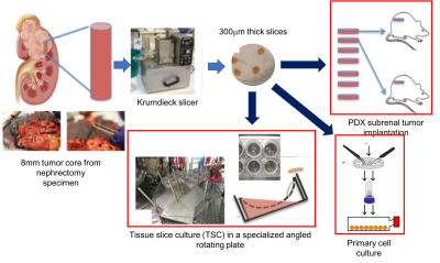 |
Characterization of metabolic adaptations of patient-derived xenografts to in vitro tissue and cell culture using high-resolution NMR
Jinny Sun, Jeffrey Hsiao, Justin Delos Santos, Robert Bok, Hongjuan Zhao, Jeremy Bancroft Brown, James Brooks, John Kurhanewicz, Donna Peehl, Renuka Sriram
In this study we investigated the metabolic changes that occurred when a renal cell carcinoma patient-derived xenograft was propagated in tissue slice culture or primary cell culture at varying pO2 levels to understand the attributes and limitations of each of the model systems for studying the metabolic underpinnings of this pathology using high resolution NMR. This data indicates drastically altered metabolism at varying pO2 and between each model system.
|
15:18
|
0509.
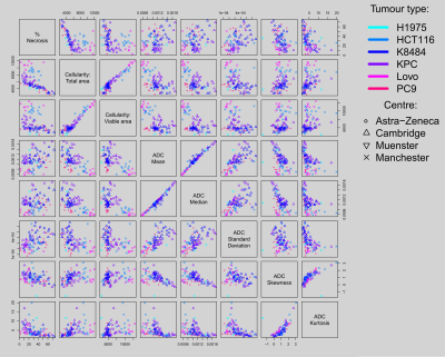 |
Does ADC better predict tumour cellularity or necrosis? A multi-centre multi-vendor study in twelve rodent tumour models
John Waterton, Dominick McIntyre, François-Xavier Blé, Sabrina Doblas, Eric Aboagye, Kathrin Heinzmann, Sandra Heskamp, James O'Connor, Sonja Schelhaas, Lydia Wachsmuth, Hervé Barjat, Cara Brodie, Dominique-Laurent Couturier, Cornelius Faber, Heather Flynn, Philippe Garteiser, Andreas Jacobs, Bernard Van Beers, Kaye Williams, Davina Honess
We studied tumour Apparent Diffusion Coefficient (ADC), necrosis and cellularity in untreated rodent tumours of twelve types from six centres using two different vendors’ equipment. Tumour types included human xenografts (conventional and patient-derived) genetically engineered mouse tumours and syngeneic rat tumours. Across this broad spectrum there was a robust inverse correlation between ADC and cellularity, and a weaker positive correlation between ADC and necrosis. ADC mean and median were the best correlates for cellularity, while ADC standard deviation and kurtosis provided the best correlates for necrosis. This work advances the biological validation of ADC as a biomarker of tumour cellularity.
|
|
