MRSI Acquisition & Reconstruction
Oral
Spectroscopy & Non-Proton MR
Wednesday, 15 May 2019
| Room 511BCEF |
15:45 - 17:45 |
Moderators: Phil Lee, Tom Scheenen |
15:45
 |
0946.
 |
Rapid High-Resolution Simultaneous Acquisition of Metabolites, Myelin Water Fractions, and Tissue Susceptibility of the Whole Brain Using "SPICY" 1H-MRSI

Yudu Li, Rong Guo, Yibo Zhao, Tianyao Wang, Ziyu Meng, Yao Li, Zhi-Pei Liang
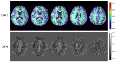
We report further advances on ultrahigh-resolution 1H-MRSI without water suppression to enable rapid simultaneous acquisition of brain metabolites, myelin water fractions (MWF) and tissue susceptibility in high spatial resolution. Building on the SPICE (SPectroscopic Imaging by exploiting spatiospectral CorrElation) subspace imaging framework, we extend the SPICE data acquisition scheme with several novel features, including the use of ultrashort-TE (~1.6 ms), very-short-TR (~160 ms), and variable density sampling of (k, t)-space. We reconstruct the spatial distributions of brain metabolites, MWF, and tissue susceptibility using model-based reconstruction methods that incorporate learned spatiospectral features. Experimental results have been obtained which demonstrate that in a single 5-min scan, we can obtain metabolites in a nominal resolution of 2.0×2.4×3.0 mm3 and QSM/MWF in a nominal resolution of 1.8×1.8×1.8 mm3.
|
15:57
 |
0947
 |
Learning Nonlinear Low-Dimensional Models for MR Spectroscopic Imaging Using Neural Networks
Video Permission Withheld
Yahang Li, Xi Peng, Fan Lam
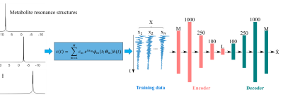
Low-dimensional subspace models have recently been developed for fast, high-SNR MRSI, by effectively reducing the degrees-of-freedom for the imaging problem. However, low-dimensional linear subspace models may be inadequate in capturing more complicated spectral variations across a general population. This work presents a new approach to model general spectroscopic signals, by learning a nonlinear low-dimensional representation. Specifically, we integrated the well-defined spectral fitting model and a deep autoencoder network to learn the low-dimensional manifold where the high-dimensional spectroscopic signals reside, and applied this learned model for denoising and reconstructing MRSI data. Promising results have been obtained demonstrating the potential of the proposed method.
|
16:09
|
0948.
 |
Using an AC/DC Coil to Improve the Line Width and Lipid Suppression for measuring 2HG in Glioma Patients at 3T
Bernhard Strasser, Nicolas Arango, Jason Stockmann, Borjan Gagoski, Bijaya Thapa, Xianqi Li, Wolfgang Bogner, Julia Small, Daniel Cahill, Tracy Batchelor, Jorg Dietrich, Lawrence Wald, Jacob White, Elfar Adalsteinsson, Ovidiu Andronesi
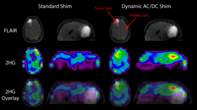
An AC/DC 32-channel coil, which can detect signal while simultaneously alter the B0-field, was used to improve lipid inversion and the spectral linewidth for measuring 2HG. Two patients and two volunteers were measured with a spiral-based 3D sequence once with the standard scanner shim only, and once with the additional shimming of the integrated B0/Rx coil. Data quality was strongly improved, leading to improved 2HG-detection in one patient, and a decreased variability of 2HG in tumor-void areas. This can reduce the likelihood of false-positive 2HG detection. Quantification of other metabolites was additionally improved by the AC/DC shim.
|
16:21
|
0949.
 |
Dephasing Optimization Through Coherence Order Pathway Selection (DOTCOPS): Validation and Inclusion of Phase Cycling
Karl Landheer, Christoph Juchem
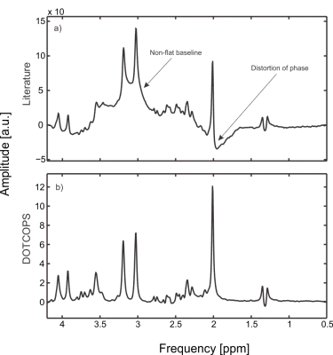
Modern magnetic resonance spectroscopic (MRS) pulse sequences frequently overlook the issue of unwanted coherence pathways as source of numerous spectral artifacts. A novel and robust algorithm which only requires the input of the desired coherence pathway was developed to efficiently dephase all unwanted coherence pathways for any MRS pulse sequence. Experiments were performed on phantoms and healthy volunteers comparing crusher schemes obtained from the literature with those obtained from the optimization algorithm for sLASER and MEGA-sLASER. The results demonstrate that the effects of unwanted coherences can be drastically reduced through the implementation of an optimized crusher and phase cycling scheme.
|
16:33
|
0950.
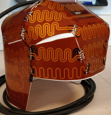 |
Next generation Crusher Coil for suppressing extra cranial lipid signals at 7 Tesla
Erik Huijing, Luca van Dijk, Aidin Ali Haghnejad, Lisan Morsinkhof, Dennis Klomp, Peter Luijten, Jannie Wijnen, Alex Bhogal
We previously presented a crusher coil for suppression of extra-cranial lipids in brain Magnetic Resonance Spectroscopic Imaging (MRSI). To improve crushing performance and the reproducibility of the production of crusher coils, a new crusher coil was designed, created and tested. By doubling the amount of wires and improving their spatial distribution around the coil (consistent meandering), we could achieve efficient lipid suppression with as little as 30% of the power, used with the proof of principle coil. The presented design is easy to reproduce, repair and modify due to a modular build.
|
16:45
|
0951.
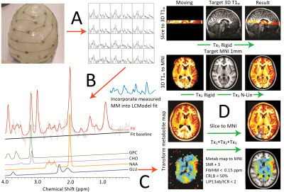 |
Preliminary detailed in-vivo brain atlas of metabolic and macromolecular distributions in humans using 7T 1H Magnetic Resonance Spectroscopic Imaging
Alex Bhogal, Tommy Broeders, Mirte Edens, Sahar Nassirpour, Paul Chang, Peter Luijten, Dennis Klomp, Christiaan Vinkers, Jannie Wijnen
This study amalgamated current state-of-the-art MRSI developments to create a detailed and reliable in vivo metabolic atlas of the healthy human brain. We used an ultra-high field 7T MR scanner to acquire high resolution, short echo-time (TE) MRSI data along with an external crusher coil for hardware based extra-cranial lipid signal suppression. Advanced post-processing and data reconstruction techniques were used in conjunction with stringent data filtering based on several quality assurance metrics, while registration to the MNI152 standard atlas allowed us to generate high-resolution metabolite/macromolecule ratio maps.
|
16:57
|
0952.
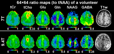 |
Ultra-high-resolution brain MRSI at 7T and 9.4T – A direct comparison
Gilbert Hangel, Philipp Moser, Bernhard Strasser, Michal Považan, Eva Hecková, Lukas Hingerl, Stanislav Motyka, Stephan Gruber, Beáta Bachratá, Benedikt Poser, Christopher Wiggins, Sahar Nassirpour, Paul Chang, Siegfried Trattnig, Wolfgang Bogner
Despite the successful demonstration of fast ultra-high resolution MRSI at 7T and 9.4T, a direct comparison has been lacking. This study fills this gap by measuring the same FID-MRSI protocol in the same volunteer group at both field strengths within a short time frame. Our results show overall similar quality measures across field strengths, with more quantifiable metabolites but also more prevalent spectral artefacts at 9.4T.
|
17:09
|
0953.
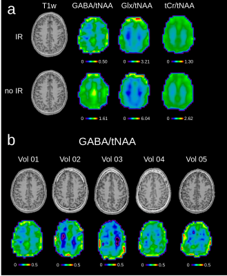 |
Whole-slice mapping of GABA and GABA+ at 7T via adiabatic MEGA-editing, real-time instability correction, and concentric circle readout
Philipp Moser, Lukas Hingerl, Bernhard Strasser, Michal Považan, Gilbert Hangel, Ovidiu Andronesi, Andre van der Kouwe, Stephan Gruber, Siegfried Trattnig, Wolfgang Bogner
In vivo detection of γ-aminobutyric acid (GABA) and glutamate (Glu), both major neurotransmitters in the human brain, benefits from the higher sensitivity at ultra-high field (7T) compared to lower field strengths. However, strong B0/B1+ inhomogeneities and chemical shift displacement errors as well as subject motion and frequency drifts can significantly impair the experiment. An adiabatic MEGA-editing scheme was developed and incorporated into a real-time corrected B1+-insensitive MRSI sequence, which enabled whole-slice metabolic imaging of neurotransmitters in the human brain with unprecedented high-resolution at 7T and allowed a comprehensive assessment of regional GABA levels without co-edited macromolecule contamination.
|
17:21
|
0954.
 |
Accelerated Spectral-Editing MRSI using Subspace Modeling, Multi-Slab Acquisition and 3D CAIPIRINHA Undersampling
Chao Ma, Debra Horng, Paul Han, Shuang Hu, Kexin Deng, Kui Ying, Georges El Fakhri
Spectral-editing MRSI allows reliable and selective detection of many important metabolites, e.g., GABA and 2-HG, by eliminating all uncoupled resonances in the J-difference spectrum. Despite significant advances in fast MRSI sequences and constrained image reconstruction, spectral-editing MRSI is still limited by its long acquisition time and low spatial resolution. Recently, a subspace-based approach has been proposed to accelerate spectral-editing MRSI, reporting encouraging phantom and in vivo results. In this work, we propose to further accelerate the subspace-based spectral editing MRSI using (i) multi-slab acquisition to maximize the time efficiency of long-TR spin-echo acquisition and (ii) a 3D (2D spatial + 1D spectral) CAIPIRINHA (Controlled Aliasing in Parallel Imaging Results in Higher Acceleration) scheme for sparse sampling of the (k,t)-space. We evaluate the performance of the proposed method using simulation, phantom, and in vivo studies.
|
17:33
|
0955.
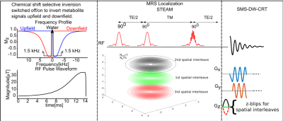 |
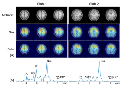
Density-Weighted Concentric Ring Trajectory using simultaneous multi-slice (SMS) acceleration: 3D Metabolite-cycled Magnetic Resonance Spectroscopy Imaging at 3 T
Pingyu Xia, Xin Shen, Xiaopeng Zhou, Mark Chiew, Albert Thomas, Ulrike Dydak, Uzay Emir
In this study, we proposed a novel simultaneous multi-slice (SMS) density weighted (DW) concentric ring trajectory (CRT) metabolite-cycling Magnetic Resonance Spectroscopy Imaging (MRSI) sequence to alleviate some conventional MRSI drawbacks, e.g. long acquisition time, eddy current artifacts, and side lobe artifacts. The sequence was tested on 5 healthy subjects, showing the feasibility of acquiring three slices of high-quality water-only and metabolite spectra simultaneously with a resolution of 5mm X 5mm X 10mm within 20 minutes.
|
|
