Perfusion & Permeability
Oral
Contrast Mechanisms
Wednesday, 15 May 2019
| Room 511BCEF |
13:30 - 15:30 |
Moderators: Patricia Figueiredo, Jun Hua |
13:30
 |
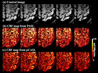
0841.
 |
Improving temporal resolution of 3D Arterial Spin Labeling perfusion imaging by combining CAIPIRINHA encoding and spatio-temporal TGV reconstruction

Stefan Spann, Xingfeng Shao, Danny Wang, Christoph Aigner, Matthias Schloegl, Kristian Bredies, Rudolf Stollberger
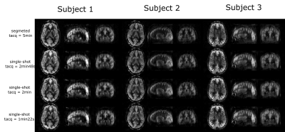
Pseudo-continuous arterial spin labeling combined with 3D segmented readouts is recommended for acquiring ASL perfusion data. However, the total number of k-space encodings limits the trade-off between motion sensitivity and image blurring. To tackle this problem we implemented an accelerated 3D-GRASE sequence with a time-dependent 2D-CAIPIRINHA sampling pattern to increase the temporal incoherence between averages or PLDs. High quality images can be gained from the under-sampled time series by a variational image reconstruction approach with total-generalized-variation (TGV) regularization in space and time. This allows acquisition of single-shot 3mm isotropic ASL data with whole brain coverage within 1min22sec.
|
13:42
|
0842.
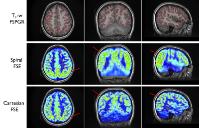 |
High-resolution whole brain ASL perfusion imaging using variably undersampled Cartesian Fast-Spin-Echo and Compressed Sensing reconstruction
Manuel Taso, Li Zhao, David Alsop
3D whole-brain ASL is typically performed using either Stack-of-Spirals FSE or GRASE. However, those readouts suffer from significant image blurring and off-resonance sensitivity. Hence, Cartesian encoding would be highly desirable because of its robustness and high image quality, but acquisition times remain prohibiting. We therefore report the implementation of an accelerated 3D-FSE sequence using variable-density Poisson-disk undersampling to provide redundant k-space center sampling while varying the outer pseudo-random sampling and Parallel-Imaging Compressed-Sensing reconstruction to provide high quality high-resolution whole brain ASL perfusion images.
|
13:54
|
0843.
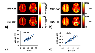 |
Comparison of MR Fingerprinting ASL with Gd-based DSC MRI: validation and direct parametric mapping with deep learning
Pan Su, Peiying Liu, Yang Li, Ye Qiao, Jun Hua, Doris Lin, Jay Pillai, Argye Hillis, Hanzhang Lu
MR Fingerprinting (MRF)-based Arterial-Spin-Labeling (ASL) has been recently proposed as a new approach to measure multiple hemodynamic parameters in a single scan. However, the previous implementation of MRF-ASL lacks the comparison with clinical standard techniques such as dynamic-susceptibility-contrast (DSC). Therefore, in this work, we validated MRF-ASL by comparing with DSC MRI. The results showed that these two methods provided visually comparable and quantitatively correlated perfusion estimations. Furthermore, we sought to directly estimate DSC-equivalent parameters from the MRF-ASL raw data using a deep-learning (DL) approach. DL-derived maps show better quality and are more consistent with DSC maps, compared to dictionary-matching results.
|
14:06
|
0844.
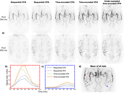 |
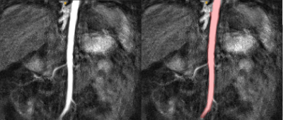
Optimization of time-encoded pseudo-continuous ASL angiography with a variable flip-angle scheme
Joseph Woods, S. Sophie Schauman, Mark Chiew, Michael Chappell, Thomas Okell
Time-encoded PCASL angiography is an attractive method for deriving some, or all, of the desired temporal information from the PCASL preparation rather than a dynamic readout. Higher flip-angles can then be used for the remaining excitation pulses in the readout, increasing the signal-to-noise ratio of the angiograms.
However, use of constant flip angles across all excitations leads to abrupt signal changes between images constructed from different time-encoding blocks. A variable flip-angle scheme is presented, which maintains a constant signal across all excitations, removes these discontinuities and ensures optimal signal levels across all images.
|
14:18
|
0845.
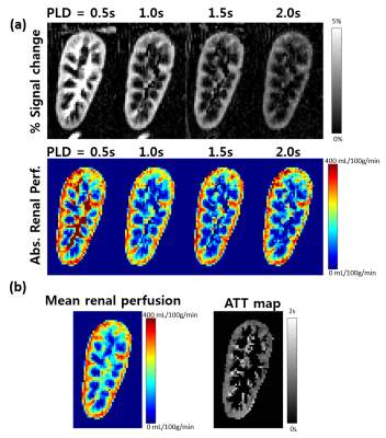 |
Assessment of renal perfusion in transplanted kidney patients using multi-delay pseudo-continuous arterial spin labeling
Hyun-Seo Ahn, Eun-Sung Kim, Hyo Sung Kwak, Sung-Hong Park
There has been no systemic evaluation for transplanted kidney patients using 3T MRI system with pseudo-continuous ASL (pCASL). In this study, we presented multi-parametric renal perfusion imaging on transplanted kidney patients using multi-delay pCASL with bSSFP readout. Influence of background suppression on temporal signal stability was investigated and two different perfusion quantification models were compared. Multi-parametric perfusion signals including mean renal perfusion and arterial transit time were measured for transplanted kidney patients. The current study would be helpful for applying multi-delay pCASL to routine clinical diagnosis of transplanted kidney patients.
|
14:30
 |
0846.
 |
Glomerular filtration rate estimation by motion-robust high spatiotemporal resolution DCE-MRI with radial VIBE and comparison with plasma clearance of 99mTc-DTPA
Sila Kurugol, Onur Afacan, Alto Stemmer, Richard Lee, Jeanne Chow, Simon Warfield
The accuracy of MRI based glomerular filtration rate (GFR) measurements using the standard Cartesian VIBE DCE-MRI is limited by motion and temporal resolution. Instead, we use motion-robust high temporal resolution radial VIBE for DCE-MRI for accurate estimation of GFR. We optimize its temporal resolution and compressed sensing reconstruction temporal regularization parameters to obtain an accurate arterial input function peak while reconstructing good quality images. We also developed a fully automated segmentation and tracer kinetic model-fitting pipeline to compute MRI-GFR. We assessed the accuracy of proposed technique to measure GFR by comparing MRI-GFR to GFR from 99mTcDTPA nuclear medicine study (NMGFR).
|
14:42
|
0847.
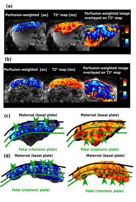 |
Joint measurement of perfusion and apparent oxygenation in the human placenta (PERFOX)
Jana Hutter, Anita Harteveld, Laurence Jackson, Suzanne Franklin, Clemens Bos, Matthias van Osch, Jonathan O'Muircheartaigh, Alison Ho, Laura McCabe, Lucy Chappell, Joseph Hajnal, Mary Rutherford, Enrico De Vita
Novel insight into placental oxygenation and perfusion was achieved by integrating velocity-selective arterial spin labeling and T2* mapping into one scan. Quantitative values can be obtained dynamically and geometrically fixed, allowing both separation of effects and joint visualization.
|
14:54
|
0848.
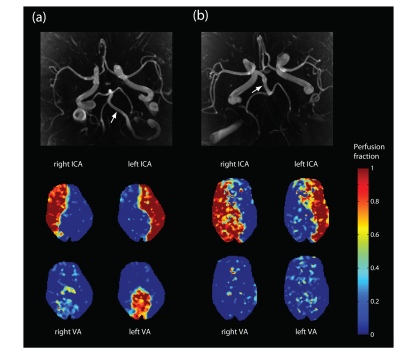 |
Quantification of cerebral perfusion fraction map using cardiac-triggered, off-resonance calibrated super-selective arterial spin labeling
Jonas Schollenberger, C. Alberto Figueroa, Luis Hernandez-Garcia
The impact of off-resonance compensation and cardiac triggering on vessel- selective ASL labeling efficiency and image quality was investigated. A strategy to calculate individual vessel’s perfusion fraction maps based on measurements of vessel-selective labeling efficiency is also presented. This work serves as a practical guideline for acquiring high quality, quantitative perfusion fraction maps with super-selective pseudo-continuous arterial spin labeling.
|
15:06
|
0849.
 |
7T high-resolution arterial spin labeling reveals layer dependent cerebral blood flow
Xingfeng Shao, Kai Wang, Danny J.J. Wang
The microcirculation of cerebral cortex has been investigated by anatomical studies in animals and human brain tissue. For the first time, we applied high resolution (0.5x0.5x1.4mm3) pulsed and pseudo-continuous ASL (pCASL) with inner-volume GRASE readout at 7T to measure CBF in the superficial, middle and deep layers of human cerebral cortex. Our results show higher perfusion in the middle layer using pCASL, while perfusion in the superficial layer of PASL may be affected by pial artery signals. In vivo imaging of layer dependent CBF opens door to investigating neurovascular coupling in a laminar fashion.
|
15:18
|
0850.
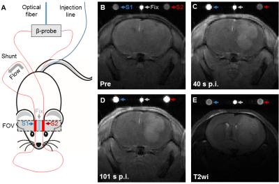 |
An Extracorporeal Circulation Mouse Model for Simultaneous Measurements of Dynamic Contrast-Enhanced Arterial Input Functions and Radiotracer Blood Concentrations
Philipp Backhaus, Florian Büther, Lydia Wachsmuth, Lynn Frohwein, Klaus Schäfers, Sven Hermann, Michael Schäfers, Cornelius Faber
Quantification of the arterial input function (AIF) in small animals is challenging in both dynamic contrast-enhanced (DCE)-MRI and in radiotracer studies. We present a novel extracorporeal circulation mouse model for DCE-measurements of the AIF in mice. The approach allows parallel measurement of tissue contrast dynamics as well as of radiotracer AIF using a β-microprobe integrated into the extracorporeal circulation.
|
|
