Novel CEST & MT Preclinical Investigations
Oral
Contrast Mechanisms
Tuesday, 14 May 2019
| Room 520A-F |
13:30 - 15:30 |
Moderators: Kannie WY Chan, Junzhong Xu |
13:30
|
0540.
 |
CEST MRI with distribution-based analysis for assessment of early stage disease activity in a multiple sclerosis mouse model

Tao Liu, Aline Thomas, Yanrong Chen, Jeff Bulte, Xiaolei Song
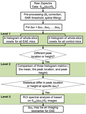
Imaging biomarkers that can detect pathological alterations earlier in multiple sclerosis (MS) progression may enable earlier intervention and improved therapeutic efficacy of available treatments. Here we assessed disease manifestations at an early stage using CEST MRI in a preclinical MS model with histogram-guided analysis. The analysis method is simple-to-execute and robust for evaluation of diseases with subtle changes that lack a priori knowledge of abnormal regions of interest (ROIs) and have multiple potentially contributing offsets. We demonstrated that CEST Z-spectra signals at 1 and 2 ppm are potential MRI biomarkers for detecting early and subtle pathological changes in EAE mice.
|
13:42
 |
0541.
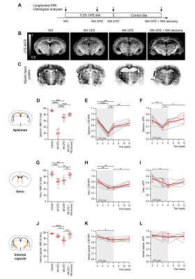 |
Ultrashort echo time magnetization transfer (UTE-MT) imaging in the cuprizone mouse model of multiple sclerosis
Caroline Guglielmetti, Tanguy Boucneau, Peng Cao, Annemie Van der Linden, Peder Larson, Myriam Chaumeil
We first evaluated the potential of ultrashort echo time magnetization transfer (UTE-MT) and MT imaging to generate high contrast images of the healthy mouse brain. Next, we conducted a longitudinal study to examine the temporal changes of UTE-MT ratio (UTE-MTR) and MTR following cuprizone (CPZ)-mediated demyelination, gliosis, and remyelination. UTE-MTR detected CPZ-induced alterations in white matter, subcortical, and cortical grey matter during demyelination, and persistent tissue microstructure changes in grey matter. Furthermore, UTE-MTR changes correlated significantly with myelin levels.
Altogether, we showed that UTE-MT imaging holds great potential to improve characterization of brain lesions in MS at clinical field strength.
|
13:54
 |
0542.
 |
Sequential and Deep Multi-Pool CEST MR Fingerprinting in In-Vivo Tumor-Bearing Mice
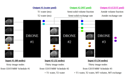
Or Perlman, Ouri Cohen, Hirotaka Ito, Hiroshi Nakashima, E. Antonio Chiocca, Matthew Rosen, Christian Farrar
Magnetic resonance fingerprinting (MRF) was recently suggested for fast and quantitative chemical exchange saturation transfer (CEST) imaging. However, for in-vivo pathologies, multiple tissue parameters will vary simultaneously, thereby reducing the schedule discrimination ability and increasing the reconstruction time. Herein, we propose the sequential utilization of three MRF acquisition schedules and their incorporation in deep-learning reconstruction networks (DRONE). The technique outputs 6 quantitative maps (water, semi-solid, and amide pool properties) with acquisition and reconstruction times of 365s and <200ms, respectively. The method was evaluated in a longitudinal brain-tumor mouse study, yielding comparable parameter values to ground-truth and traditional Z-spectrum evaluations.
|
14:06
|
0543.
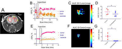 |
Hemodynamic imaging in brain tumors using dextran1-enhanced CEST MRI
Chuheng Chen, Zheng Han, Xiang Xu, Renyuan Bai, Verena Staedtke, Jiadi Xu, Linda Knutsson, Peter Zijl, Guanshu Liu
Sugar-based compounds have shown potential as biodegradable contrast agents for cancer detection. Recently, a multi-size dextran MRI approach was developed for detecting permeability-related properties in tumors. Here, we explored a small size dextran, dextran1 (MW~ 1 kD), as a new MRI contrast agent for detecting brain tumor hemodynamic properties including blood–brain barrier (BBB) breakdown and cerebral blood volume. Our results demonstrate that dextran1 has great potential in indicating BBB integrity, which is confirmed by dynamic contrast-enhanced (DCE) MRI and histology findings. Importantly, contrary to D-glucose, dextran1 does not enter the CSF, removing a source of interfering background signal that is present in dynamic glucose-enhanced (DGE) MRI.
|
14:18
|
0544.
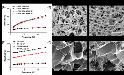 |
Imaging injectable liposomal hydrogels using CEST MRI for applications in the brain
Xiongqi Han, Jianpan Huang, Peng Xiao, Joseph Ho Chi Lai, Kannie Wai Yan Chan
Nanocarrier-loaded hydrogel shows good prospects in biomedical application. However, there is lack of noninvasive methods for real time monitoring to guide therapy. Here, we developed a CEST-nanosensor incorporated injectable hydrogel using two most commonly used hydrogel (alginate, and hyaluronan-methylcellulose (HAMC)) and liposomes. Both hydrogels showed storage modulus (G’) comparable to native brain tissue and good injectabilities. Moreover, we observed that our LipoCEST hydrogels generate unique CEST contrasts at 5 ppm (barbituric acid (BA), drug) and -3.4 ppm (liposomes) on 3T, facilitating the multicolor CEST imaging and imaging-guided therapy in brain applications.
|
14:30
|
0545.
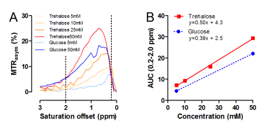 |
CryptoCEST, a promising tool for differential diagnosis and treatment monitoring of fungal brain lesions
Liesbeth Vanherp, Kristof Govaerts, Matteo Riva, Katrien Lagrou, Greetje Vande Velde, Willy Gsell, Uwe Himmelreich
Differential diagnosis of fungal brain infections from other types of lesions is challenging. We assessed the presence of fungal metabolites in Cryptococcus lesions using in vivo MR spectroscopy and CEST. Hereby, we studied the use of trehalose, a disaccharide and fungal biomarker, for endogenous contrast in CEST imaging. Using the novel CryptoCEST technique, we were able to non-invasively differentiate Cryptococcus lesions from glioma. However, no significant effect of antifungal treatment on the CEST contrast was observed. If translated to the clinic, this technique has great potential in assisting in the differential diagnosis and specific spatial localization of cryptococcal brain lesions.
|
14:42
|
0546.
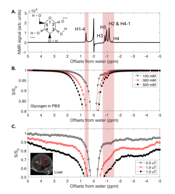 |
In vivo MRI of mouse liver glycogen based on the nuclear Overhauser enhancement (NOE) with water (glycoNOE)
Yang Zhou, Xiang Xu, Jiadi Xu, Lin Chen, Yuguo Li, Peter van Zijl, Nirbhay Yadav
Glycogen is abundant in different tissues and plays a central role in glucose homeostasis. We propose a new method for imaging glycogen using the nuclear Overhauser enhancement (NOE) between glycogen and water. We mapped the glycogen NOE signal in the Z-spectral range around -1 ppm from water, both in phantoms and in mouse liver in vivo. For validation, we performed a fasting protocol and glucagon infusions. Glycogen levels were greatly reduced after 24 hours fasting and after the intraperitoneal injection of glucagon (100 ml, 1mg/ml). This glycoNOE MRI has the potential for non-invasive imaging of liver glycogen levels in vivo.
|
14:54
|
0547.
 |
Detection of abnormal glucose uptake and metabolism in perinatal hypoxia using glucoCEST MRI
Tsang-Wei Tu, Chao-Hsiung Hsu, Tirone Johnson, Paul Wang, Joseph Scafidi
Perinatal brain injury, such as perinatal hypoxia from chronic lung disease, results in devastating, neurologic impairment. The immediate and long-term effects on brain energy metabolism of glucose – a major source of energy for the brain - are not known. Previous studies have shown that perinatal brain injury from perinatal hypoxia results in long-term decreases in neuronal oxidative metabolism of glucose and decreased synthesis of N-acetylaspartate. In this study, we utilized dynamic glucoCEST enhancement MRI to investigate the pattern of glucose uptake and metabolism between the mice of normoxic and hypoxic treatment.
|
15:06
|
0548.
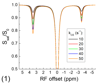 |
Saturation transfer of relayed nuclear Overhauser enhancement: its relationship with the chemical exchange
Tao Jin, Julius Chung, Seong-Gi Kim
In saturation transfer experiments, the Nuclear Overhauser enhancement (NOE) is often detected from mobile macromolecules and is usually assumed through an exchange-relayed pathway. However, the relationship of such relayed-NOE signals with the chemical exchange is still unclear. We reported simulation, phantom and in vivo studies to examine the dependence of relayed-NOE signal on the chemical exchange.
|
15:18
|
0549.
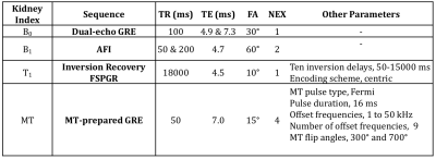 |
Quantitative Magnetization Transfer Detects Renal Fibrosis in Swine Kidney with Renal Artery Stenosis
Kai Jiang, Christopher Ferguson, John Woollard, James Krier, Xiangyang Zhu, Lilach Lerman
Quantitative magnetization transfer (qMT) was used on swine kidneys with renal artery stenosis (RAS). The reproducibility of qMT at 1.5 and 3.0 T was tested in two RAS pigs and its utility in measuring renal fibrosis investigated in seven pigs at 3.0 T by comparing to histology. The Henkelman-Ramani equation was used to derive the bound pool fraction f. Comparable f values were obtained at 1.5 and 3.0 T. Measured f also correlated well with renal fibrosis by histology. In conclusion, qMT is reproducible at different field strengths and can be used to measure renal fibrosis in swine with RAS.
|
|
