Relaxometry: Measuring, Understanding & Using
Oral
Contrast Mechanisms
Thursday, 16 May 2019
| Room 511BCEF |
16:00 - 18:00 |
Moderators: Martina Callaghan, Richard Dortch |
16:00
 |
1209.
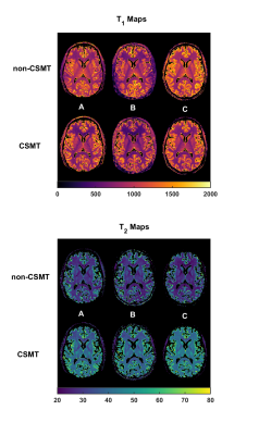 |
Multi-Vendor Validation of Controlled Saturation Magnetization Transfer (CSMT) for VFA T1 and T2 mapping

Rui Pedro A G Teixeira, Radhouene Neji, Tobias Wood, Ana Baburamani, Shaihan Malik, Jo Hajnal
White matter T1 values have been reported to range from 699ms-1735ms1. The recently introduced Controlled Saturation Magnetization Transfer (CSMT)2 qMRI approach suggests this might be due to MT effects. In this work employ the CSMT framework on 3 major MR manufacturers and demonstrate it allows a decrease in variability of T1/T2 estimation both in phantom and in-vivo.
|
16:12
|
1210
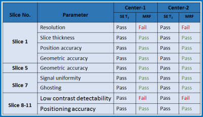 |
Repeatability Measurements of Magnetic Resonance Fingerprinting Metrics using ACR and ISMRM/NIST Phantoms: A Multi-Center Study
Video Permission Withheld
Amaresha Konar Shridhar, Guido Buonincontri, Vaios Hatzoglou, Maggie Fung, Pedro Gomez, Rolf Schulte, Kavya Prasad, Michael Liu, Miriam Klein, Sairam Geethanath, Lawrence Schwartz, Amita Shukla-Dave, Sachin Jambawalikar
Magnetic Resonance Fingerprinting (MRF) provides multiple quantitative imaging (QI) metrics within a single MR acquisition. The quantitative images must be standardized to generate MR protocols for acquisition of biomarkers. In this study both qualitative and quantitative repeatability experiments have been performed using ACR and ISMRM/NIST system phantoms at three collaborative centers (2 USA, 1 Europe) to evaluate imaging data consistency and the inherent reliability (Coefficient of Variation) of MRF. Results of both qualitative and quantitative study has shown reliability of MRF method.
|
16:24
|
1211.
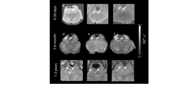 |
Iron-Induced MR Contrast in Human Locus Coeruleus: A Cautionary Tale of Misleading Post Mortem MRI Results
Evgeniya Kirilina, Charlotte Lange, Carsten Jäger, Tilo Reinert, Kerrin Pine, Thomas Lohmiller, Siawoosh Mohammadi, Tobias Streubel, Malte Brammerloh, Anneke Alkemade, Birte Forstmann, Andreas Herrler, Alexander Schnegg, Markus Morawski, Nikolaus Weiskopf
MR contrast mechanisms in human locus coeruleus were studied combining high-resolution post mortem MRI, histology, ion-beam microscopy, and electron paramagnetic resonance. We demonstrate that the main source of MR contrast in formalin fixed LC is paramagnetic iron accumulated in noradrinergic neurons. However, we show that MR contrast in LC drastically changes during the first six months of tissue fixation. We assign these changes to iron been scavenged by neuromelanin and the change of its paramagnetic state. The results have major consequences for MRI of the locus coeruleus, demonstrating a fundamental change rather than the commonly known gradual changes in contrast due to formalin fixation.
|
16:36
|
1212.
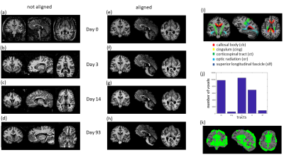 |
Longitudinal assessment of relaxation and magnetization transfer saturation rates during formalin fixation across fiber pathways of the human brain
Tobias Streubel, Mohammad Ashtarayeh, Herbert Mushumba, Sebastian Papazoglou, Klaus Püschel, Siawoosh Mohammadi
We longitudinally investigated the effect of brain tissue fixation, using 4% paraformaldehyde, on three potential quantitative myelin MRI markers across different white matter fiber pathways of the human brain: longitudinal (R1) and apparent transverse (R2*) relaxation rates and magnetization transfer (MT) using the quantitative multi-parameter mapping (MPM) protocol. To better understand the temporal evolution of the fixation process within the whole brain and its influence on MRI parameters, we monitored the temporal evolution of the fixation process of a whole human post-mortem brain using the same MPM protocol at 15 time-points (one unfixed, in-situ MRI scan and 14 ex-vivo MRI scans) at different stages of the fixation process (days 1-93).
|
16:48
 |
1213.
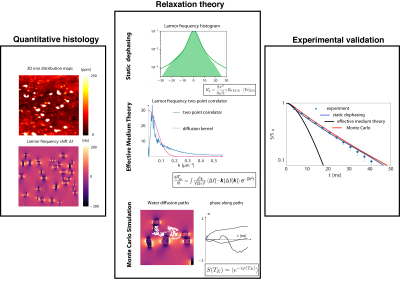 |
A Biophysical Model of Iron-Induced Relaxation in Human Substantia Nigra for Early Stage Parkinson Diagnostic
Malte Brammerloh, Charlotte Lange, Tilo Reinert, Carsten Jäger, Filippos Gavriilidis, Kerrin Pine, Isabel Weigelt, Robert Trampel, Enrico Reimer, Markus Morawski, Thomas Arendt, Nikolaus Weiskopf, Evgeniya Kirilina
We present a biophysical model that quantitatively describes gradient and spin echo relaxation in substantia nigra induced by iron accumulated the neuromelanin of dopaminergic neurons. This model was informed by 3D quantitative microscopic iron distributions obtained from classical histology and Proton-Induced X-ray Emission. It was validated by comparison to quantitative MRI on post mortem human brain tissue. We demonstrate that the total iron content of DN can be extracted analytically from relaxometry in nigrosomes. This results provides an important step toward a highly desired early stage biomarker for detecting dopaminergic neurons depletion in Parkinson's disease.
|
17:00
|
1214.
 |
Image contrast at 0.55T
Adrienne Campbell-Washburn, Daniel Herzka, Peter Kellman, Alan Koretsky, Robert Balaban
In addition to having cost advantages, low-field MRI may offer new imaging opportunities by virtue of the physical attributes. Here we report T1, T2 and T2* tissue parameters and contrast agent relaxivities for a custom high performance 0.55T MRI system. We found that T1 was 29% shorter at 0.55T compared 1.5T, whereas T2 was lengthened by 33% and T2* was lengthened by 54% at 0.55T. Marketed small molecular weight gadolinium-based contrast agents demonstrate similar relaxivity at 0.55T and 1.5T, indicating similar dosing requirements, and larger molecular complex agents had increased T1 relaxivity at 0.55T, indicating potential advantages at low-field.
|
17:12
|
1215.
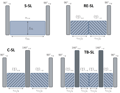 |
A totally balanced spin lock preparation module for accurate and artifact-free T1?-mapping
Maximilian Gram, Daniel Gensler, Anton Xu, Peter Nordbeck, Wolfgang Rudolf Bauer, Peter Michael Jakob, Michael Seethaler
To date, one of the main challenges of accurate and artifact-free T1ρ-mapping is the sensitivity of the required spin lock preparation module against field imperfections. Already established methods are sensitive to B0 and B1 field inhomogeneities. In this work, we present a novel spin lock preparation module that aims to be totally balanced, meaning that every pulse is being compensated by a correspondent pulse with opposite phase. Our new method proves to be highly robust to both types of field inhomogeneities. The superiority over common methods is demonstrated by Bloch simulations and measurements of a glucose phantom at 7.0T.
|
17:24
|
1216.
 |
Free Breathing R2* Mapping Using Three-Dimensional Self-Gating Motion-Compensated Stack-of-Radial MRI
Xiaodong Zhong, Tess Armstrong, Marcel Nickel, Stephan Kannengiesser, Li Pan, Vibhas Deshpande, Holden Wu
R2* mapping is an emerging biomarker to evaluate iron overload. Due to its relative insensitivity to motion, free-breathing 3D stack-of-radial imaging is a useful technique in patient populations with breath-hold difficulties. However, its performance for R2* quantification is still under investigation. This study proposes a new 3D self-gating motion-compensated free-breathing stack-of-radial MRI method for R2* quantification. Preliminary in vivo results demonstrated good agreement of R2* mapping in the liver with the proposed method compared to the reference results of breath-hold Cartesian MRI.
|
17:36
|
1217.
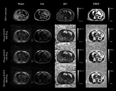 |
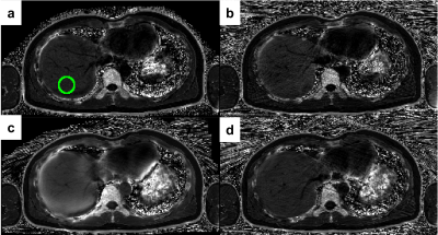
Free-Breathing Water, Fat, and Iron Quantification in the Abdomen Using Radial Multi-Echo Acquisition and Respiratory-Resolved Model-Based Reconstruction
Manuel Schneider, Thomas Benkert, Eddy Solomon, Dominik Nickel, Matthias Fenchel, Berthold Kiefer, Andreas Maier, Hersh Chandarana, Kai Tobias Block
A novel method for free-breathing fat/iron quantification with integrated R2* correction is presented, which is based on radial stack-of-stars 3D GRE acquisition with multiple echoes. An iterative model-based approach with TV as sparsifying transformation is used for the reconstruction. Results from full k-space data, retrospectively undersampled data, and motion-resolved processing are compared to a Cartesian reference technique in 5 clinical patients. The proposed method provides comparable results while eliminating the requirement to perform acquisitions during breath-holding, which poses a significant advantage for patients with reduced breath-hold capability such as pediatric, sick, or elderly patients.
|
17:48
|
1218.
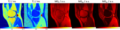 |
Click Your Fat Away: Rapid Synthetic Knee MR Imaging with Switchable Fat and Magnetization-Transfer Contrast
Tom Hilbert, Jan Fritz, Patrick Omoumi, José Marques, Emilie Mussard, Jean-Philippe Thiran, Reto Meuli, Tobias Kober
Synthetic MRI is increasingly used in clinical practice to supplement or replace conventional MRI, due to the ability to generate both a multitude of MR images with weighted contrasts and quantitative T1, PD and T2 maps, which is of high interest in musculoskeletal imaging. To further broaden the application of synthetic image contrasts, we developed a method which allows switching between images with and without fat signal as well as magnetization-transfer weighting. The method includes three pulse sequences to estimate quantitative maps for the calculation of a variety of contrasts and requires a total acquisition time of 6:46 min.
|
|
