Multidimensional Signal Encoding Decoding
Oral
Acquisition, Reconstruction & Analysis
Thursday, 16 May 2019
| Room 710B |
13:45 - 15:45 |
Moderators: Teresa Correia, Jonathan Tamir |
13:45
|
1175.
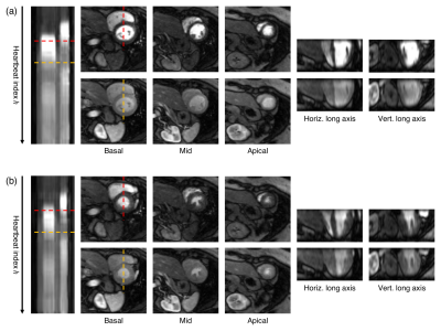 |
Respiratory motion compensated Multitasking for 3D myocardial perfusion without breath-holds, ECG, or multiple boluses

Anthony Christodoulou, Nan Wang, Jaime Shaw, Xiaoming Bi, Yibin Xie, Christopher Nguyen, Debiao Li
Quantitative myocardial perfusion MRI is potentially powerful for diagnosing coronary artery disease. However, It is challenging to accurately measure dynamic contrast enhancement throughout the whole moving heart. Most perfusion methods employ some combination of ECG-triggering, breath-holds, dual-bolus acquisition, and multislice 2D acquisition, leading to difficult workflows and limited spatial coverage. Here we describe a non-ECG, free-breathing, single-bolus, 3D alternative which employs the MR Multitasking framework to perform low-rank tensor imaging. We add 3D respiratory motion compensation to this framework to eliminate the need for breath-holds. This new method is demonstrated and evaluated in healthy volunteers.
|
13:57
 |
1176.
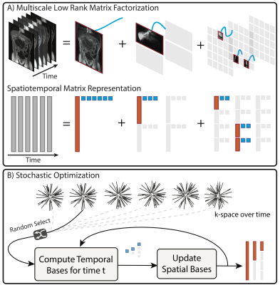 |
Extreme MRI: Super-High-Res Dynamic Volumetric MRI from Continuous Non-Gated Acquisition
Frank Ong, Xucheng Zhu, Peder Larson, Joseph Cheng, Shreyas Vasanawala, Michael Lustig
The goal of this work is to recover transient dynamics in 3D dynamic MRI by reconstructing images with near-millimeter spatial resolution and sub-second temporal resolution without gating. This setting poses two major challenges: extreme undersampling and extreme computational/memory cost. To achieve this “extreme MRI”, we propose two innovations: explicit multi-scale low rank matrix factorization to regularize the problem and reduce memory usage, and stochastic optimization to reduce computation. We demonstrate the feasibility of the proposed method in DCE imaging acquired with 3D cones trajectory and lung imaging acquired with 3D UTE radial trajectory.
|
14:09
|
1177.
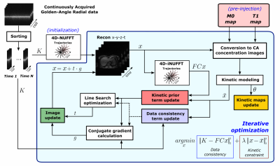 |
A kinetic-guided compressed sensing approach for DCE-MRI reconstruction
Michele Scipioni, Niccolo Fuin, Julie Price, Onofrio Catalano, Ciprian Catana
In this work, we propose a reconstruction method for free-breathing DCE-MRI, that combines golden-angle radial sampling, parallel imaging and tracer kinetic (TK) modeling in an iterative reconstruction approach. We introduce a new model in which information coming from TK modeling are treated as a priori knowledge, assisting image reconstruction. The proposed approach is compared with Total Variation Compressed Sensing reconstruction achieving comparable denoising effect in the spatial domain, but improved temporal fidelity and TK modeling.
|
14:21
|
1178.
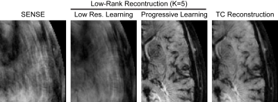 |
High Resolution DSC Perfusion Imaging Using Low Rank Reconstruction with Progressive Learning
Julia Velikina, Alexey Samsonov
Single shot echo-planar imaging, traditionally used for dynamic susceptibility contrast (DSC) perfusion-weighted MRI (PWI), is not efficient for high resolution imaging due to T2* signal decay and poor compatibility with advanced reconstruction techniques. Spiral acquisition samples k-space more efficiently and is amenable to advanced reconstructions necessary for imaging at higher resolution and for multi-echo imaging. However, for high accelerations, spirals lack necessary k-space center training data. We enable low-rank-based reconstruction for multi-echo spiral acquisition by introducing progressive learning of temporal models. Our approach is demonstrated for multi-echo spiral DSC PWI with high spatial/temporal resolution (1.1x1.1mm/1.1s).
|
14:33
 |
1179.
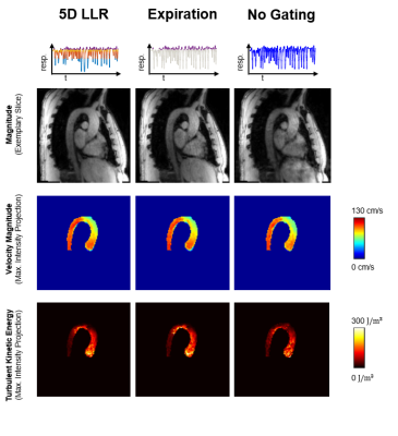 |
Multi-Point 5D Flow MRI - Accelerated Cardiac- and Respiratory-Motion Resolved Mapping of Mean and Turbulent Velocities in 4 Minutes
Jonas Walheim, Hannes Dillinger, Sebastian Kozerke
This work presents a framework for respiratory motion-resolved multi-point 4D Flow MRI (MP 5D Flow MRI) of mean and turbulent velocities in 4 minutes using a combination of Cartesian Golden angle undersampling, data-driven motion detection and locally low-rank imaging reconstruction. In an imaging study with 9 volunteers, 7-point 5D Flow MRI was compared to a standard, navigator-gated 4-point 4D Flow MRI parallel imaging protocol. Results demonstrate that flow fields from MP 5D Flow MRI in end-expiration agree well with 4D Flow MRI data while scan time was reduced by a factor of 4.5. In addition, MP 5D Flow MRI provides higher accuracy for low velocities and allows assessing TKE over a larger dynamic range compared to 4D Flow MRI.
|
14:45
|
1180.
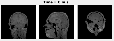 |
Prospective 3D+t non-rigid motion estimation at high frame-rate from highly undersampled k-space data: validation and preliminary in-vivo results
Niek Huttinga, Tom Bruijnen, Cornelis van den Berg, Peter Luijten, Alessandro Sbrizzi
We previously presented a framework to reconstruct 3D non-rigid motion-fields from highly undersampled k-space data by exploiting the lower-dimensional nature of motion-fields. Here, a quantitative comparison with a well-established image-based motion estimation is performed on 3D abdomen images. To also exploit temporal compressibility the framework is extended to reconstructions of spatio-temporal non-rigid motion-fields at high frame-rate, and validation is performed on 2D+t abdomen cine-acquisitions. Additionally, a dedicated non-Cartesian 3D trajectory was employed to prospectively acquire highly undersampled k-space data, and reconstruct 3D+t head motion at 16Hz. High quality respiratory/head motion-fields are obtained and the framework outperforms the fully-sampled image-based motion estimation even for undersampling up to 8x.
|
14:57
|
1181.
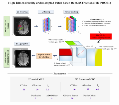 |
High-Dimensionality Undersampled Patch-Based Reconstruction (HD-PROST) for Accelerated Multi-Contrast Magnetic Resonance Imaging
Aurelien Bustin, Gastao Cruz, Olivier Jaubert, Karina Lopez, René Botnar, Claudia Prieto
In magnetic resonance imaging, multiple contrasts are exploited to extract clinically relevant tissue parameters and pathological tissue changes. Multi-contrast acquisitions find important applications in parameter mapping (e.g. T1 and T2 mapping) and magnetic resonance fingerprinting. However, these acquisitions lead to long scan times since multiple images with different contrasts need to be acquired. In this study, we present a new reconstruction technique, termed as High-Dimensionality undersampled Patch-based RecOnSTruction (HD-PROST), for highly accelerated 2D and 3D multi-channel multi-contrast MRI.
|
15:09
|
1182.
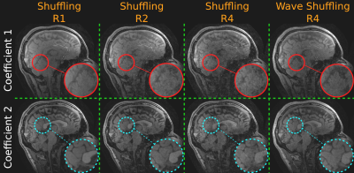 |
Rapid, Time-Resolved Brain Imaging with Multiple Clinical Contrasts using Wave-Shuffling.
Siddharth Iyer, Daniel Polak, Congyu Liao, Steve Cauley, Berkin Bilgic, Kawin Setsompop
Previously, we presented Wave-Shuffling: a technique that combines random-sampling and the temporal low-rank priors of T2-shuffling with the sinusoidal gradients of Wave-CAIPI to achieve highly-accelerated, time-resolved acquisitions. In this work, we optimize and apply Wave-Shuffling to T2-SPACE and MPRAGE to achieve rapid, 1 mm-isotropic resolution, time-resolved imaging of the brain where we recover a time-series of clinical contrast. We also present preliminary explorations into Joint-Contrast Wave-Shuffling to boost signal-to-noise ratio and reconstruction-conditioning at high accelerations.
|
15:21
|
1183.
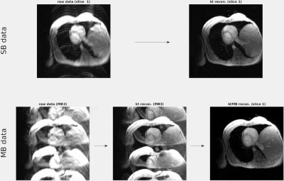 |
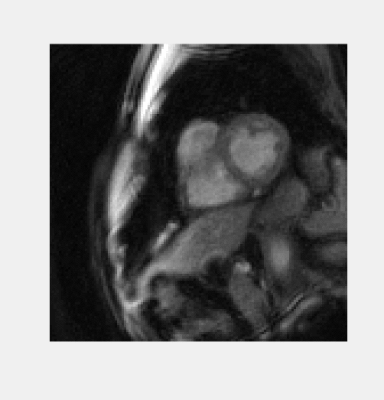
Autocalibrated multiband CAIPIRINHA with k-t-acceleration: Towards the complete spatio-temporal coverage of the heart motion in one single breath-hold
Giulio Ferrazzi, Jean Pierre Bassenge, Johannes Mayer, Clarissa Wink, Alexander Ruh, Michael Markl, Bernd Ittermann, Tobias Schaeffter, Sebastian Schmitter
Recent developments in cardiac multiband CAIPIRINHA with autocalibration allow the simultaneous acquisition and reconstruction of multiple slices in one breathhold. Nevertheless, applications such as high resolution multi-slice cine imaging, and multi-slice three-directional velocity encoding of the myocardium, are still hindered by excessive breathhold duration.
In this study, we combine autocalibrated multiband CAIPIRINHA with k-t-acceleration to substantially accelerate data acquisition. When applied to gradient-echo and phase-contrast imaging, our technique allowed cardiac cine acquisitions at high spatial resolution (1.1mm), and three-directional velocity encoding of the myocardium, to be acquired in one breath-hold of 18 heart-beats at 2 or 3 slices, simultaneously.
|
15:33
|
1184.
 |
Self-calibrating through-time spiral GRAPPA for flexible real-time imaging
Dominique Franson, Jesse Hamilton, Mark Griswold, Nicole Seiberlich
A self-calibrating through-time spiral GRAPPA method is presented that uses the same undersampled scan for both the accelerated data and the calibration data. This method can be used to capture at least one heartbeat worth of data at 12 slice positions in less than one minute, in a free-breathing and ungated scan.
|
|
