Quantitative Parameter Mapping
Oral
Acquisition, Reconstruction & Analysis
Monday, 13 May 2019
| Room 520A-F |
16:00 - 18:00 |
Moderators: Philipp Ehses, Rahel Heule |
16:00
|
0307.
 |
MANTIS: Model-Augmented Neural neTwork with Incoherent k-space Sampling for accelerated MR parameter mapping

Fang Liu, Li Feng, Richard Kijowski
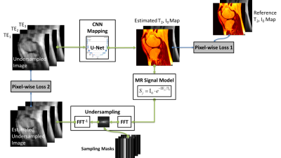
The purpose of this work was to develop and evaluate a novel deep learning-based reconstruction framework called Model-Augmented Neural neTwork with Incoherent k-space Sampling (MANTIS) for accelerated MR parameter mapping. Our approach combines end-to-end CNN mapping with k-space consistency using the concept of cyclic loss to further enforce data and model fidelity. Incoherent k-space sampling is used to improve reconstruction performance. A physical model is incorporated into the proposed framework, so that the parameter maps can be efficiently estimated directly from undersampled images. The performance of MANTIS was demonstrated for T2 mapping of the knee joint. Our study demonstrated that the proposed MANTIS framework represents a promising approach for efficient MR parameter mapping. MANTIS can potentially be extended to other types of parameter mapping with appropriate models.
|
16:12
 |
0308.
 |
Three-Dimensional Whole Brain Simultaneous T1, T2, and Apparent Diffusion Coefficient Mapping Using MR Multitasking
Sen Ma, Anthony Christodoulou, Christopher Nguyen, Fei Han, Nan Wang, Yibin Xie, Debiao Li
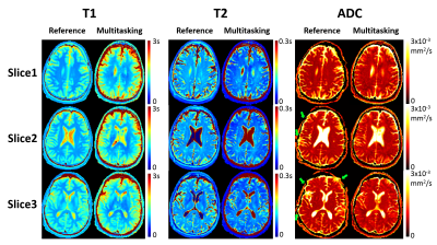
Three-dimensional, multi-parametric quantitative mapping of relaxation and diffusion parameters is desirable for many clinical imaging application, including the diagnosis and follow-up assessment of tumors. Traditionally, this is performed by separate scans which are time-consuming. We propose a novel Multitasking framework to achieve 3D whole-brain simultaneous T1/T2/ADC mapping in ~9min. The underlying multidimensional image is modeled as a low-rank tensor, with a time-resolved phase correction technique to compensate for the phase inconsistency induced by pulsatile motion during diffusion preparation. T1/T2/ADC measurements in healthy volunteers agree with reference methods, yielding intraclass correlation coefficients > 0.80 for both gray and white matter.
|
16:24
|
0309.
 |
Magnetization transfer (MT) of human brain at 7T in the context of a 3D multi-parameter mapping protocol
Hampus Olsson, Mads Andersen, Jimmy Lätt, Ronnie Wirestam, Gunther Helms
3D multi-gradient echo MRI can be used to estimate T1, T2*, PD and the magnetization transfer (MT), which is increasingly used for multi-parametric mapping (MPM) of human brain. The increased polarization at 7T compared to lower B0 allows for increased spatial resolution or reduced scan times. However, SAR restrictions imposed on the MT pulse and B1 inhomogeneity pose challenges. In this work, we propose a protocol for MPM of human brain at 7T with special attention paid to eliminating bias when mapping MTsat while obtaining submillimeter isotropic spatial resolution in under 12 minutes with acceptable SNR.
|
16:36
 |
0310.
 |
Echo Planar Time-Resolved Imaging (EPTI) with subspace constraint and optimized k-t trajectory
Zijing Dong, Fuyixue Wang, Timothy Reese, Berkin Bilgic, Kawin Setsompop
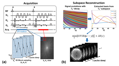
Echo planar time-resolved imaging (EPTI) is a multi-contrast quantitative imaging technique, which achieved fast acquisition of distortion- and blurring-free images at multiple echo times (TE). To improve the SNR and accuracy of EPTI at high-accelerations, in this study, we developed a subspace-constrained reconstruction for EPTI and proposed new k-t sampling trajectories to take advantage of this reconstruction. The subspace reconstruction is also augmented with phase-cycling to extract high-resolution phase data, without need of high-resolution B0 calibration scan. Using the proposed approach, whole-brain 1.1mm-isotropic multi-echo images, and T2* and B0 maps are reconstructed from 3D-EPTI data acquired within 50 seconds.
|
16:48
|
0311.
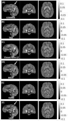 |
Loss Adaptive Dipole Inversion (LADI): A novel data driven approach for quantitative susceptibility mapping
Srikant Kamesh Iyer, Brianna Moon, Rishab Kumar, Walter Witschey
This abstract presents a novel data driven approach for high quality QSM reconstructions without the use of complex and computationally intensive reconstruction models. The purpose of this approach is to develop a reconstruction technique which does not depend on the use of spatial priors from the magnitude image to remove artifacts and reduce blurring of edges. In our proposed formulation, the data fidelity term is updated based on the deviation of the estimated susceptibility map from the measured local field. With the proposed fidelity-loss adaptive reconstruction formulation, removal of artifacts was achieved without causing smoothing of sharp features.
|
17:00
|
0312.
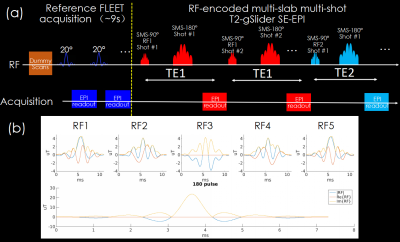 |
T2-gSlider: rapid high resolution T2 mapping with generalized SLIce Dithered Enhanced Resolution and model-based reconstruction
Xiaozhi Cao, Congyu Liao, Siddharth Iyer, Hongjian He, Kawin Setsompop, Jianhui Zhong, Berkin Bilgic
To obtain rapid high isotropic resolution whole-brain T2 maps, a T2 generalized Slice-dithered enhanced resolution (T2-gSlider) acquisition/reconstruction framework is proposed. An accelerated RF-encoded multi-slab, multi-shot SE-EPI acquisition with variable TEs was developed to obtain high SNR acquisitions with reduced TR. A structured low-rank constraint was applied to reconstruct highly undersampled multi-shot data and achieve robust reconstruction for each slab. A Bloch simulated subspace shuffling model was utilized for T2quantification and incorporated into gSlider reconstruction to further accelerate the acquisition. The proposed framework is demonstrated to enable whole-brain 1mm isotropic T2 mapping in ~40 seconds.
|
17:12
|
0313.
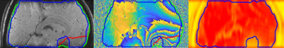 |
Towards robust – and accurate – QSM in cortical and sub-cortical regions of the human brain at 9.4T: the influence of masking
Gisela Hagberg, Elisa Tuzzi, Joana Loureiro, Thomas Ethofer, Rolf Pohmann, Jonas Bause, Pascal Martin, Marina Pavlova, Marc Himmelbach, Anja Zeller, Christoph Laske, Andreas Fallgatter, Klaus Scheffler
Quantitative susceptibility mapping (QSM) targets a fundamental MR-parameter but is problematic due to the presence of a strong background and local field variations. These may cause multiple phase wraps which are particularly prominent at high fields and long echo-times. We propose automated tissue masking excluding brain areas with excessive phase wraps and show how this approach can improve the quality of QSM. Performance was evaluated with regard to iron quantification in subcortical and cortical areas, and was compared with R2* maps in the same 21 subjects aged 19-56y and literature values.
|
17:24
|
0314.
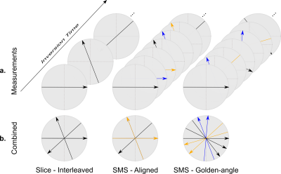 |
Model-based reconstruction for simultaneous multi-slice T1 mapping using single-shot inversion-recovery radial FLASH
Xiaoqing Wang, Sebastian Rosenzweig, Nick Scholand, H. Christian M. Holme, Martin Uecker
Recent advances in real-time MRI and model-based reconstructions have enabled single-slice T1 mapping within a single inversion recovery. To allow fast multi-slice T1 mapping, this work employs radial simultaneous multi-slice (SMS) schemes and develops an SMS model-based reconstruction approach for high-resolution multi-slice T1 mapping based on single-shot inversion recovery FLASH. In comparison to conventional multi-slice approaches, the proposed SMS model-based reconstruction achieves high resolution (0.75 x 0.75 x 5 mm3) T1 maps for three slices of the brain within 4 s with a higher precision and a better preservation of image details.
|
17:36
|
0315.
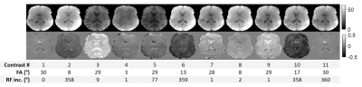 |
Whole-brain 3D multi-parametric quantitative extraction at 7T using parallel transmission Universal Pulses
Lisa Leroi, Vincent Gras, Ludovic de Rochefort, Mathieu Santin, Romain Valabrègue, Franck Mauconduit, Denis Le Bihan, Nicolas Boulant, Alexandre Vignaud
Performing simultaneous quantitative MRI at ultra-high field is challenging, as B0 and B1 heterogeneities, and Specific Absorption Rate increase with field strength. In this work, Quantitative Imaging using Configuration States is successfully applied in vivo at 7T using calibration-free parallel transmission Universal Pulses to retrieve 3D whole-brain M0, flip angle, T1 and T2 maps in a clinically-relevant time. The method relies on the acquisition of multiple contrasts with spoiled SSFP sequence by varying flip angle and radiofrequency spoiling in a limited and optimized number of sets. Quantification of the physical parameters was then performed by fitting acquired data to the Bloch-Torrey equation.
|
17:48
|
0316.
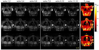 |
Single Breath-Hold T2 Quantification utilizing a Spiral Multi-Contrast TSE Sequence
Naoharu Kobayashi, Michael Garwood
A single breath-hold multi-contrast TSE sequence with spiral k-space sampling is introduced for T2 quantification. Sparsity constraint image reconstruction was applied to reconstruct undersampled datasets acquired under single breath-holding. The feasibility of the proposed sequence and image reconstruction were tested in brain and thoracic imaging for normal volunteers. The proposed method achieved T2 quantification of the thoracic region without clear cardiac motion artifacts.
|
|
