Microstructure: Approaching Cellular Complexity
Oral
Diffusion
Monday, 13 May 2019
| Room 511BCEF |
08:15 - 10:15 |
Moderators: Valerij Kiselev, Chantal Tax |
08:15
 |
0056.
 |
Estimating compartment- and cell-specific microscopic anisotropy in the human brain using double-diffusion encoding spectroscopy at 7T

Chloé Najac, Henrik Lundell, Marjolein Bulk, Hermien E. Kan, Andrew G. Webb, Itamar Ronen
Double diffusion encoding spectroscopy (DDES) offers the unique ability to estimate compartment and cell-specific micro-anisotropy (μFA) in vivo. Recently, this method allowed the estimation of intracellular micro-anisotropy in the white matter. Here, we propose to measure the μFA in different diffusion weighting settings to explore intra- and extracellular μFA in both white (WM) and grey matter (GM). We show that intracellular μFA is similar in both WM and GM, but extracellular space in WM is significantly more anisotropic compared to GM.
|
08:27
|
0057.
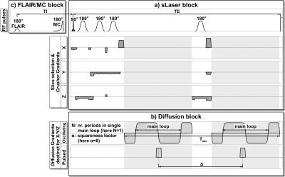 |
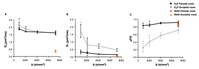
MRS Extended by Oscillating Diffusion Gradients as a Probe for Investigation of Human Brain Tissue Microstructure
André Döring, Roland Kreis
Pulsed diffusion and oscillating diffusion gradients implemented in a semi-Laser sequence for measuring at long and short diffusion times were tested in a phantom and applied in vivo. Metabolite diffusion constants measured in human gray matter in 6 healthy volunteers at short diffusion times were significantly higher than those determined at long diffusion times, suggesting an enhanced sensitivity to diffusion on the cellular and subcellular level.
|
08:39
|
0058.
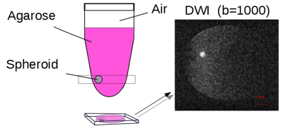 |
Oscillating-gradient diffusion-weighted MRI provides accurate cell radii in tumor spheroids
Marcel Kettelmann, Stephan Niland, Mirjam Gerwing, Markus Wick, Sascha Koehler, Lydia Wachsmuth, Moritz Wildgruber, Johannes Eble, Cornelius Faber
Diffusion weighted MRI using oscillating gradients has previously been shown to provide microstructural information on celllularity in tumors, which may serve as marker for monitoring therapy response. Which geometrical model for analysis of such data provides most reliable results, is, however, a matter of debate. Here, we used the IMPULSED approach, and show that cell radii in tumor spheroids of three different cell lines can be determined with high accuracy. In an in vivo model, however, deviations from radii determined by laser scanning microscopy were found.
|
08:51
 |
0059.
 |
In-vivo Neural Soma Imaging Using B-tensor Encoding and Deep Learning
Noemi Gyori, Christopher Clark, Iulius Dragonu, Daniel Alexander, Enrico Kaden
Diffusion MRI is successfully used to map white matter in the brain. In this work we develop a new clinically viable technique with particular focus on grey matter microstructure. To capture the heterogeneous morphology of grey matter, it is imperative to disentangle cylindrical and spherical geometries commonly attributed to neurites and neural soma. We achieve this by leveraging the latest advances in B-tensor encoding and deep learning techniques and present microstructural feature maps of neurites and neural soma in-vivo in the human brain.
|
09:03
|
0060.
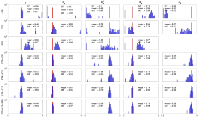 |
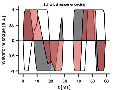
COMPARISON OF DIFFERENT TENSOR ENCODING COMBINATIONS IN MICROSTRUCTURAL PARAMETER ESTIMATION
Maryam Afzali, Chantal Tax, Cyrano Chatziantoniou, Derek Jones
Diffusion-weighted imaging provides information to study the brain microstructure. Several studies in the literature have shown that there is degeneracy in the estimated parameters for a commonly used microstructural model. B-tensor encoding is one of the strategies that has been proposed to solve the degeneracy. The combination of linear-spherical tensor encoding (LTE+STE) and linear-planar (LTE+PTE) have been utilized in previous works. In this paper, we compare different combinations of b-tensor encoding, (LTE+STE), linear-planar (LTE+PTE), planar-spherical (PTE+STE) and linear-planar-spherical (LTE+PTE+STE). We also compare the results of fit using a nonlinear least square algorithm and microstructure imaging of crossing (MIX) method. The results show that the combination of tensor encodings with MIX fitting algorithm leads to lower bias and higher precision in the parameter estimates than single tensor encoding.
|
09:15
|
0061.
 |
Beyond the Standard Model in Spinal Cord
Jonas Olesen, Noam Shemesh, Sune Jespersen
A prevalent model for the diffusion signal from neural tissue is the so-called Standard model which represents axons as “sticks”, i.e., with zero radial diffusivity. Spinal cord tissue is characterized by larger axons than the brain which was recently shown to possibly invalidate the assumption up to large diffusion times. This calls for an appropriate extension of the Standard Model. Here, results from such an endeavour with an extensive dataset from a rat spinal cord are summarized. SM parameters were reliably estimated to be unphysical indicating that the model is insufficient in spinal cord white matter.
|
09:27
|
0062.
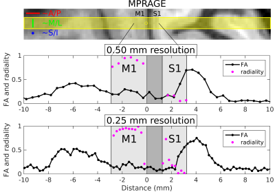 |
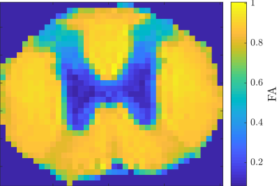
Tangential and radial diffusion in human primary somatosensory and motor cortex: evidence from in-vivo line-scan acquisitions at 7T with 250–500 micron radial resolution
Mukund Balasubramanian, Robert Mulkern, Stephan Maier, Jonathan Polimeni
Eight healthy volunteers were scanned at 7T using a line-scan diffusion sequence with each line prescribed perpendicularly to primary somatosensory (S1) and motor (M1) cortex, and with 250–500 micron resolution along the line. We observed tangential diffusion in S1 and radial diffusion in M1, consistent with prior reports, but with the high radial resolution used here enabling us to identify the deep layers of S1—where high diffusion anisotropy was seen—as the source of the tangential diffusion, with low anisotropy in the upper layers. In M1, radial diffusion with moderate anisotropy was seen at nearly all cortical depths.
|
09:39
|
0063.
 |
Multi-Dimensional Diffusion MRI of the Human Sciatic Nerve
Michael Pridmore, Filip Szczepankiewicz, Guillaume Gilbert, Brian Johnson, Carl-Fredrik Westin, Richard Dortch
Multi-dimensional diffusion MRI is a promising new tool to assess tissue microstructure. Here, we developed a high-resolution protocol that overcomes the challenges of nerve imaging in humans (e.g., the influence of fat, short-T2s) and translated the method to the sciatic nerve in the thigh. Preliminary results showed that measures of microscopic fractional anisotropy in sciatic nerve were 1) similar to reported measures in white matter and 2) were repeatable across scans in the same subject. Additional comparisons to conventional diffusion encodings of the sciatic nerve suggest that multi-dimensional diffusion MRI may yields more specific measures of nerve microstructure.
|
09:51
|
0064.
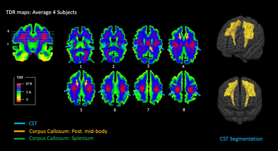 |
Temporal Diffusion Ratio (TDR): A Diffusion MRI technique to map the fraction and spatial distribution of large axons in the living human brain
Flavio Dell'Acqua, Robert Dallyn, Andrea Chiappiniello, Ahmad Beyh, Chantal Tax, Derek Jones, Marco Catani
This work presents a new approach to mapping the fraction and spatial distribution of large axons (d>3μm) across white matter (WM) in the living human brain. By collecting high b-value (b=8000 s/mm2) diffusion MRI data at two diffusion times on a Connectome scanner, we were able to generate a new contrast specific to the characteristic signal decay of large axons at different diffusion times. Using this approach, we were able to identify and discriminate consistently some of the major WM pathways expected to carry the largest axons within the human brain. WM characterisation using TDR can offer important and practical clinical applications.
|
10:03
|
0065.
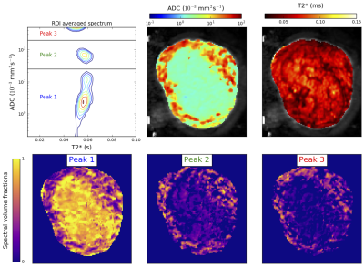 |
Combined Diffusion-Relaxometry MRI to Identify Dysfunction in the Human Placenta
Paddy Slator, Jana Hutter, Marco Palombo, Laurence Jackson, Eleftheria Panagiotaki, Alison Ho, Lucy Chappell, Mary Rutherford, Joseph Hajnal, Daniel Alexander
We demonstrate simultaneous diffusion-relaxometry in the in-vivo human placenta. Two MRI measures widely used for characterizing the placenta, T2* relaxometry and diffusion, are combined into a single scan. We estimate the T2*-ADC spectrum, which enables study of the coupling between these complementary MR contrasts by disentangling joint effects. This gives new potential for improved characterisation of placental dysfunction compared to single contrast MRI and/or ultrasound, and hence could inform improved evaluation of pregnancy complications.
|
|
