Fiber Orientations & Tractography
Oral
Diffusion
Monday, 13 May 2019
| Room 511BCEF |
13:45 - 15:45 |
Moderators: Fulvia Palesi |
13:45
 |
0162.
 |
Mapping of fibre-specific relaxation and diffusivities in heterogeneous brain tissue

João P. de Almeida Martins, Chantal M. W. Tax, Sarah E. Mailhiot, Filip Szczepankiewicz, Maxime Chamberland, Carl-Fredrik Westin, Derek K. Jones, Daniel Topgaard
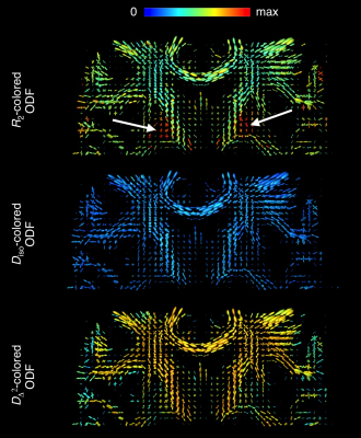
While diffusion MRI tractography has provided important insights on the human brain connectome, fibre-tracking through heterogeneous voxels has proven to be a challenging endeavour. Recently, we devised MRI acquisition- and processing methods to resolve sub-voxel heterogeneity with nonparametric 5D relaxation-diffusion distributions where contributions from distinct tissues are separated while circumventing the use of limiting assumptions. The separation between tissue-signals provides a clean mapping of nerve fibres that can then be used as an input in fibre-tracking algorithms. Additionally, values of relaxation rates and diffusivities are estimated for each distinct fibre bundle, potentially giving tract-specific information on chemical composition and microstructure.
|
13:57
 |
0163.
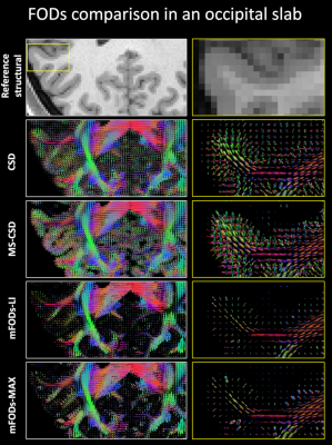 |
Estimation of multiple fiber orientation distributions (mFODs) from diffusion MRI data using spherical deconvolution
Alberto De Luca, Fenghua Guo, Alexander Leemans
Fiber orientation distributions (FODs) of white matter (WM) are commonly estimated from brain diffusion MRI data with spherical deconvolution (SD) approaches. Typically, only WM is considered to be anisotropic in SD, relying on suboptimal isotropic modeling of grey matter (GM). Here we present a general framework to reconstruct multiple anisotropic FODs (mFODs) from multiple response functions, allowing for the investigation of anisotropy in GM. The estimated mFODs were evaluated on a dataset from the HCP project with five response functions generated with the diffusion kurtosis and NODDI approaches, and their performances compared to state-of-the-art SD approaches.
|
14:09
|
0164.
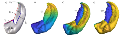 |
Diffusion MRI in the unfolded hippocampus
Uzair Hussain, Jordan DeKraker, Corey Baron, Ali Khan
The hippocampus is of high interest to the research community due to its involvement in many neurological disorders. However, in-vivo imaging, particularly diffusion weighted imaging, is challenging due to the hippocampus’ complicated curved geometry and small size. We address these challenges with an approach that ‘unfolds’ the hippocampus into a thin sheet. This allows migration of the diffusion data into this unfolded hippocampus, which enables visualization of microstructural sensitive diffusion parameters in a space where hippocampal subfields can be readily distinguished.
|
14:21
|
0165.
 |
Towards validation of diffusion MRI tractography: bridging the resolution gap with 3D Polarized Light Imaging
Abib Alimi, Samuel Deslauriers-Gauthier, Rachid Deriche
Three-dimensional Polarized Light Imaging (3D-PLI) is an optical approach presented as a good candidate for validation of diffusion Magnetic Resonance Imaging (dMRI) results such as orientation estimates (fiber Orientation Distribution Functions) and tractography. We developed an anlytical approach to reconstruct fiber ODFs from 3D-PLI datasets. From these fODFs, here we compute brain fiber tracts via dMRI-based probabilistic tractography algorithm. Reconstructed fODFs at different scales proves the ability to bridge the resolution gap between 3D-PLI and dMRI, demonstrating, therefore, a great promise to validate diffusion MRI tractography thanks to multi-scale fiber tracking based on 3D-PLI.
|
14:33
|
0166.
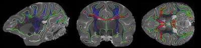 |
On the intrahemispheric connectivity of the monkey: a diffusion tractography and tract tracing analysis
Gabriel Girard, Roberto Caminiti, Alexandra Battaglia-Mayer, Etienne St-Onge, Karen Ambrosen, Simon Eskildsen, Kristine Krug, Tim Dyrby, Maxime Descoteaux, Giorgio Innocenti, Jean-Philippe Thiran
In this work, we compare diffusion tractography with neuronal retrograde tract tracing of the frontal, cingulate and parietal areas of the monkey. We analyze the agreements between the tractography and the tracing for connected and not connected regions. We report an accuracy of 0.71 across all pairs of regions, with twice the number of true positive than false positive connections. Some regions show accuracy higher than 0.80, while other regions show accuracy lower than 0.6. A further analysis of the location of false positive and false negative connections will help understand the limitations and improve diffusion tractography algorithms.
|
14:45
|
0167.
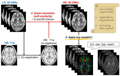 |
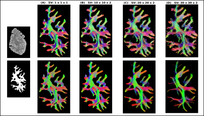
SUper-REsolution TRACTography (SURE-TRACT) pipeline using self-similarity between diffusional and anatomical images
Hong Hsi Lee, Ying Chia Lin, Gregory Lemberskiy, Benjamin Ades-aron, Steven Baete, Fernando Boada, Els Fieremans, Dmitry Novikov
Here, we propose a model-free, self-similarity based SUper-REsolution TRACTography (SURE-TRACT) pipeline to increase the resolution of diffusion weighted images (DWIs) by translating the high spatial frequency details from the co-registered high-resolution anatomical image of the same subject. The generated high-resolution DWIs enable to identify fiber tracks and estimate biophysical parameters with greater anatomical detail. Validating our pipeline using Human Connectome Project data, we showed that the SURE-TRACT pipeline resolves partial volume effects, and is more flexible to different acquisition protocols than other recent machine-learning based algorithms.
|
14:57
|
0168.
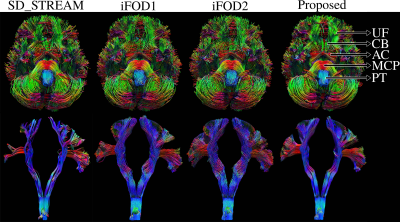 |
A novel fiber-tracking algorithm using parallel transport frames
Dogu Baran Aydogan, Yonggang Shi
White matter fiber-tracking algorithms have remarkably improved during the last two decades. However, multiple validation studies warn about the reliability and reproducibility of results, and point out to the need for better algorithms. In propagation based tracking, connections are typically modeled as piece-wise linear segments. In this work, we propose a novel propagation based probabilistic tracker using parallel transport frames which is capable of generating geometrically smooth curves. Moreover, our tracker has a mechanism to reduce noise related propagation errors. Our experiments on FiberCup and Human Connectome Project data show visually and quantitatively superior results compared to three algorithms in MRtrix3.
|
15:09
|
0169
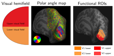 |
Mapping short association fibres in the human visual system with ultra high resolution and high sensitivity diffusion MRI
Video Permission Withheld
Fakhereh Movahedian Attar, Evgeniya Kirilina, Daniel Hänelt, Luke Edwards, Kerrin Pine, Nikolaus Weiskopf
Short association fibres connect proximal cortical areas over short distances. These fibres are highly underrepresented in the current MRI-derived human brain connectome. We combined sub-millimetre resolution diffusion MRI, acquired with a 300 mT/m gradient system and high sensitivity coil for imaging the occipital cortex, with fMRI-driven retinotopic maps of V1/V2. These maps were used to identify the short V1-V2 connections in the human visual processing stream. V1-V2 connectivity was in agreement with previously reported anatomical and functional connectivities. Our results provide an important step towards the construction of a more complete MRI-derived human brain connectome via robust short fibre mapping.
|
15:21
|
0170.
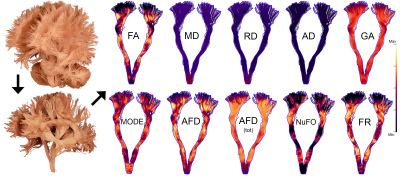 |
Metrics that Matter: Improved statistical power to detect differences in tissue microstructure through dimensionality reduction
Maxime Chamberland, Erika Raven, Sila Genc, Kate Duffy, Greg Parker, Chantal Tax, Maxime Descoteaux, Derek Jones
Various diffusion metrics have been proposed for characterising tissue microstructure. However, it is unclear which metric best captures individual microstructural differences. One possible approach is to explore all available metrics. However, this increases the chance of Type I error and makes interpretation difficult. Using data-reduction approaches, we identified two principal components that capture 85% of the variance in diffusion measurements. The first captures properties related to hindrance, while the second reflects tissue complexity. We demonstrate the utility of this approach by showing significant correlations with age of these new metrics, whereas little to no effects were observed with any individual metric.
|
15:33
|
0171.
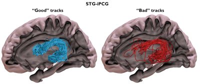 |
Improving the inter-subject reproducibility of diffusion MRI connectivity analysis by controlling the bundle reliability of individual streamlines at the group-level connectome.
Brian Silverstein, Eishi Asano, Yasuo Nakai, Ayaka Sugiura, Min-Hee Lee, Jeong-Won Jeong
Identifying true positives from diffusion weighted imaging (DWI)-based tractography is not trivial, and no universal gold standard has yet been developed. In this study, we introduce a method which utilizes the group-level streamline pathway distribution to determine inter-subject reproducibility of streamline bundles. At the participant level, streamlines that do not correspond to reproducible bundles are removed, resulting in improved reproducibility and increased fidelity when identifying differences between pathways. Additionally, we utilized electrical stimulation-based cortico-cortical evoked potentials to assess how well reliable bundles reflect underlying connectivity. Cleaned structural connectivity data was found to better correlate with electrophysiological connectivity.
|
|
