Microstructure: Validation Using Complementary Methods
Oral
Diffusion
Tuesday, 14 May 2019
| Room 520A-F |
15:45 - 17:45 |
Moderators: Els Fieremans, Markus Nilsson |
15:45
|
0647.
 |
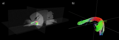
Validating axonal directionality with 3D X-ray scattering

Marios Georgiadis, Dmitry Novikov, Zirui Gao, Manuel Guizar-Sicairos, Lin Yang, Shirish Chodankar, Jelle Veraart, Ben Ades-Aron, Choong Heon Lee, Sunglyoung Kim, Piotr Walczak, Jiangyang Zhang, Aileen Schroeter, Markus Rudin, Els Fieremans
Diffusion MRI (dMRI) is sensitive to neuronal alignment, yet its directional signal does not depend only on the fiber orientation distribution function (fODF). Current validation methods for measuring and modeling the ODF have major limitations. Small-angle X-ray scattering (SAXS) produces directly structural signal, specific to myelin’s repetitive structural arrangement. We apply 3D scanning SAXS on mouse brain sections, retrieve fODFs and compare against dMRI-derived axonal directionality. We also apply SAXS-tensor tomography to mouse spinal cord, produce tractography maps and correlate dMRI- and SAXS-TT-derived fODF parameters. These demonstrate SAXS’s potential for providing novel microstructural insights and structurally validating dMRI-derived fODFs.
|
15:57
 |
0648.
 |
X-ray microcomputed tomography as a natively isotropic, nondestructive, 3D validation dataset for diffusion MRI
Scott Trinkle, Sean Foxley, Narayanan Kasthuri, Patrick La Rivière
In this work, we present the use of synchotron x-ray microcomputed tomography (microCT) as a validation dataset for diffusion tensor imaging (DTI). DTI data were acquired of a post-mortem mouse brain. After metal staining, synchrotron microCT data of the sample were acquired, with 1.2 μm isotropic resolution across the whole brain. Orientation distribution functions were calculated from the microCT data using structure tensor analysis, and tractography was performed on the anterior commissure tract. Comparisons with tractography results from the diffusion MRI data show good agreement.
|
16:09
 |
0649.
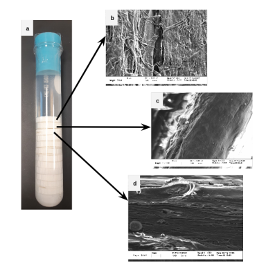 |
Microstructural characterization and validation of a 3D printed phantom for diffusion MRI
Farah Mushtaha, Tristan Kuehn , John Moore , Corey Baron, Ali Khan
Validating diffusion MRI (dMRI) representations and models of brain tissue is challenging because there is no reference ground-truth for in vivo scans. We propose a form of 3D printed phantoms as a flexible paradigm for investigating and validating microstructural indices and crossing fibres that is reproducible with inexpensive materials. As a proof of concept, we obtained multishell dMRI data in samples with varying crossing angles and printing parameters, and investigated the performance of constrained spherical deconvolution to extract diffusion parameters that accurately describe the crossing fibre bundles. We also investigated the effect of printing parameters on the phantoms’ microstructural anisotropy.
|
16:21
|
0650.
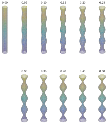 |
Exploring the effect of varying axonal shape on the transverse diffusion inside EM-reconstructed axons using 3d Monte Carlo simulations
Hong Hsi Lee, Els Fieremans, Dmitry Novikov
Diffusion inside axons is restricted and thus non-Gaussian, with diffusion MRI (dMRI) signal strongly sensitive to the shape of the confining axon. This sensitivity is confounded by the coarse-graining of the diameter/shape variation along the fiber during the diffusion time. Here, we analytically relate dMRI metrics to the axonal shape, and validate our theory using 3d Monte-Carlo simulations in beaded cylinders and realistic axons reconstructed from electron microscopy images of the mouse brain white matter. Our simulation results show that the intra-axonal space has a non-trivial kurtosis transverse to axons. Its value is different from that in a perfectly straight cylinder, and needs to be considered in axonal diameter measurements (e.g., spinal cord, strong gradients, intra-axonal metabolites).
|
16:33
|
0651.
 |
Validating MR axon diameter mapping using confocal microscopy
Jelle Veraart, Daniel Nunes, Els Fieremans, Dmitry Novikov, Noam Shemesh
Axon diameter mapping using diffusion MRI in the rat corpus callosum was validated using confocal microscopy with a staining for neurofilaments. When confounding factors such as extra-axonal water and dispersion are addressed, the effective MR axon radii are in good quantitative agreement with histology. However, using MRI, we are limited to the estimation of a single metric representing the entire distribution, which has shown to be dominantly sensitive to the largest axons in the voxel volume of interest.
|
16:45
|
0652.
 |
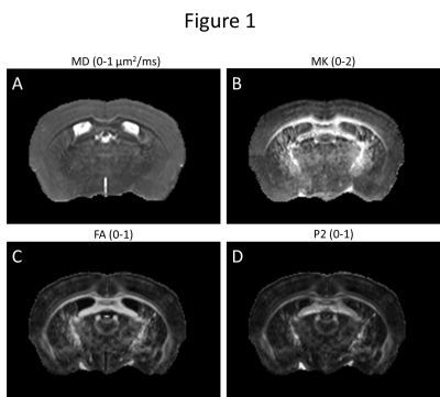
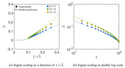
Histological validation of the brain cell body imaging with diffusion MRI at ultrahigh field
Marco Palombo, Daniel Nunes, Daniel Alexander, Hui Zhang, Noam Shemesh
Biophysical modelling of diffusion-weighted MRI (DW-MRI) data can help to gain more insight into brain microstructure. However, models need to be validated. This work validates a recently-developed technique for non-invasive mapping of brain cell-body (soma) size/density with DW-MRI, by using ultrahigh-field DW-MRI experiments and histology of mouse brain.
Predictions from numerical simulations are experimentally confirmed and brain’s maps of MR-measured soma size/density are shown to correspond very well with histology. We provide differential contrasts between cell layers that are less expressed in tensor analyses, leading to novel complementary contrasts of the brain tissue. Limitations and future research directions are discussed.
|
16:57
|
0653.
 |
The BigMac dataset: ultra-high angular resolution diffusion imaging and multi-contrast microscopy of a whole macaque brain
Amy Howard, Saad Jbabdi, Alexandre Khrapitchev, Jerome Sallet, Greg Daubney, Jeroen Mollink, Connor Scott, Nicola Sibson, Karla Miller
Diffusion MRI has the ability to reveal the complex connectivity of the human brain. However, the link between the diffusion signal and the underlying tissue microstructure remains elusive. To drive diffusion MRI validation, we present BigMac: a unique dataset which combines ultra-high angular resolution diffusion MRI with microscopy throughout an adult macaque brain. With this dataset we ask how under-sampling q-space biases our reconstruction of the ‘true’ diffusion profile. Our results indicate that the error associated with interpolating under-sampled data decays exponentially with the angular resolution of sampling, and that high angular resolution is necessary to characterise acutely crossing fibres.
|
17:09
|
0654.
 |
CHENONCEAU: towards a novel mesoscopic (100/200µm) post mortem human brain MRI atlas at 11.7T
Justine Beaujoin, Alexandros Popov, Raïssa Yebga Hot, Fabrice Poupon, Jean-François Mangin, Christophe Destrieux, Cyril Poupon
Ultra-high field MRI combined with strong gradients gives access to ex-vivo anatomical and diffusion MRI datasets at the mesoscopic scale. This wok presents the unique CHENONCEAU dataset acquired on an entire left hemisphere (and soon on an entire brain) at 11.7T with ultra-high resolutions (100, 150, 200µm) thus paving the way to the creation of a novel mesoscopic MRI atlas of the human brain structure, connectivity and cytoarchitecture.
|
17:21
|
0655.
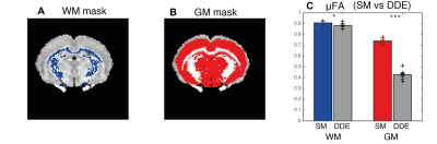 |
The two-compartment diffusion “standard model” misestimates microscopic anisotropy in-vivo
Rafael Neto Henriques, Sune Jespersen, Noam Shemesh
Several microstructural models have been proposed to increase the specificity of diffusion MRI. However, improper model assumptions can compromise the accuracy of model estimates. Here, we compared model-independent metrics extracted from double diffusion encoding (DDE) with the metrics arising from the current (two-compartment) diffusion “standard model” (SM) in in-vivo rat brains. Our results revealed that SM produces overestimated microscopic anisotropy for both white and grey matter. These findings question the validity of SM and calls for future developments of more accurate models.
|
17:33
|
0656.
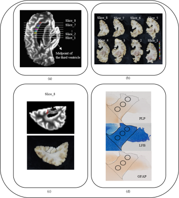 |
Validation of diffusion models: a post-mortem MRI and histology study
Zihan Zhou, Qiqi Tong, Hongjian He, Keqing Zhu, Hui Lu, Lei Zhang, Qiuping Ding, Laura Jonkman, Jianhui Zhong
The Diffusion Kurtosis Imaging (DKI) model can successfully characterize non-Gaussian diffusion. In turn, the White Matter Tract Integrity (WMTI) model is proposed to be based on the DKI model to further characterize the intra- and extra-axonal compartments. However, the accuracy with which model parameters reflect the underlying tissue characteristics has not been tested. Here, we compared two MRI model metrics using a unique combined post-mortem MRI and histopathology approach. Preliminary results show that AWF, De,⊥De,⊥and
MD from MRI correlate strongly with myelin fraction and that De,∥De,∥ from
MRI correlates strongly with astrocyte fraction.
|
|
