fMRI: Preclinical
Oral
fMRI
Thursday, 16 May 2019
| Room 511BCEF |
08:15 - 10:15 |
Moderators: Kai-Hsiang Chuang, Cornelius Faber |
08:15
 |
1050.
 |
Awake and behaving mouse fMRI in Go/No-go task.

Wenjing Chen, Zhe Han, Xifan Chen, Kaiwei Zhang, Chengyu Li, Zhifeng Liang
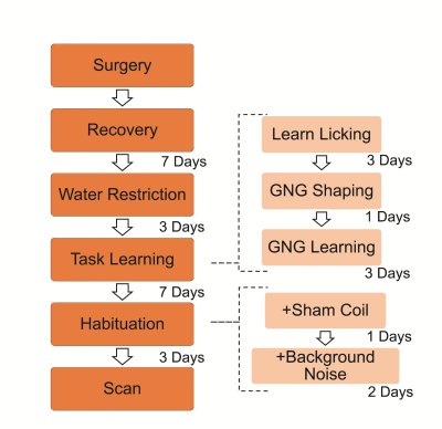
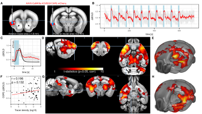
The current study describes a novel awake and behaving mouse fMRI paradigm, which enables the functional imaging of mice brain during an olfaction based go/no-go (GNG) task. This novel paradigm incorporates a MR compatible behavioral apparatus (olfactometer, licking detection and water delivery) and awake head fixation mechanism, all specifically designed to be used with a cryogenic coil at high field. High-resolution functional images uncovered the large-scale networks that are differentially recruited in the hit vs. correct-rejection trials, with greatly limited motion artefacts. This method paved the way for future whole-brain, systematic mapping of cognitive processes in mice.
|
08:27
 |
1051.
 |
Triple network activity regulation mediated by the insular cortex in the mouse brain.
Francesca Mandino, Ling Yun Yeow, Chai Lean Teoh, Heidi Foo, Renzhe Bi, Jiayi Zhang, Nathaniel Low, Tricia Lim, John Gigg, Olivo Malini, Yu Fu, Joanes Grandjean
The triple-network model is a contemporary theoretical framework derived from empirical neuroimaging data to explain a wide range of observations stemming from multiple psychopathologies. Central to the model are interactions between the salience, default-mode, and central executive networks. The insula area is a central node of the salience network. Using acute photostimulation, we report evidence to support the existence of a triple-network system within the mouse brain. Further, by using sustained optogenetic neuromodulation, we show that inhibition of the insular area acts on homotopic functional connectivity but fails to affect wider interactions within triple-network system in a resting-state setting.
|
08:39
|
1052.
 |
Thalamic low frequency activity contributes to resting-state cortical interhemispheric MRI functional connectivity
Xunda Wang, Alex T. L. Leong, Russell W Chan, Ed X. Wu
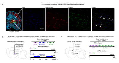
The brain consists of numerous interconnected parallel and hierarchical networks subserving sensory, behavioral and cognitive functions. Resting-state functional MRI (rsfMRI) connectivity has helped study the complex brain-wide functional networks. Yet, less is known about the role of thalamus in rsfMRI connectivity. Utilizing optogenetic excitation and pharmacological inactivation to manipulate the neural activity of somatosensory thalamocortical neurons, we demonstrate that thalamus contributes to rsfMRI connectivity within and beyond its sensory modality, likely through the recruitment of interhemispheric low frequency neural oscillations at all cortical layers. Our work highlights the thalamus as a pivotal structure underpinning rsfMRI connectivity observations.
|
08:51
|
1053.
 |
Ultrahigh spatial and temporal resolution fMRI with implanted CMOS-based planar microcoil at 14.1T
Marlon Pérez-Rodas, Jonas Handwerker, Michael Beyerlein, Hellmut Merkle, Rolf Pohmann, Jens Anders, Klaus Scheffler
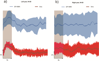
Compared to electrophysiological or optical recording of brain activity, fMRI has a rather low spatial and temporal resolution. Here, we propose the use of implanted microcoils for studying animal brain activity in-vivo with ultra high sensitivity compared to conventional coils. A fully integrated CMOS NMR transceiver containing an on-chip-microcoil, integrated amplifiers and a demodulator is used to acquire ultra-localized signal (10nl) at ultrahigh temporal resolution (5ms) showing unprecedented high speeds and spatial resolutions of the BOLD response.
|
09:03
|
1054.
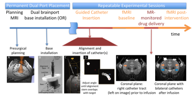 |
MR-Guided Pharmacological Intervention: Creating Causal Capabilities for fMRI
Walter Block, Rasmus Birn, Miles Olsen, Samuel Hurley, Ethan Brodsky, Abigail Rajala, Caitlynn Filla, Alan McMillan, Andrew Alexander, Rick Jenison, Luis Populin
Neuroscience is in need of precise interventional tools that alter local neural dynamics while monitoring whole brain network activity. We demonstrate methods to guide catheters to deliver and monitor pharmacologic alteration of a local brain region in anaesthetized Rhesus monkeys while monitoring changes in resting state functional connectivity MRI (rs-fcMRI) throughout all brain networks. Expected and unexpected alterations in rs-fcMRI after unilateral and bilateral infusions of inhibitory agents in the limbic system are provided. The approach shows promise for using the alterations to compute effective connectivity through fMRI.
|
09:15
|
1055.
 |
Temporal dynamics of mouse BOLD fMRI in the sensory pathway
Won Beom Jung, Hyun-Ji Shim, Seong-Gi Kim
Ultra-high field fMRI allows us to detect the functional sensory pathway including thalamic nuclei. Although hemodynamic response function is determined spatially by the vascular architecture and temporally by the evolution of hemodynamic changes, fMRI signals may provide insight into tracking sequential neural processing. Here, we investigated the spatiotemporal evolution of functional sensory pathways during forepaw stimulation in anesthetized mice using ultra-high field BOLD fMRI.
|
09:27
|
1056.
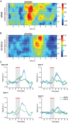 |
Line-scanning diffusion fMRI reveals a rapid-onset (<200 ms) component
Daniel Nunes, Noam Shemesh
Diffusion fMRI (dfMRI) has been proposed as a more direct means for mapping neural activity more accurately than BOLD fMRI. However, the origin of dfMRI signals is still an ongoing debate. Here, we developed a line-scanning dfMRI technique achieving very high temporal resolution (100 ms), and measured activity in the forelimb S1 upon rat forepaw stimulation. Our results show a rapid-onset (<200ms) dfMRI component that was not found in BOLD fMRI. Upon inducing hypercapnia, the fast dfMRI component was nearly unaffected while the slower dfMRI component was substantially modulated, suggesting a potentially neural origin for the former.
|
09:39
|
1057.
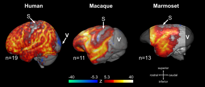 |
Using BOLD fMRI to map anesthesia-induced burst suppression in humans and non-human primates
Nikoloz Sirmpilatze, Jürgen Baudewig, Judith Mylius, Daniel Golkowski, Andreas Ranft, Rüdiger Ilg, Susann Boretius
Deeply anesthetized and comatose states are often accompanied by a distinctive pattern of electroencephalographic activity, called “burst suppression”. This pattern’s underlying mechanism and functional significance remain largely unknown. In this work we demonstrated that burst suppression can be detected in fMRI data, without the need for accompanying measures of neural activity. We then used this fMRI approach to perform whole-brain mapping of burst suppression in anesthetized human volunteers and non-human primates. We found that burst suppression involves the same set of brain areas across primate species, and is mostly absent in primary visual and somatosensory areas.
|
09:51
|
1058.
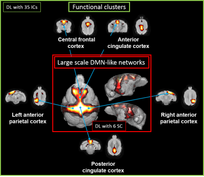 |
Multilevel functional organization of the mouse lemur primate brain
CLEMENT GARIN, NACHIKET ABHAY NADKARNI , JEAN-LUC PICQ, SALMA BOUGACHA , MARC DHENAIN
Resting state networks have been characterized in numerous mammals covering human, non-human primates, dogs, rabbits and rodents, though only ever at single semi-arbitrary levels of complexity. In humans, resting state networks analyses have been extended to extracting networks of varying complexity, representing different levels of a possible “functional hierarchy”. We performed the first study of “functional hierarchy” in animals. We focused on the gray mouse lemur (Microcebus murinus), a small primate attracting increased attention as a model for cerebral and age-related disorders.
|
10:03
|
1059.
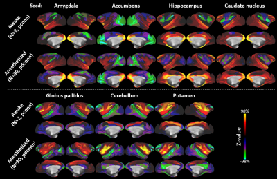 |
Subcortico-centric view of macaque neocortical organization investigated using resting-state fMRI
Joonas Autio, Atsushi Yoshida, Takayuki Ose, Kantaro Nishigori, Masataka Yamaguchi, Masahiro Ohno, Chihiro Yokoyama, David Van Essen, Matthew Glasser, Takuya Hayashi
An important aspect to understand evolutionary differences across primate species is through conserved subcortical circuitry and diversification of neocortical inputs. Here, we explore neocortical profiles of major subcortical structures using ‘Human Connectome Project-style’ resting-state functional MRI (rfMRI) connectivity in alert and anesthetized macaque monkeys. Our results reveal that the major subcortical “limbic and associative” structures have largely overlapping neocortical rfMRI connectivity profiles. These findings suggest important differences relative to previous reports of functional connectivity profiles in humans, and may provide a valuable clue to the evolution of human brain function and behavior in the primate lineage.
|
|
