Laminar & Columnar fMRI
Oral
fMRI
Tuesday, 14 May 2019
| Room 510A-D |
15:45 - 17:45 |
Moderators: Dimo Ivanov, Jonathan Polimeni |
15:45
 |
0608.
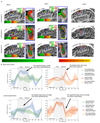 |
Measuring layer-dependent fMRI activity across layers in cognitive brain areas: challenges and capabilities

Laurentius Huber, Emily Finn, David Jangraw, Peter Bandettini
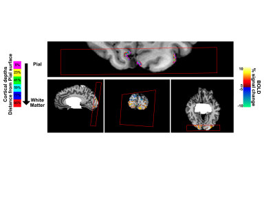
While recent advances in high-resolution fMRI allow for investigating activity across cortical layers, applications beyond the well-studied primary sensory and primary motor areas have been complicated by multiple technical challenges: lack of anatomical landmarks, complicated folding structure, weak signal, restricted task design, etc. The purpose of this study is to develop a scanning/stimulation/analysis setup that allows us to overcome these challenges. Our results suggest that with advanced imaging methodology, corresponding task design, and appropriate analysis strategies it is possible to reliably measure layer-dependent activity differences in cognitive brain areas, such as the dorsolateral prefrontal cortex.
|
15:57
 |
0609.
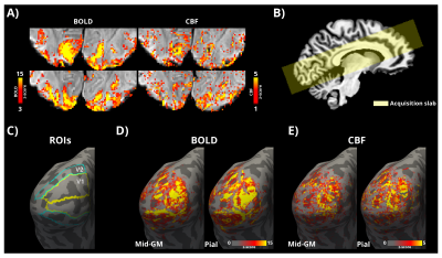 |
Laminar CBF and BOLD fMRI in the human visual cortex using arterial spin labelling at 7T
Sriranga Kashyap, Dimo Ivanov, Martin Havlicek, Benedikt Poser, Kamil Uludag
Laminar fMRI at ultra-high field is typically carried out using the BOLD contrast. Despite its unrivalled sensitivity, the BOLD contrast is limited in its spatial specificity (e.g. due to intracortical ascending and pial veins). Alternatively, the regional change in cerebral blood flow (CBF), is a quantitative measure directly related to neuronal activation. However, CBF fMRI with high spatial resolution is challenging due to the relatively lower SNR. Building on previous work, we demonstrate for the first time, sub-millimetre spatial resolution simultaneous CBF and BOLD acquisition using arterial spin labelling (ASL) for laminar fMRI at 7T.
|
16:09
|
0610.
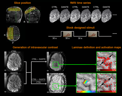 |
A Novel Intravascular Contrast for Laminar Functional MRI
Yuhui Chai, Linqing Li, Laurentius Huber, Bandettini Peter
In this work, we proposed a novel intravascular contrast for laminar specific fMRI in the human brain at 7T. This technique was shown to be sensitive to both cerebral blood volume (CBV) and flow (CBF) changes. We demonstrate that this new tool, with its highly specified functional layer profile, robust reproducibility and improved sensitivity, allows neuroscientific investigation of information flow across cortical microcircuits.
|
16:21
|
0611.
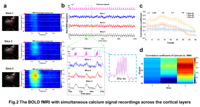 |
Concurrent intracellular calcium recordings with laminar fMRI mapping
Hang Zeng, Sangcheon Choi, Filip Sobczak, Bruce Rosen, Xin Yu
The ultra-high magnetic field strengths of 14.1T allows visualization of laminar-specific neurovascular coupling events with fMRI in living animals. Combining line-scanning fMRI and intracellular calcium signal recording, we present laminar-specific coupling features at high temporal and spatial resolution along the cortical thickness, showing strong Layer 4 fMRI correlation to the calcium signal and largely varied coupling features from adjacent cortices in the anesthetized rats.
|
16:33
|
0612.
 |
Whole brain depth-dependent task based connectivity with laminar fMRI
Daniel Sharoh, Tim van Mourik, Lauren Bains, Katrien Segaert, Kirsten Weber, Peter Hagoort, David Norris
Laminar resolution, functional magnetic resonance imaging (lfMRI) is a noninvasive technique with the potential to distinguish top-down and bottom-up signal contributions on the basis of depth-dependent interactions with distal regions. Hitherto, lfMRI has not been used to investigate whole-brain distributed networks nor complex cognitive tasks. We show here that lfMRI can reveal whole-brain directed networks during word reading. We identify language critical regions based on their association with the top-down signal stream and herewith establish lfMRI for the non-invasive assessment of directed connectivity.
|
16:45
|
0613.
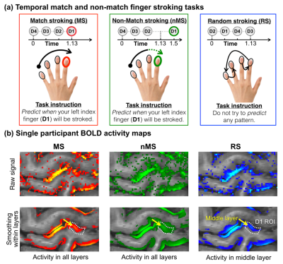 |
Increased activity in superficial and deep layers of human S1 for prediction error
Yinghua Yu, Laurentius Huber, Yuhui Chai, David Jangraw, Arman Khojandi, Jiajia Yang, Peter Bandettini
Sensory processing in humans is thought to rely on a predictive model of the environment. And these predictions are constantly optimized to minimize future sensory prediction errors. However, the neural microcircuits underlying this prediction error model are still poorly understood. Here, we used an index finger prediction task that consists of sequential finger-stroking in high-resolution (0.71mm) BOLD and VASO fMRI at 7T to investigate how the prediction error activity changes across layers in the human primary somatosensory cortex (S1). We found that prediction error activity is stronger in superficial and deep layers rather than the middle layers of S1.
|
16:57
|
0614.
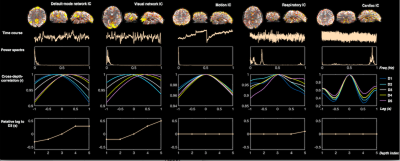 |
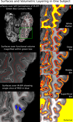
De-noising high-resolution fMRI data using cortical depth-dependent analysis
Jingyuan Chen, Anna Blazejewska, Nina Fultz, Bruce Rosen, Laura Lewis, Jonathan Polimeni
In this study, we exploit cortical depth-dependent information to de-noise high-resolution fMRI data. We have proposed a novel de-noising pipeline that can automatically differentiate neural and non-neural fluctuations according to cross-cortical-depth delay patterns. We also show that by excluding voxels intersecting the pial surface can reduce physiological effects and improve neuronal/spatial specificity.
|
17:09
|
0615.
 |
Robust detection of layer-specific activities in the human LGN
Yazhu Qian, Zihao Zhang, Jing An, Danny Wang, Peng Zhang
The lateral geniculate nucleus (LGN) of the thalamus is the main relay station of retinal input to the visual cortex. It plays important roles in perception and cognition, and has close relationships with several eye and brain diseases. The current study showed that high resolution BOLD fMRI at 7T can reliably distinguish eye layers as well as magnocellular and parvocellular layer activities of the human LGN.
|
17:21
|
0616.
 |
Probing spatio-temporal information during attention modulations in humans with sub-second sampling of cortical depth dependent BOLD fMRI signals at 7T
Luca Vizioli, Alexander Bratch, Sudhir Ramanna, Kamil Ugurbil, Essa Yacoub
Temporal dynamics of the BOLD signal have been recently exploited with fast TR fMRI. However, the ability to concurrently retain sub-millimeter spatial resolution along with high temporal resolution (e.g. =< ~600 ms TR), with sufficient SNR efficiency, has been elusive. Such a data set could provide unprecedented access to studying the human brain non-invasively. In this work, we push the spatio-temporal limits of 7T fMRI and investigate the possibility of exploiting BOLD temporal dynamics as a function of cortical depth, using spatio-temporal multi-voxel pattern analysis during attention modulations.
|
17:33
|
0617.
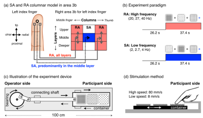 |
High-resolution fMRI maps of columnar organization in human primary somatosensory cortex
Jiajia Yang, Laurentius Huber, Yinghua Yu, Yuhui Chai, Arman Khojandi, Peter Bandettini
More than half a century ago, animal studies revealed that area 3b of the primary somatosensory cortex (S1) receives cutaneous input from slow-adapting (SA) and rapidly adapting (RA) cutaneous receptors in a columnar manner. However, there have been no direct observations of columnar origination in the human SI. Here, for the first time, we investigate the columnar organization in the human S1 by using advanced high-resolution (0.7mm) fMRI at 7T. We find that the human area 3b is columnar organized with alternating SA and RA columnar preferences. We find that SA selective stimulation evokes activity in the middle layers only.
|
|
