fMRI Acquisition Methods
Oral
fMRI
Thursday, 16 May 2019
| Room 520A-F |
13:45 - 15:45 |
Moderators: Jun Hua, Kamil Uludag |
13:45
 |
1165.
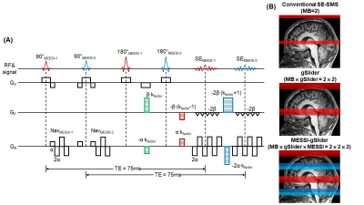 |
Accelerated spin-echo fMRI using Multisection Excitation by Simultaneous Spin-echo Interleaving (MESSI) with ‘complex-basis’ RF-encoded generalized SLIce Dithered Enhanced Resolution Simultaneous Multi-Slice (MESSI-gSlider-SMS)

SoHyun Han, Congyu Liao, Mary Kate Manhard, Jonathan R. Polimeni, Kawin Setsompop
High spatiotemporal resolution spin-echo fMRI is challenging as it requires a long echo time to generate BOLD-contrast, resulting in longer repetition times. We propose a new technique, Multisection Excitation by Simultaneous Spin-echo Interleaving (MESSI), which utilizes the dead time in long TE acquisitions to improve the slice-coverage of SE-fMRI. For further accelerations, we combine the MESSI with both ‘complex-basis’ RF-encoded gSlider and conventional-SMS. Compared with standard SE-EPI acquisition with the same TR, the proposed MESSI-gSlider acquisition shows comparable tSNR but with eight-fold increase in slice-coverage. This method should be beneficial for applications requiring high spatiotemporal resolution SE-fMRI with whole-brain coverage.
|
13:57
|
1166.
 |
Novel alpha-180 SE based LINE-scanning method (SELINE) for laminar-specific fMRI
Sangcheon Choi, Hang Zeng, Rolf Pohmann, Klaus Scheffler, Xin Yu
Laminar-specific functional magnetic resonance imaging (fMRI) opens new possibility for studying the neuronal circuitry and functional connectivity of cortex. FLASH based line-scanning method was proposed with high temporal and spatial resolution to better characterize the fMRI onset time across the cortical layers by combining 2 saturation RF pulses. However, the imperfect RF saturation performance led to poor boundary definition of the ROI from the cortex. In this work, we propose an α-180° SE based line-scanning (SELINE) method, solving this problem. We will improve this method to better understand the distinct laminar-specific neuronal circuitry and functional connectivity.
|
14:09
|
1167.
 |
Full-FOV, Whole-brain, Half-millimetre Resolution fMRI at 7T using Accelerated multi-band EPIK with TR-external Phase Correction
Seong Dae Yun, N. Jon Shah
Ultra-high spatial resolution fMRI can identify brain activations with precise spatial localisation. There have been numerous attempts to achieve a sub-millimetre resolution in fMRI by using reduced- or full-FOV imaging. Although the reduced-FOV scheme can achieve more enhanced resolution than the full-FOV scheme, the restricted FOV often limits its use for more general functional studies. This work aims to present a novel half-millimetre resolution fMRI method which can also provide full-FOV and whole-brain coverage. The method was developed based on EPIK and a TR-external EPI phase correction scheme. Here, the above configuration was employed for exemplary finger-tapping fMRI at 7T.
|
14:21
|
1168.
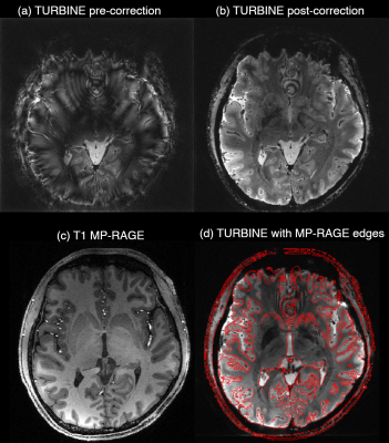 |
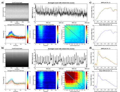
Ultra-high spatial resolution TURBINE fMRI at 7T
Nadine Graedel, Karla Miller, Mark Chiew
Ultra-high spatial resolution fMRI often uses 3D-EPI for data acquisition. However, 3D-EPI can be susceptible to artefacts from inter-shot variability, such as motion and physiological noise. Here, we present an improved hybrid radial-Cartesian 3D EPI sampling strategy, TURBINE, to generate ultra-high spatial resolution fMRI data at conventional scan times at 7T. TURBINE enables intrinsic correction of global and z-dependent shot-to-shot phase variations, and self-navigated motion correction. Using these features, along with a temporally regularized reconstruction, we demonstrate robust BOLD activation in 0.67mm isotropic resolution (16mm slab, TRvol = 2.32s) and 0.8x0.8x2.0mm (whole-brain, TRvol = 2.4s) acquisitions.
|
14:33
|
1169.
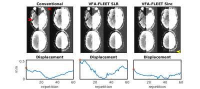 |
High-resolution segmented-accelerated EPI using Variable Flip Angle FLEET with tailored slice profiles
Avery Berman , Thomas Witzel, William Grissom , Daniel Park , Kawin Setsompop, Jonathan Polimeni
New evidence suggests that fMRI has spatial specificity at scales far below current voxel sizes, but encoding limits preclude single-shot EPI at sufficient spatial resolution. Segmented EPI can help overcome these limits, but is well-known to be temporally unstable. Here we propose a reordering of the EPI segments, known as FLEET, combined with variable progression of flip angles to maximize the image signal level and a tailored RF pulse design to maintain compatible slice profiles. We demonstrate that this approach provides stable segmented EPI acquisitions with negligible ghosting, and when combined with acceleration can provide submillimeter fMRI acquisitions at 3T.
|
14:45
|
1170.
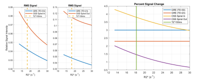 |
Comparison of Oscillating Steady State to GRE BOLD for fMRI
Shouchang Guo, Douglas Noll
The combination of a quadratic phase sequence with balanced gradients leads to an oscillating steady state (OSS) MRI signal with an average signal amplitude that is 2-3 times the Ernst angle GRE imaging. With signal phase varies with off-resonance making the resultant signal T2*-weighted and thus, suitable for BOLD fMRI. In this work, simulations of changes to the tissue T2*, as seen in fMRI, are carried out for OSS and compared to GRE. The OSS method is compared to GRE for high-resolution fMRI studies, demonstrating substantially higher activation counts as well as temporal SNR (tSNR).
|
14:57
|
1171.
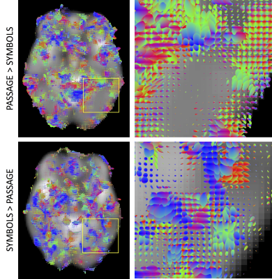 |
Task-based High Angular Resolution Functional Imaging (tHARFI) shows directional contrast of BOLD signal
Kurt Schilling, Katherine Aboud, Hakmook Kang, Zhaohua Ding, Adam Anderson, Laurie Cutting, John Gore, Bennett Landman
We present a method to detect and quantify correlated BOLD signals in brain white matter using task-based high angular resolution functional imaging (tHARFI). This technique measures the anisotropic component of these signals, highlighting directional differences between two different task states. The ability to quantify these correlated functional signals in white matter may improve our ability to delineate functional circuits in the brain, and complement other modalities to better understand structural and functional relationships in the brain.
|
15:09
|
1172.
 |
Geometric Distortions and Signal-To-Noise Ratio of Conventional and Inner Fields-of-View for T2*-Weighted Echo-Planar Imaging of the Spinal Cord
Ying Chu, Jürgen Finsterbusch
Geometric distortions and signal-to-noise ratios (SNR) of T2*-weighted echo-planar imaging (EPI) of the spinal cord are compared for conventional and inner-FOV acquisitions based on 2D-selective RF (2DRF) excitations. For conventional acquisitions, the required FOV increases with the in-plane object size yielding more pronounced distortions, prolonged echo times (TEs), and reduced SNR. For inner-FOV acquisitions, the FOV is small and independent of the object size yielding only minor distortions. The 2DRF pulse duration must be adapted for larger object sizes resulting in slightly prolonged TEs but overall TEs remain shorter and SNR values are larger than for conventional acquisitions.
|
15:21
|
1173.
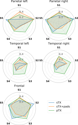 |
Resting-state fMRI at UHF: Optimizing BOLD sensitivity by using plug-and-play parallel transmission at 7T
Vincent Gras, Benedikt Poser, Xiaoping Wu, Raphaël Tomi-Tricot, Nicolas Boulant
The 7T Human Connectome Project (HCP) resting-state fMRI (RS-fMRI) protocol employs slice-accelerated multiband (MB) EPI with a total of 10-fold acceleration (MB 5 and in-plane 2). Such highly accelerated acquisitions provide exquisite temporal resolution at good sensitivity in most cortical regions, but quickly become very SNR starved in transmit field (B1+) deprived regions. This can be mitigated using parallel RF transmission (pTX), which however usually comes at the expense of time-consuming calibrations and pulse computations. This work shows experimentally with HCP-style RS-fMRI scans that a plug-and-play alternative for B1+ mitigation using pTX is possible with multiband Universal Pulses.
|
15:33
|
1174.
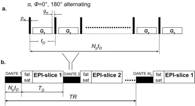 |
DANTE Prepared EPI for Fast Whole Brain VASO Detection at 3T
Linqing Li, Laurentius Huber, Yuhui Chai, Sean Marrett, Peter Bandettini
Vascular space occupancy (VASO) fMRI may provide more quantifiable and localized hemodynamic measures of brain activitythan conventional BOLD measurements. Due to application of inversion recovery pulse, conventional approach for VASO measurements suffers from low imaging efficiency, high SAR and interference of inflow and CBF effect. We demonstrated that DANTE prepared single echo or multi-echo EPI can be used to acquire robust full brain multi-slice VASO activation with benefit of high image efficiency, low SAR without complication of inflow and CBF effect.
|
|
