fMRI: Physiology
Oral
fMRI
Tuesday, 14 May 2019
| Room 516C-E |
13:30 - 15:30 |
Moderators: Jean Chen, Ian Driver |
13:30
 |
0530.
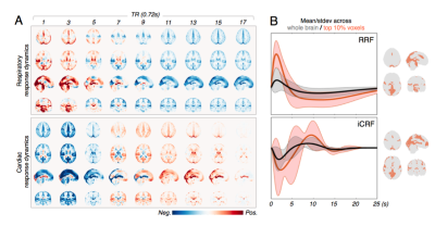 |
Resting-State “Physiological” Networks

Jingyuan Chen, Laura Lewis, Catie Chang, Nina Fultz, Ned Ohringer, Bruce Rosen, Jonathan Polimeni
In this abstract, we offer a systemic characterization of the spatiotemporal patterns of fMRI signals subsequent to slow fluctuations in respiratory volume and heart rate. We show that these slow physiological dynamics contain structured network patterns that are somewhat consistent across individuals. We also show that global signal regression (GSR) may introduce anti-correlating patterns of the physiological dynamics to the final observations of functional connectivity.
|
13:42
|
0531.
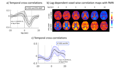 |
Contribution of sympathetic vasoconstriction to the fMRI global signal during autonomic arousals
Pinar Ozbay, Catie Chang, Dante Picchioni, Hendrik Mandelkow, Miranda Chappel-Farley, Peter van Gelderen, Jacco de Zwart, Jeff Duyn
Recent work has suggested that the fMRI global signal (GS) in part may result from activity carried by the extensive sympathetic innervation of the extraparenchymal arteries of the brain. One of the pathways that potentially triggers sympathetic activity is the phenomenon of subcortical (autonomic) arousal, which is closely associated with the K-complex, a signature event observed in scalp EEG. In this work, we analyzed previously acquired data during sleep, and showed a strong association between fMRI GS, K-complexes, and photoplethysmography (PPG) from the finger skin, a proxy for sympathetic activity. Since sympathetic activity may be variable and elicited by a variety of stimuli during both sleep and wake, it likely plays an important, while largely overlooked role in most fMRI experiments.
|
13:54
|
0532.
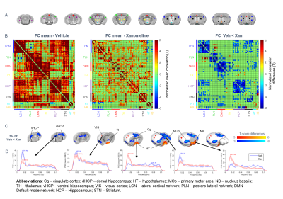 |
Pharmacological stimulation of cholinergic activity alters brain-wide spontaneous fMRI network dynamics
Daniel Gutierrez-Barragan, Carola Canella, Alberto Galbusera, Stefano Panzeri, Alessandro Gozzi
We recently demonstrated that resting-state fMRI (rsfMRI) network dynamics can be mapped with voxel-resolution in the mouse, and that oscillatory state transitions govern rsfMRI network dynamics in the resting mouse brain. Here we show that pharmacological modulation of cholinergic activity strongly affects the spatio-temporal dynamics of rsfMRI state fluctuations, encompassing novel spatial topographies and altered spatio-temporal dynamics. Our results implicate ascending cholinergic activity in the generation of oscillating fMRI states in the resting brain.
|
14:06
|
0533.
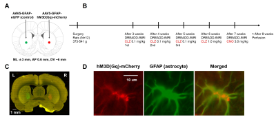 |
Modulation of resting-state functional MRI signal by astrocyte
Akira Sumiyoshi, Satoshi Ikemoto, Elliot Stein, Yihong Yang, Hanbing Lu
Although resting-state functional MRI (rs-fMRI) is extensively used to study brain circuitry, recent animal studies suggest a non-neuronal origin of the rs-fMRI signal. We hypothesized that astrocytes may play an important role in rs-fMRI signal. We used chemogenetic technology to selectively activate astrocytes (increasing Ca2+ levels) and recorded rs-fMRI signals in lightly anesthetized rats. Chemogenetic activation of astrocytes following 0.1 mg/kg of clozapine injection induced signal intensity changes and reduced functional connectivity. These in vivo results are consistent with previous brain slice studies, confirming a potentially important role of astrocytes in rs-fMRI signals.
|
14:18
 |
0534.
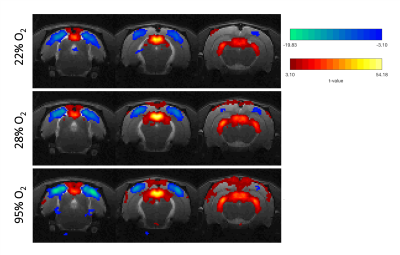 |
Negative BOLD responses in the rat visual pathway
Rita Gil, Francisca Fernandes, Clémence Ligneul, Noam Shemesh
Negative BOLD responses (NBRs) in the rat visual cortex (VC) are reported, for the first time, upon high frequency visual stimulation. So far, in rats, only an attenuation of the positive BOLD response (PBR) in VC had been reported with increase of the stimulus frequency up to 10-12Hz1,2. Here, experiments with very high sensitivity thanks to a cryoprobe operating at 9.4T, reveal NBRs in VC and how they are modulated by hyperoxia (already reported for PBRs3,4). Results suggest the possibility that the neurovascular couplings operating under PBRs and NBRs might not be the same.
|
14:30
|
0535.
 |
Characterization of BOLD hemodynamic response function in subcortical and cortical regions of human brain at 3T and 9.4T
Jung Hwan Kim, Amanda Taylor, Marc Himmelbach, Gisela Hagberg, Klaus Scheffler, David Ress
Measurement of blood oxygen level dependent hemodynamic response function (BOLD HRF) can be used to evaluate neurovascular coupling and understand underlying physiology (e.g. oxygen uptake and blood flow). However, there is dearth of understanding subcortical neurovascular coupling, which is critical for brain health. Here, we characterize human subcortical HRFs at both 3T and 9.4T and compare them with HRFs in visual cortex.
|
14:42
|
0536.
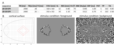 |
Testing temporal dependence of spatial specificity in BOLD fMRI at 7T: comparing short versus long stimulus duration
Anna Blazejewska, Michael Bernier, Shahin Nasr, Jonathan Polimeni
The spatial specificity of the BOLD-fMRI response has previously been shown to vary systematically across cortical depths, with highest specificity found in voxels furthest from the pial surface, while highest sensitivity of the response is found in voxels closest to the pial vessels. Analogous trade-offs between spatial specificity and sensitivity can be appreciated in the temporal evolution of the BOLD-fMRI response. Here we investigate spatial specificity of the response to both short-duration and long-duration stimuli as they evolve in time using high-resolution BOLD-fMRI at 7T, and demonstrate that BOLD-fMRI response exhibits high specificity both at early and late time points.
|
14:54
|
0537.
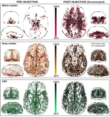 |
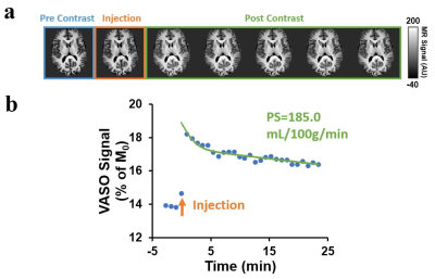
Prospects for improving neuronal specificity of fMRI with Ferumoxytol: an evaluation of vascular segmentation and cortical depth-dependent analysis
Michaël Bernier, Jingyuan Chen, Ned Ohringer, Nina Fultz, Rebecca Leaf, Olivia Viessmann, Laura Lewis, Lawrence Wald, Jonathan Polimeni
Ferumoxytol, a superparamagnetic iron oxide nanoparticle, is commonly used as an intravenous treatment for anemia, but has been recently employed as a blood-pool contrast agent for MRI. Here we evaluated Ferumoxytol as a tool to improve the neuronal specificity of fMRI using it for both improved vascular segmentation and for CBV-weighted functional contrast. We employed multi-echo gradient recalled echo (ME-GRE) acquisitions with functional imaging pre and post injection, performed vascular extraction/segmentation, and report apparent quantitative CBV changes surrounding vessels as a function of echo-time. This work demonstrates the possibility of high-resolution CBV mapping, gray- and white-matter angiography, and cortical depth-dependent analyses with this contrast agent.
|
15:06
|
0538.
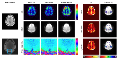 |
Hyperoxia calibrated fMRI for dynamic mapping of CMRO2 responses using MR-based measurements of whole-brain Yv: Comparison to standard calibration models and measurement of CMRO2 response to finger tapping
Erin Englund, Maria Fernandez-Seara, Ana Rodriguez-Soto, Hyunyeol Lee, John Detre, Zachary Rodgers, Felix Wehrli
The magnitude of neurometabolic responses to stimuli can be quantified using calibrated fMRI. Here we compare existing calibration models to Yv-based calibration. The Yv-based calibration model measures whole-brain venous oxygen saturation in the superior sagittal sinus along with BOLD changes and ASL-measured CBF changes to derive maps of the calibration constant, M, in response to a hyperoxia stimulus. M-maps were compared between the proposed and existing calibration models. Then, relative CMRO2 changes in response to a finger tapping task were computed based on the derived M-maps. A ~19% increase in CMRO2 in the motor cortex was observed.
|
15:18
|
0539.
 |
Non-contrast assessment of BBB permeability using WEPCAST MRI: validation with contrast-agent based method
Zixuan Lin, Jinsoo Uh, Yang Li, Dengrong Jiang, Hanzhang Lu
Water-extraction-with-phase-contrast-arterial-spin-tagging (WEPCAST) MRI was recently proposed for non-contrast assessment of blood-brain barrier (BBB) permeability to water. However, this approach has not been directly compared with contrast-based method. In this study, we used Gd-based MRI method to validate the WEPCAST MRI. Two methods provide consistent results, suggesting that WEPCAST MRI has a potential to become an alternate to clinical contrast-based BBB permeability assessment.
|
|
