fMRI: Multimodal
Oral
fMRI
Monday, 13 May 2019
| Room 710A |
13:45 - 15:45 |
Moderators: Shella Keilholz, Jeroen Siero |
13:45
 |
0202.
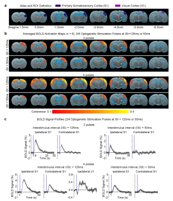 |
Neural activity temporal pattern dictates the long-range brain-wide propagation pathways: An optogenetic fMRI study

Alex T. L. Leong, Xunda Wang, Celia M. Dong, Russell W. Chan, Ed X. Wu
The current overarching challenge in neuroscience is to establish an integrated understanding of the brain networks, particularly the spatiotemporal patterns of neural activities that give rise to functions and behavior. fMRI provides the most versatile neuroimaging platform for mapping large-scale activities in vivo. However, on its own, many important details of the underlying network activities remain unresolved. Here, we employed fMRI in combination with pulsed optogenetic stimulation paradigms to probe the spatiotemporal dynamics and functions of the well-defined topographically-organized somatosensory thalamo-cortical network. We reveal unique long-range propagation pathways that are dictated by distinct neural activity temporal patterns initiated from the thalamus.
|
13:57
 |
0203.
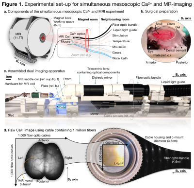 |
Spanning spatiotemporal scales with simultaneous mesoscopic calcium imaging and functional MRI
Evelyn Lake, Xinxin Ge, Xilin Shen, Peter Herman, Fahmeed Hyder, Jessica Cardin, Michael Higley, Dustin Scheinost, Xenophon Papademetris, Michael Crair, R Todd Constable
Neuroscience interrogates brain function across multiple spatiotemporal scales. Yet, most research is confined to one spatiotemporal milieu limiting translation of knowledge across scales. Here we span spatiotemporal scales having built a custom apparatus and analytical framework for simultaneous wide field mesoscopic Ca2+ imaging of the entire cortex and fMRI at 11.7T in mice. We describe the new hardware/software, and present three findings: there is correspondence between spontaneous fluctuations in the magnitude of Ca2+ and fMRI evoked responses, Ca2+ and fMRI connectivity metrics are stable throughout an imaging session, and there is correspondence between Ca2+ and fMRI spontaneous activity patterns.
|
14:09
|
0204.
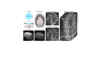 |
Multimodal awake mouse imaging: from two-photon microscopy to BOLD-fMRI
Michèle Desjardins, Kilic¸ Kivilcim, Martin Thunemann, Céline Mateo, Dominic Holland, Christopher Ferri, Jonathan Cremonesi, Boaqiang Li, Qun Cheng, Kimberly Weldy, Payam Saisan, David Kleinfeld, Takaki Komiyama, Thomas Liu, Robert Bussell, Eric Wong, Miriam Scadeng, Andrew Dunn, David Boas, Sava Sakadžic, Joseph Mandeville, Richard Buxton, Anders Dale, Anna Devor
Functional Magnetic Resonance Imaging (fMRI) in awake behaving mice is well positioned to bridge the detailed cellular-level view of brain activity, which has become available due to recent advances in microscopic optical imaging and genetics, to the macroscopic scale of human noninvasive observables. Here, we demonstrate Blood Oxygen Level Dependent (BOLD) fMRI in awake mice implanted with chronic transparent cranial ''windows'', compatible with two-photon microscopy, optical imaging, and optogenetic light stimulation. We thus provide a proof of feasibility for multimodal imaging approaches in awake mice, which in the future can be extended to behavioral studies and biomedical applications.
|
14:21
|
0205.
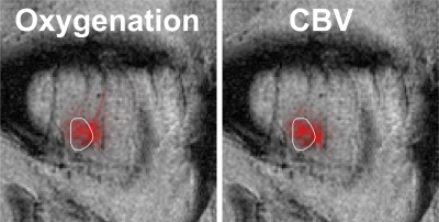 |
Ultra high field BOLD measurements combined with simultaneous determination of blood oxygenation and blood volume by optical imaging
Rebekka Bernard, Klaus Scheffler, Rolf Pohmann
To disentangle the different parameters contributing to the BOLD effect, a combined setup for intrinsic optical imaging and ultra high field fMRI in rats was designed, using a magnetic field compatible, high sensitivity camera and professional optical components. By illumination of the brain surface with light in four different wavelengths, oxygenation and CBV was observed concurrently with fMRI during forepaw stimulation. Simultaneous measurement of those parameters can help to better understand the BOLD effect and to add additional value to both optical imaging and fMRI experiments.
|
14:33
|
0206.
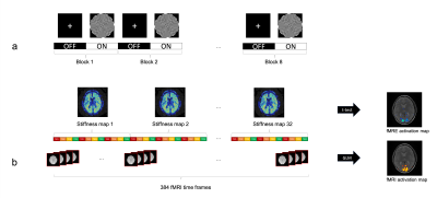 |
Simultaneous fMRE and fMRI measures the viscoelastic and BOLD responses of the human brain to functional activation in the visual cortex
Patricia Lan, Kevin Glaser, Richard Ehman, Gary Glover
Here, we demonstrate a novel multi-modal method to simultaneously acquire robust fMRE and fMRI activation maps. A block paradigm of 24s ON (flashing checkerboard at 10Hz) and 24s OFF (fixation cross) was used and images were acquired with a single-shot spin-echo EPI MRE sequence. Our results show that tissue stiffness within the visual cortex increases 6-12% with visual stimuli. Furthermore, the fMRE and fMRI activation maps agree and overlap spatially within the visual cortex, providing convincing evidence that fMRE is possible in the cortex.
|
14:45
|
0207.
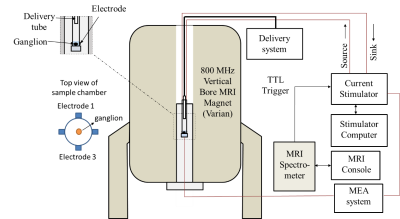 |
Functional Magnetic Resonance Electrical Impedance Tomography of Aplysia abdominal ganglion
Fanrui Fu, Munish Chauhan, Rosalind Sadleir
Functional Magnetic resonance electrical impedance tomography (fMREIT) has the potential to directly image neural activity. In our tests, we used the Aplysia abdominal ganglion (AAG) as a neuronal activity source. Potassium chloride (KCl) was used to modulate neuronal activity. Analysis of magnitude images, subtracted MREIT phase images and Laplacian of MREIT Bz images was performed to evaluate the difference in MREIT images with and without neuronal activity.
|
14:57
|
0208.
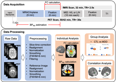 |
Baseline Striatal Dopamine Binding Potential Predicts Functional Connectivity to Ventral Tegmental Area in Control but not in MDD: A Simultaneous [11C] Raclopride PET-fMRI Study
Xue Zhang, Fuyixue Wang, J. Paul Hamilton, Jingyuan Chen, Ian Gotlib, Mehdi Khalighi, Gary Glover
Our previous work has indicated a significant connection between dopamine release/binding and fMRI activation during reward processing in healthy controls (CTL), but not in major depressive disorder (MDD). It motivates us to explore whether there is a similar disrupting effect in the coupling of resting-state fMRI and baseline dopamine binding potential in MDD. By conducting a simultaneous [11C] Raclopride PET and fMRI study, we obtained significant correlations between striatal dopamine binding potential and VTA-striatum functional connectivity in CTL, but not in MDD, indicating that the decoupling of dopaminergic system and striatum may play a vital role in the pathophysiology of MDD.
|
15:09
|
0209.
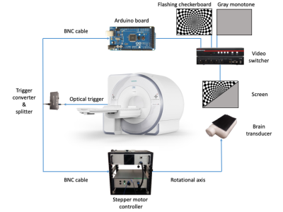 |
Spatial-temporal dynamics of the visual cortex stiffness driven by a flashing checkerboard stimulus
Jose de Arcos, Daniel Fovargue, Radhouene Neji, Sam Patz, Ralph Sinkus
In this work we explore the relationship between localized stiffness changes in the visual cortex and the stimulus switching frequency using a novel functional MRE system (fMRE) capable of probing stiffness changes in the human brain driven by a 12Hz flashing checkerboard stimulus. We measured the mechanical response (fMRE) and BOLD response (fMRI) of the brain to slow (36.96s) switching speeds at timescale typical for classical fMRI experiments and compare the results to faster switching speeds (840ms) where the hemodynamic response (HDR) is entirely saturated, and hence cannot follow anymore the input signal. Combining this with our previous results we observe a characteristic behavior of stiffness changes: for switching speeds where the HDR is saturated the stiffness is higher during the OFF state, while we observe the opposite for switching speeds where the HDR can follow the stimulus. Both differed in baseline to control scans (OFF/OFF).
|
15:21
|
0210.
 |
Simultaneous Measurement of functional MRI and MRS by Fast Non-water Suppressed Keyhole MR Spectroscopy Imaging
Xin Shen, Pingyu Xia, Masoumeh ?Moghadam, Jamie Near, Xiaopeng Zhou, Mark Chiew, Ulrike Dydak, Uzay Emir
The non-water suppressed magnetic resonance spectroscopic imaging (MRSI) sequence with concentric k-space trajectory was proposed to measure functional MRI and MRSI signals simultaneously. A right-hand finger-tapping task was performed at 3T MRI scanner to test the simultaneous hemodynamic and neurochemical measurements at human primary motor cortex. The results showed a significant overlap between T2* and metabolite (glutamate) changes.
|
15:33
|
0211.
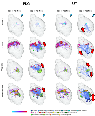 |
Imaging the Influence of Central Amygdala Neuronal Circuits on Nociception: a Combined Approach of Optogenetics and fMRI
Isabel Wank, Pinelopi Pliota, Silke Kreitz, Wulf Haubensak, Andreas Hess
Optogenetics has proven to be a highly useful tool to delineate the function of distinct proteins. Here, this approach was combined with fMRI to activate in-vivo selectively two interacting, but supposedly opposing, neuronal circuits of the central lateral amygdala (CEl). A classical fMRI paradigm was chosen to study the influence of the activation of either PKCδ- or somatostatin-expressing neurons on central pain processing, and to identify involved brain networks or areas. PKCδ was found to act preferentially anti-nociceptive by controlling via thalamus higher-order brain regions. Somatostatin on the other hand was shown to interact very closely with brainstem regions, controlling in a “bottom-up”-fashion thalamus, limbic system and cortex.
|
|
