MR-Guided Intervention
Oral
Interventional MRI
Wednesday, 15 May 2019
| Room 513D-F |
15:45 - 17:45 |
Moderators: Ali Özen, Ehud Schmidt |
15:45
|
0966.
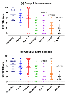 |
Comparing imaging changes and pain response in patients with intra- or extra-osseous bone metastases treated palliatively with magnetic resonance guided high intensity focused ultrasound (MRgHIFU)

Sharon Giles, Matthew Brown, Ian Rivens, Martin Deppe, Merel Huisman, Young-Sun Kim, Gail ter Haar, Nandita deSouza
Imaging changes and pain relief were compared in 21 patients with intra- versus extra-osseous bone metastases treated palliatively with magnetic resonance guided high intensity focused ultrasound (MRgHIFU). Thermal dose volumes measured on proton resonance frequency shift (PRFS) thermometry were significantly larger in the extra-osseous group. Intra-osseous lesions showed focal non-enhancement by Day 30, and patients had better pain response to MRgHIFU than those with extra-osseous lesions. By Day 30, 67% (6/9) patients with intra-osseous lesions were responders, compared with 33% (4/12) of patients with extra-osseous lesions. In neither group was pain response indicated by non-enhancement on Gd-T1W imaging.
|
15:57
|
0967.
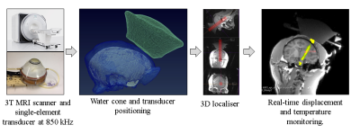 |
Real-time in-vivo tissue temperature and displacement measurements in the brain for MR-guided HIFU treatment and comparison to in-silico focused ultrasound simulation.
Valéry Ozenne, Charlotte Constans, Pierre Bour, Mathieu Santin, Harry Ahnine, Romain Valabrègue, Stephane Lehéricy , Jean-François Aubry, Bruno Quesson
MR-guided High Intensity Focus Ultrasound is an appealing technology in neurosurgery. For such application, accurate targeting and monitoring are crucial. A recent sequence allowing simultaneous measurements of temperature and displacement measurements is used to identify in real-time both the focal point by Acoustic Radiation Force Intensity (ARFI) and verify the absence of heating during ARFI sonication. The method has been evaluated in vivo in a non-human primate under anesthesia with a single-element transducer. A comparison with in-silico focused ultrasound simulation is also provided.
|
16:09
|
0968.
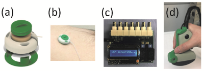 |
Combining MR and ultrasound imaging, through sensor-based probe tracking
Bruno Madore, Cheng-Chieh Cheng, Frank Preiswerk
Sensors attached to the skin can ‘spy’ on product ultrasound scanners, allowing the position and orientation of the ultrasound probe to be inferred. These MR-compatible sensors can emit and/or receive ultrasound waves, they are small in size (about 3x3x1cm) and are easily fixed to the torso. In transmit-receive mode they can monitor physiological motion, and in receive-only mode they can monitor the motion of an ultrasound probe. The sensors can accompany subjects to serial MRI and ultrasound imaging exams and act as a common denominator between the two, allowing ultrasound images to be placed in the spatial context of MRI.
|
16:21
|
0969.
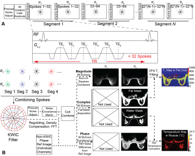 |
A Variable Flip Angle Golden-Angle-Ordered 3D Stack-of-Radial Sequence for Combined PRF/T1 Monitoring of MR-Guided HIFU Ablation
Le Zhang, Tess Armstrong, Xinzhou Li, Holden Wu
Proton resonance frequency (PRF) shift is widely used for MR temperature mapping, but fails in adipose tissues. T1 measurement offers an alternative temperature mapping method in adipose tissues. Combined PRF-T1temperature mapping has been evaluated for Cartesian MRI, but there is a lack of research for non-Cartesian techniques. In this work, we propose a new 3D stack-of-radial technique for combined PRF-T1 MR temperature mapping. Results from non-heating in vivo scans demonstrate good stability of T1 and PRF measurements. Data from ex vivo high-intensity focused ultrasound (HIFU) ablations demonstrate good agreement between both PRF and T1 mapping compared to temperature probes.
|
16:33
 |
0970.
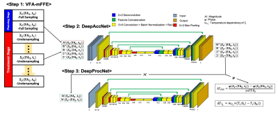 |
Real-Time T1/PRF-Based MR Thermometry Using Deep Learning and VFA-mFFE for Guidance of HIFU Treatment
Jong-Min Kim, You-Jin Jeong, Han-Jae Cheong, Jae-Won Yoo, Jeong-Hee Kim, Chulhyun Lee, Chang-Hyun Oh
MR temperature mapping of adipose and aqueous tissues is crucial in ensuring the safety and efficacy of HIFU treatment in regions of the body where the adipose and aqueous tissues are treated. We suggest a simultaneous and real-time temperature mapping method for adipose and aqueous tissues by using deep learning and multi-echo fast field echo with variable flip angle. Additionally, an image reconstruction method to obtain the temperature maps from the undersampled data obtained in the treatment stage is proposed by additionally using the high-resolution image obtained in the planning stage.
|
16:45
|
0971.
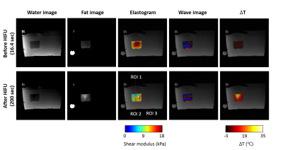 |
Simultaneous fat-referenced PRFS thermometry and MR Elastography for the monitoring of thermal ablation
Kisoo Kim, Elodie Breton, Afshin Gangi, Jonathan Vappou
Coupled MR Elastography (MRE) and Proton Resonance Frequency Shift (PRFS) thermometry have been proposed for the monitoring of thermal ablations. Fat-referenced PRFS thermometry based on water-fat separation allows correcting for errors caused by field drift. We propose a new approach for combined MRE and fat-referenced PRFS thermometry, allowing for improved thermometry while keeping the acquisition time unchanged. This is obtained by varying simultaneously TE and MRE phase offsets for water-fat separation and MRE, respectively. Feasibility of the method was demonstrated in a water/fat phantom during a High Intensity Focused Ultrasound ablation experiment, in which elasticity and temperature changes were monitored.
|
16:57
 |
0972.
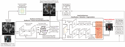 |
Real-Time Needle Detection and Segmentation using Mask R-CNN for MRI-Guided Interventions.
Xinzhou Li, Steven Raman, David Lu, Yu-Hsiu Lee, Tsu-Chin Tsao, Holden Wu
Real-time needle tracking for MRI-guided interventions is challenging due to variations in the needle features and the contrast between the needle and surrounding tissue. Mask region-based convolutional neural network (R-CNN) is a powerful deep-learning technique for object detection and segmentation in natural images, which has the potential to overcome these challenges. In this study, we train the Mask R-CNN model using annotated intra-procedural images from MRI-guided prostate biopsy cases and real-time images from MRI-guided needle insertion in a phantom. Mask R-CNN achieved accurate needle detection and segmentation in real time (~80 ms/image), which has the potential to improve MRI-guided interventions.
|
17:09
|
0973.
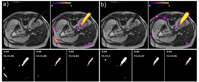 |
Deep Learning Based Needle Localization on Real-Time MR Images of Patients Acquired During MR-guided Percutaneous Interventions
Jonathan Weine, Elodie Breton, Julien Garnon, Afshin Gangi, Florian Maier
Automatic localization of needles in real-time images can facilitate MR-guided percutaneous interventions. It enables automatic slice repositioning and targeting support and, thus, allows for faster workflows. Systematic acquisition of training data for deep learning tasks in the context of interventional MRI can be difficult due to the fact, that treatment quality must not be impaired. Therefore, we investigated whether images of porcine animal experiments can be used to train deep learning algorithms for needle artifact segmentation in human datasets. Results show that transfer is feasible at 1.5T. Additional fine tuning using small amounts of human data further reduces the error.
|
17:21
|
0974.
 |
Real-time catheter tracking for cardiac MR-Thermometry during RF-ablation
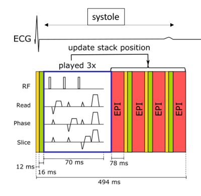
Pierre Bour , Valéry Ozenne, Marylène Delcey , Tom Lloyd, Rainer Schneider, Thomas Pohl, Wadie Ben-Hassen, Pierre Jais, Bruno Quesson
MR-thermometry in the heart has been developed over the last decade for RF-ablation monitoring to predict ablation outcomes. This study investigates a new approach to compensate respiratory motion. A catheter tracking module was integrated in a single-shot gradient echo echo-planar imaging sequence to dynamically position automatically the stack-of-slices on the catheter tip. As compared to the literature the proposed method uses local displacements at the vicinity of the catheter without prior knowledge relative to the displacement of the heart during respiration. In this study we have investigated the feasibility of this approach in vivo during RF-ablation in sheep.
|
17:33
|
0975
 |
Cervical-Cancer Imaging for brachytherapy planning employing an endo-vaginal array that includes an enhanced forward-looking coil
Video Permission Withheld
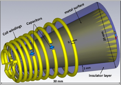
Akbar Alipour, Eric Meyer, Hassan Elahi, Sarah Fink, Henry Halperin, Akila Viswanathan, Ehud Schmidt
Advanced cervical-cancer spreads from the cervix to the posterior-endometrium and vaginal walls. Improving imaging SNR in this region shortens scan-times required for precisely localizing live-tumor locations before high-dose-rate (HDR) interstitial radiation-therapy. Current endo-vaginal coils focus on vaginal-wall imaging and are in-effective for posterior-endometrium/cervix imaging. This study designs and builds an endo-vaginal array which contains sideways-looking vaginal-wall, and forward-looking cervix/posterior-endometrium elements. The forward-looking “spiral”-coil is metallic-backed, pushing the RF-lobe upwards, enlarging its FOV. The array was designed with electromagnetic simulation and tested in phantoms and swine. Forward-looking SNR over a 3x3x3 cm3 FOV was ~4-8 times that of the spine-array.
|
|
