RF Arrays & the Elements Within
Oral
Engineering
Tuesday, 14 May 2019
| Room 710A |
08:15 - 10:15 |
Moderators: Boris Keil, Debra Rivera |
08:15
 |
0430.
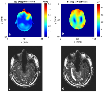 |
Toward Human Head Imaging at 10.5T Using an Eight-Channel Transmit/Receive Array of Bumped Fractionated Dipoles

Alireza Sadeghi-Tarakameh, Angel Torrado-Carvajal, Russell Lagore, Sean Moen, Xiaoping Wu, Gregor Adriany, Gregory Metzger, Lance DelaBarre, Kamil Ugurbil, Ergin Atalar, Yigitcan Eryaman
Ultra-high field (UHF)-magnetic resonance imaging (MRI) provides numerous benefits such as significant increase in signal-to-noise ratio (SNR), however, increasing the field strength, the local specific absorption rate (SAR) starts to become a limiting factor. Utilizing a TxArray coil with improved SAR performance may provide a good solution for this issue. In this study, we designed and built an eight-channel transmit/receive array of bumped fractionated dipoles for head imaging at 10.5T. We established a good agreement between simulations and experiments in order for RF safety validations. In addition, a cadaver head was imaged using pTx pulses to evaluate the imaging performance.
|
08:27
|
0431.
 |
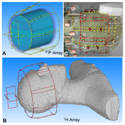
Design and implementation of a combined sodium-loop proton-dipole transceiver array for body imaging at 10.5 Tesla
M. Arcan Erturk, Russell Lagore, Edward Auerbach, Naoharu Kobayashi, Gregor Adriany, Kamil Ugurbil, Gregory Metzger
A multinuclear combined sodium-proton transceiver array consisting of 8 proton-dipoles and 8 sodium-loops for imaging the body at 10.5T was designed and implemented. Performance of the array was compared against 3 other designs using numerical simulations with the goal of achieving multinuclear imaging capabilities while minimizing the losses typically associated with such dual-tuned coils. The first 10.5T sodium images were obtained inside a saline-filled torso-size phantom with the implemented coil.
|
08:39
 |
0432.
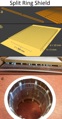 |
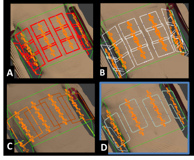
Split-Ring-Resonator shield improves SAR efficiency and homogeneity of birdcage antenna.
Carel van Leeuwen, Stanislav Glybovski, Peter Luijten, Cornelis van den Berg, Alexander Raaijmakers
A Split-Ring-Shield is a metamaterial structure that operates as a magnetic shield and thereby increases the penetration of the B1-field of a dipole antenna into a phantom. However, due to increased sample resistance it cannot be used effectively with a conventional birdcage. As a solution, a multi-transmit birdcage coil is presented, which can be used with the SRS. Simulations indicate a 10% increase in SAR-efficiency compared to a conventional birdcage. This setup was constructed and preliminary images were obtained.
|
08:51
|
0433.
 |
Double-Tuned 31P/1H Human Head Array with High Performance at Both Frequencies for Chemical Shift Spectroscopic Imaging (CSI) at 9.4T.
Nikolai Avdievich, Loreen Ruhm, Johanna Dorst, Anke Henning
SNR enhancement at ultra-high field (UHF, >7T) is very critical for X-nuclei imaging. X-nuclei CSI of a human brain is potentially valuable for diagnostics of many diseases. CSI benefits from a whole-brain coverage and a high transmit performance not only at X- but also at 1H-frequency. It is rather difficult to optimize the DT-array at both frequencies at the same time. Therefore, often the performance of the X-nuclei portion of the RF array coil is optimized while the 1H-array performance is compromised. In this work, we developed a novel DT-array, which provides good performance at both 31P and 1H frequencies.
|
09:03
|
0434.
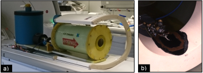 |
A Robust Cryogenic RF Coil (88K) for In-vivo Hyperpolarized 13C MRI of Rats
Juan Diego Sánchez Heredia, Rafael Baron, Esben S. Szocska Hansen, Daniel H. Johansen, Vitaliy Zhurbenko, Christoffer Laustsen, Jan H. Ardenkjær-Larsen
We report the performance of a cryogenic RF receive-only coil for 13C imaging of small animals. It is experimentally demonstrated 2-fold SNR improvement in comparison to a room temperature coil in immediate vicinity of the sample. The self-developed cryostat employed for coil cooling shows thermal stability within 5 h of use for 6 L of LN2, which can be extended up to 12 h if more LN2 is added. The Q88K/Q290K ratio of the unloaded coil is 550/285.
|
09:15
|
0435.
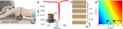 |
Dielectric resonator for targeted breast MRI at 3T
Alena Shchelokova, Anna Mikhailovskaya, Viacheslav Ivanov, Ivan Sushkov, Irina Melchakova, Elizaveta Nenasheva, Alexey Slobozhanyuk, Anna Andreychenko, Andrew Webb
A novel concept for targeted MRI that can be directly integrated into an existing clinical system is proposed and demonstrated for 3T breast imaging. A practical demonstration of the concept features a dielectric resonator, based on a composite material with a very high permittivity that is electromagnetically coupled to the birdcage body coil. In vivo breast imaging with the proposed resonator showed 70% higher signal-to-noise ratio and reduced motion artefacts, while the peak spatial SAR was reduced by a factor-of-eight on average, compared to standard birdcage body coil transmission and four-element array receive.
|
09:27
|
0436.
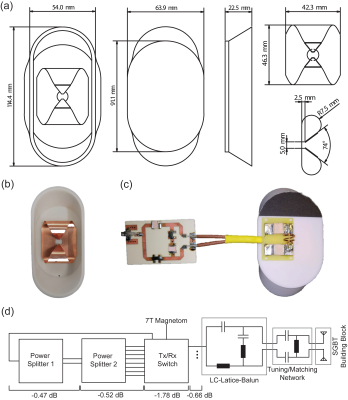 |
Compact Self Grounded Bow Tie Antenna Resonator for Cardiac MRI at 7.0 Tesla
Thomas Eigentler, Laura Boehmert, Andre Kuehne, Daniel Wenz, Eva Oberacker, Haopeng Han, Lukas Winter, Thoralf Niendorf
A compact dielectric resonator antenna array was developed for cardiovascular MRI at 7.0T MRI. The antenna building block is based on the concept of a self-grounded bow-tie (SGBT) antenna placed inside a resonator cavity filled with deuterium oxide. This approach ensures light-weight design and affords high-density RF arrays, which constitutes a major advantage over current state-of-the-art electric dipole configurations. The proposed high-density SGBT transceiver array provides ample parallel imaging and real time imaging capabilities. It contributes to the technological basis for the future clinical assessment of parallel transmit techniques designed for ultrahigh field cardiac MR.
|
09:39
|
0437.
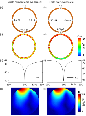 |
Over-overlapped Loop Arrays
Ming Lu, John Gore, Xinqiang Yan
Loops are usually overlapped by approximately 10% in area to minimize the mutual inductive coupling. Due to geometrical constraints, the size of each loop has to be reduced to increase the numbers of coils in an array. In this work, we propose a simple loop array design that achieves over-overlap as well as low reactive/resistive coupling. A 40% overlap was chosen in this study. Numerical results show that the proposed over-overlapped loop arrays could increase the performance of parallel imaging and SNR.
|
09:51
|
0438.
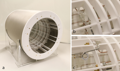 |
An eight-channel array coil for zero echo time imaging
Manuela Rösler, Markus Weiger, David Brunner, Romain Froidevaux, Thomas Schmid, Klaas Pruessmann
With dedicated short-T2 techniques such as zero echo time (ZTE) imaging not only rapidly decaying signal from the sample but also from the RF coil (plastic housing, glue, solder flux etc.) is detected. To enable experimental examination of parallel imaging opportunities, a 1H-reduced eight channel array was designed. ZTE images of short-T2* rubber samples and PETRA images of human head could be acquired.
|
10:03
|
0439.
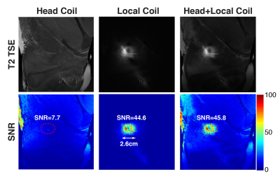 |
Development of a Local Pituitary Coil for Assessment of Pituitary Microadenomas
Jiahao Lin, Rock Hadley, Ling Li, Robb Merrill, Marvin Bergsneider, Robert Candler, KyungHyun Sung
Pituitary microadenomas are difficult to detect due to their small size, and the surgeon is sometimes forced to multiply slice to explore the pituitary gland – at considerable risk of damaging this essential master hormone gland. In this study, we design and evaluate a local pituitary coil that allows placing it in direct proximity to the pituitary gland. The local pituitary coil had increased signal-to-noise ratio by a factor of six, in comparison with a commercial 20-channel head/neck coil, and had a sufficient coil coverage for the pituitary microadenomas at around 1cm depth from the coil.
|
|
