|
2nd Hour
Poster: Frontiers of Neuro Techniques
Power Pitch Poster
Neuro
Monday, 13 May 2019
Power Pitch Theater B - Exhibition Hall
17:00 - 18:00
| |
|
Plasma # |
|
0237.
 |
16 |
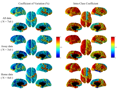
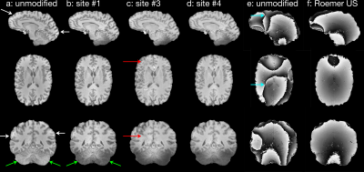
Robustness of PSIR segmentation and R1 mapping at 7T: a travelling head study

Olivier Mougin, William Clarke, Ian Driver, Catarina Rua, Andrew Morgan, Susan Francis, Keith Muir, Adrian Carpenter, Chris Rodgers, Richard Wise, David Porter, Stuart Clare, Richard Bowtell
Ultra-high magnetic field (7T) MRI scanners can provide high spatial resolution images and excellent contrast for classifying brain tissue, but robustness of tissue segmentation and R1 quantification across sites is key for the implementation of multi-site studies. Here, we present a subset of the main UK7T travelling-head study, focusing on harmonized T1-weighted images (0.7mm3 isotropic resolution) acquired on six subjects across three 7T sites, with five repeats at one site. The aim of this study is to assess the harmonisation and robustness of the MP2RAGE sequence and PSIR reconstruction across sites, by focusing on segmentation reproducibility and T1 estimation.
|
|
0238.
 |
17 |
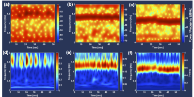
Separation of Cardiac- and Respiratory-driven CSF Motions under Free Breathing based on Realtime Phase Contrast Imaging and S-Transform
Kagayaki Kuroda, Tetsuya Tokushima, Satoshi Yatsushiro, Nao Kajiwara, Tomohiko Horie, Hideki Atsumi, Mitsumori Matsumae
To separately visualize respiratory- and cardiac-driven motions of intracranial cerebrospinal fluid (CSF) under free breathing, CSF velocity distribution in 6 healthy volunteers and 3 hydrocephalus patients were acquired with asynchronous real time phase contrast (PC). Spectrograms of CSF velocity waveform as well as ECG and respiratory signals were obtained by Stockwell Transform (ST), in which the length of a Gaussian window length was adaptively changed according to the time-varying frequency of the signals. Comparison with the conventional short-term Fourier transform (STFT) with fixed length window revealed that separation of respiratory and cardiac components of CSF motion was possible with ST.
|
 |
0239.
 |
18 |
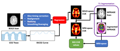
CVR-MRICloud: an automated online tool for the processing of cerebrovascular reactivity (CVR) MRI data
Zachary Baker, Yang Li, Peiying Liu, Yue Li, Michael Miller, Susumu Mori, Hanzhang Lu
Cerebrovascular reactivity (CVR) has recently become a focus for many labs. However, CVR calculation has always required at least some degree of manual intervention. Therefore, our lab has developed CVR-MRICloud, a free, online, fully automated CVR processing pipeline. Our pipeline returns CVR maps, relative bolus arrival time maps, and region-of-interest CVR values. The maps are given in their original space as well as standardized MNI space. The pipeline has been shown to procure results corresponding to accepted CVR processing techniques that rely on manual intervention. This pipeline has potential to streamline other researchers’ acquisition of CVR values in subjects.
|
|
0240
 |
19 |
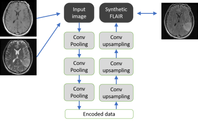
Comparing neural networks for synthesizing FLAIR images from T1WI and T2WI
Video Permission Withheld
Takashi Abe, Yuki Matsumoto, Yuki Kanazawa, Yoichi Otomi, Maki Otomo, Moriaki Yamanaka, Mihoko Kondo, Saya Matsuzaki, Ariunbold Gankhuyag, Enkhamgalan Dolgorsuren , Oyundari Gonchigsuren, Masafumi Harada
- We checked the performances of different convolutional encoder decoder (CED), one of the neural networks when synthesizing FLAIR using T1WI and T2WI. With the shallow CED, the resolution was good but the contrast was poor, and when the CED became deeper, contrast became better, but the resolution became worse. Next we also added “skip-connection” to CED, but the image quality was not improved with the Inception(GoogLeNet)-like parallel skip-connection, and the image quality improved with the ResNet-like serial skip-connection; that was a mixture of shallow and deep CED, and resembled the structure of U-net.
|
|
0241.
 |
20 |
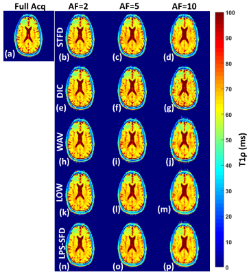
Performance comparison of compressed sensing algorithms for accelerating T1? mapping of Human Brain
Rajiv Menon, Marcelo Zibetti, Ravinder Regatte
3D-T1ρ mapping sequences are useful MRI methods in various neuropathologies but its data acquisition requires long scan times. We compared the performance of 5 compressive sensing (CS) algorithms with acceleration factors (AF) up to 10. We evaluated image quality and T1ρ estimation errors as a function of AF. Six healthy volunteers were recruited and they underwent T1ρ imaging of the whole brain with full Cartesian acquisition. Assessment of image reconstruction and T1ρ estimation errors in this study show that the CS method using spatial and temporal finite differences as a regularization function performs the best for accelerating T1ρ quantification in the brain.
|
|
0242.
 |
21 |
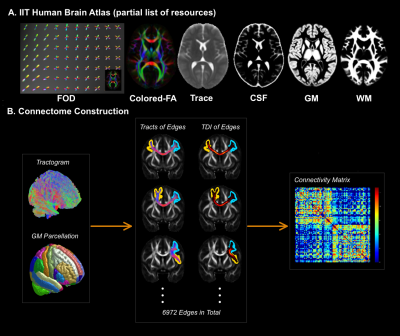
Enhancement and Evaluation of the White Matter Connectome of the IIT Human Brain Atlas
Xiaoxiao Qi, Shengwei Zhang, Konstantinos Arfanakis
In the IIT Human Brain Atlas project we have developed anatomical as well as state-of-the-art diffusion tensor and high angular resolution diffusion imaging (HARDI) templates, probabilistic gray matter labels, and probabilistic connectivity-based white matter labels for the young adult brain. The purpose of this work was two-fold: a) to enhance the white matter connectome of the IIT Human Brain Atlas through an improved tractography strategy, appropriate filtering of streamlines, and use of more precisely defined gray matter labels, and b) to evaluate how representative the new connectome is of young adult participants of the Human Connectome Project.
|
|
0243.
 |
22 |
Single-shot Diffusion-weighted Spiral Imaging in the Brain on a Clinical Scanner
Peter Börnert, Holger Eggers, Kay Nehrke, Peter Koken, Jan Groen, Suthambhara Nagaraj, Johan van den Brink, Silke Hey
Single-shot diffusion-weighted imaging is predominantly performed with echo planar imaging today. Spiral imaging allows shorter echo times and thus promises higher signal-to-noise ratio, but is sensitive to various system imperfections. While previous work resorted to using a field camera for this reason, this work demonstrates the feasibility of single-shot diffusion-weighted spiral imaging in the brain on a clinical scanner without extra hardware for field monitoring. Good image quality was generally achieved in volunteers for different diffusion gradient directions up to high b-values using the demand trajectory for gridding, parallel imaging for acceleration, and static main field inhomogeneity mapping for deblurring.
|
|
0244.
 |
23 |
The developing Human Connectome Project (dHCP): fetal acquisition protocol
Anthony Price, Lucilio Cordero-Grande, Emer Hughes, Suzanne Hiscocks, Elaine Green, Laura McCabe, Jana Hutter, Giulio Ferrazzi, Maria Deprez, Thomas Roberts, Daan Christiaens, Eugene Duff, Vyacheslav Karolis, Shaihan Malik, Mary Rutherford, David Edwards, Joseph Hajnal
The developing Human Connectome Project seeks to map connectivity in the human brain based on structural, functional and diffusion MRI data acquired from over 1000 subjects (neonatal and fetal). A dedicated acquisition protocol has been developed to efficiently image fetal brain in utero. We describe here the methods and parameters being used alongside initial pre-processing steps. The acquisition protocol has been tuned to complement the neonatal data already collected while adapting to the difficult challenges of imaging the fetal brain in utero. It has been deployed to image over 145 fetuses to date with a success rate of ~90%.
|
|
0245.
 |
24 |
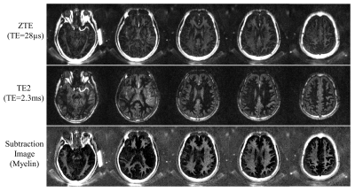
Inversion Recovery Zero Echo Time (IR-ZTE) Imaging for Direct Myelin Detection in Human Brain
Hyungseok Jang, Michael Carl, Yajun Ma, Yanjun Chen, Saeed Jerban, Eric Chang, Jiang Du
In MRI, direct myelin imaging is challenging due to the short T2* decay (less than 0.5ms) and very low proton density. In the literature, it has been reported that ultrashort echo time (UTE) imaging can directly capture the fast decaying myelin signal. To further enhance the dynamic range, adiabatic inversion recovery (IR) preparation can be utilized so that the white matter signal can be suppressed. Moreover, dual echo UTE imaging scheme can suppress the remaining gray matter signal. In this study, we explore the feasibility of IR prepared zero echo time (IR-ZTE) imaging for direct myelin imaging in the human brain.
|
|
0246.
 |
25 |
Planar rosette spectroscopic imaging at 7T
Jullie Pan, Chan Moon, Victor Yushmanov, Claud Schirda, Frank Lieberman, Hoby Hetherington
To make spectroscopic imaging clinically feasible, rapid and robust acquisitions with high SNR are necessary. We develop and apply rosette spectroscopic imaging at 7T using a 8x2 transceiver array and high degree B0 shimming to acquire rapid (<3min) whole plane brain studies at 0.7cc to 0.3cc resolution. To achieve high spectral bandwidth with moderate gradient demands, two temporal interleaves are used. We demonstrate the performance of this acquisition in controls and tumor patients, with use of regression statistics for determination of abnormality.
|
|
0247.
 |
26 |
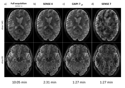
Whole brain sub-mm resolution T2* weighted anatomy imaging in less than 2 minutes
Arjan Hendriks, Federico D’Agata, Tim Schakel, Liesbeth Geerts, Dennis Klomp, Natalia Petridou
T2* weighted imaging can be used to study both normal and pathological tissue. These images are commonly obtained using traditional gradient echo sequences which can lead to long scan times that are problematic particularly in a clinical setting. 3D EPI offers a faster alternative with scan times on the order of few minutes. Here, the scan time of T2* weighted 3D EPI scans is further reduced with a shot selective 2D CAIPI acquisition pattern. Whole brain T2* weighted anatomical scans with a resolution of 0.5 mm isotropic were acquired in 1:27 minutes. This holds promising perspectives for future applications in routine examinations.
|
 |
0248.
 |
27 |
Reduced Regional Cerebral Venous Oxygen Saturation is a Risk Factor for Cognitive Impairment in Hemodialysis Patients: A Susceptibility-weighted Image Mapping Study
Chao Chai, Huiying Wang, Jinping Li, Jinxia Zhu, Xianchang Zhang, E. Mark Haacke, Shuang Xia, Wen Shen
The purpose of this study was to noninvasively evaluate the changes of regional cerebral venous oxygen saturation (rSvO2) in hemodialysis patients using quantitative susceptibility-weighted image mapping (SWIM) and then to investigate the relationship between rSvO2, clinical risk factors, and neuropsychological testing results. The results suggest that cerebral rSvO2 is reduced in hemodialysis patients and that this reduction may correlate with neurocognitive dysfunction. Hematocrit, iron, glucose, and pre- and post-dialysis DBP were independent risk factors for reduced cerebral rSvO2.
|
|
0249.
 |
28 |
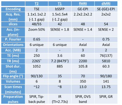
The UK7T Network’s Harmonized Neuroimaging Protocols
William Clarke, Olivier Mougin, Ian Driver, Catarina Rua, Andrew Morgan, Stuart Clare, Susan Francis, Richard Wise, Adrian Carpenter, Christopher Rodgers, Keith Muir, Richard Bowtell
The UK7T Network is a consortium of 7-tesla-MRI capable sites in the United Kingdom, operating with three different scanner models, manufactured by two MR vendors. The Network has established a harmonized set of anatomical and functional MRI protocols for standardized neuroimaging across currently available human 7T scanners.
Here we make these harmonized protocols available to the community, along with example data, and describe the need for manual calibration to achieve harmonization across sites.
|
|
0250.
 |
29 |
High-resolution MR imaging of human brain with multi-echo integrated SSFP
Huilou Liang, Kaibao Sun, Zhentao Zuo, Jing An, Yan Zhuo, Danny J.J. Wang, Rong Xue
Balanced SSFP (bSSFP) has been used in structural and functional MRI, but always suffered from banding artifacts. While phase cycling was widely used to reduce banding artifact, it would take more scan time. Recently, integrated SSFP (iSSFP) was introduced to acquire banding-free images in shorter scan time than phased-cycled bSSFP. In this work, multi-echo iSSFP was further developed to improve SNR and acquire ultrahigh-resolution images with moderate scan time at 7T. Phantom and in vivo experiments demonstrated that the combined image produced by weighted averaging of multi-echo iSSFP showed obvious SNR and contrast improvement and inherited the characteristics of bSSFP.
|
|
0251.
 |
30 |
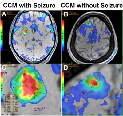
Quantitative Susceptibility Mapping: In Vivo Biomarkers for Cerebral Cavernous Malformations Related Epilepsy
Li Ma, Chunxue Wu, Shuo Zhang, Zongze Li, Lizhi Xie, Xiaolin Chen, Xun Ye, Hao Wang, Yuanli Zhao, Shuo Wang, Jizong Zhao
Hemosiderin deposits surrounding the cerebral cavernous malformations (CCMs) had been proposed to be associated with the pathogenesis of CCM-related epilepsy (CRE). An increased perilesional and extralesional iron deposition were found in CCMs with epilepsy through susceptibility maps of quantitative susceptibility mapping (QSM). To investigate novel biomarkers for the in vivo and longitudinal evaluation of CCM lesions with epilepsy, this study was to explore the iron quantity in CCMs patients with CRE using quantitative susceptibility mapping.
|
|
